1.General information
Erysipelas, erysipelas - these and similar terms are used mainly in Slavic languages; in medical Latin and most Western European languages, the original ancient Greek name erysipelas was fixed, i.e. "red skin" In the old days there were also synonyms “holy fire”, “Saint Anthony’s fire” or, in Russian, “Antonov’s fire”, although some dictionaries define this term as one of the designations for gangrene.
Erysipelas is an acute skin infection (or recurrent exacerbation of a chronic one), the hallmarks of which are swelling and erythema - a bright red, “fiery” hue of the affected area. In general, erysipelas is one of the most common human infectious diseases; the localization of inflammation can be very different, however, due to the peculiarities of the anatomical structure, the disease often manifests itself on the skin of the face, primarily in the soft tissues of the external nose (vestibule, wings, nasal walls): a high concentration of sebaceous glands, capillaries and nerve endings facilitates the penetration and activation of the pathogen .
A must read! Help with treatment and hospitalization!
We care about your health
Epithelial tumors
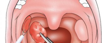
Papillomas. There are papillomas of the vestibule and the nasal cavity. The first ones are gray in color, dense, villous surface, small in size, not malignant. It is believed that papillomas are caused by viruses from the family of papillomoviruses types 6, 11 and 16. Papillomas of the nasal cavity primarily affect the mucous membrane. They often recur after surgical removal. With anterior rhinoscopy or endoscopy, papillomas are gray-white in color, soft in consistency, single or multiple, bleed when touched, and resemble cauliflower. Treatment of nasal papillomas is predominantly surgical or cryodestruction. Surgically removed material should be sent for histological examination due to the possibility of malignant degeneration (transitional cell carcinoma). Another problem in the treatment of these patients is the frequent recurrence of papillomas. To prevent it, it is recommended to prescribe interferon drugs - domestic Laferon or foreign intron A. The method of treatment with Laferon consists of inhalation administration at the beginning of therapy (1 million Laferon is diluted in 20 ml of 0.9% NaCl solution). Inhalations continue for the first 10 days, then Laferon is prescribed intramuscularly once a day, at a dose of 1 million units, for 20 days. Some clinics determine the titers of antibodies to it to monitor the presence of the virus in the blood serum. When the levels increase above 1:200 in the blood of patients, antiviral drugs (Zoverax or others) are prescribed. Papilloma viruses also infect the cervix, which leads, in less pathogenic types, to the occurrence of condylomas.
Inverted papilloma. Called due to the ability of squamous epithelium to invaginate in the form of a wide ribbon into the connective tissue, these tumors have destructive growth, recur and become malignant. They are also known as columnar cell papillomas.
Adenoma is a benign tumor of the glandular epithelium of the nasal mucosa. It mainly affects the lower parts of the nasal cavity on a broad base with a smooth surface. Characterized by slow growth. The tumor is erysipelas-gray in color, no more than 2 cm in size, and is accompanied by rapid growth, germination into adjacent tissues, and changes in the histological structure. Its treatment is surgical. The removed material should be examined histologically.
Nonepithelial tumors
Hemangioma is a tumor developing from blood vessels, similar in nature to developmental defects. What it has in common with other tumors is rapid growth, during which hemangioma destroys surrounding tissue and causes a cosmetic and sometimes functional defect. In 1863, Vikhrov published a macro-microscopic classification of hemangiomas, dividing them into capillary, cavernous and racemose (branched). Subsequently, this principle was included in many classifications.
Today, this classification is used, which includes additional data on tumor growth, its spread, blood supply characteristics and relationship with large blood vessels.
True hemangiomas: 1) capillary (simple): exophytic (venous and arterial type); 2) cavernous: mucous-submucosal; diffuse (spreading into depth); 3) branched (angiodysplasia); 4) mixed (angiofibromas, hemlymphangiomas, branched with cavernous, branched with Barre-Mason glomusangiomas, etc.).
Diagnosis of tumors in the nose
- Routine external examination of the patient. Palpation of the tumor.
- Anterior and posterior rhinoscopy.
- Examination of the nasal cavity and paranasal sinuses using an endoscope. Diaphanoscopy.
- Comprehensive X-ray examination.
- CT and MRI.
- Cytological examination of discharge obtained during puncture, sinus lavage, as well as nasal discharge.
- Histological examination of pieces of the neoplasm.
Treatment of nasal tumors
- Surgical removal of the tumor (if it is of significant size with preliminary embolization of the vessels feeding the tumor).
- Cryodestruction of the tumor.
- Sclerosis therapy.
- Radiation therapy in combination with surgical removal (for tumor malignancy).
Bleeding polyps of the cartilaginous part of the nasal septum are red, round in shape with a smooth surface. They are characterized by frequent bleeding. Treatment is surgical.
Fibromas, myomas, lipomas are rarely found in the cavity and paranasal sinuses. Angiofibroma, fibromyoma, osteofibroma, adenofibroma, neurofibroma, histiocytoma may occur. The treatment of these tumors is surgical.
Osteomas. They are most common among benign tumors of the paranasal sinuses, in people 20-40 years old. According to some authors, osteomas are localized in the area of bone sutures in 50% of patients. Histologically, three forms of osteomas are distinguished: compact (ivory), spongy and compact-spongy. Osteomas are extremely slow growing and predominantly asymptomatic. They are often discovered incidentally during X-ray or CT scanning. With large tumors or localization in the main sinus, a headache may be observed; with osteomas of the frontal sinus, exophthalmos, diplopia, and visual impairment may be observed.
Treatment of osteomas is exclusively surgical. The sooner it is carried out, the better success will be achieved. Mandatory histological examination of the removed tumor. Sometimes, with compact osteomas of the main sinus, when there is a risk of serious complications (damage to the cavernous sinus, internal carotid artery), surgical intervention should be carried out jointly with neurosurgeons.
Chondromas. They develop from the remains of the primordial cartilaginous skeleton and can be attributed to developmental anomalies. Tumors are characterized by slow expansive growth with penetration into the orbit or cranial cavity. They often recur and, in addition, can become malignant. Osteochondromas can occur in the nasal cavity. Neurogenic tumors
Paragangliomas (glomus tumors, chemodectomas) grow from small cell groups of the nasal mucosa. Rarely found in the nasal cavity. The tumor has infiltrative growth, often recurs, and malignantly degenerates. In such cases, it often grows into the skull and brain.
Tumor-like lesions
Tumor-like diseases are described in the relevant sections of the reference book, but fibrous dysplasia deserves special attention.
Fibrous dysplasia. Refers to tumor-like lesions of the nose and paranasal sinuses and is a defect in the formation of osteogenic mesenchyme. Characterized by the replacement of bone with fibrous tissue. Some authors consider the disease to be a developmental anomaly of unknown origin. Diagnosis of the disease is radiological but mainly histological confirmation is the main motive for choosing treatment. This lesion is also known as fibrous osteodystrophy, osteitis fibrosa. The bones of the upper jaw, main and frontal bones are predominantly degenerated. Treatment is surgical. The affected bone areas are removed. Relapses are possible. Sometimes such patients are treated in dental hospitals. V.
Malignant tumors of the nose and paranasal sinuses
The incidence of malignant tumors is steadily increasing by 2.6-3.0% per year. Malignant tumors of the nose and paranasal sinuses account for about 0.5% of cancer incidence statistics. In 2002, per 100 thousand population of Ukraine, the incidence of malignant tumors of this localization was 0.59, 287 patients were diagnosed for the first time. For comparison, in Belarus in the same year it was 0.7, 70 patients were identified. In Russia, 832 patients were detected for the first time, the incidence was 0.6. In the United States, 2,000 patients with cancer of the nasal cavity and paranasal sinuses are diagnosed annually.
Localization of tumors in the nose and paranasal sinuses, which are a system of narrow thin-walled cavities rich in nerves, blood and lymphatic vessels, promotes growth and rapid spread to adjacent formations, which significantly complicates treatment and leads in many cases to death.
Among the factors that cause malignant growth in this area are wood dust, petroleum products, professions associated with exposure to potent compounds - benzenes, acids, alkalis, nickel ores, varnishes and smoking. Among the factors that contribute to the growth of these tumors, chronic diseases are also noted - rhinitis, polypous sinusitis, ethmoiditis, frontal sinusitis, viral agents. The Epstein-Bar virus is considered the etiological factor of malignant lymphomas, papillomovirus types 11, 16, leads to the occurrence of transitional cell carcinoma and papillomas in the area of the nasal epithelium.
In most cases, the disease affects people of working age. Early clinical manifestations in people with tumors of this localization are insignificant and therefore the disease is more often detected in stages III – IV, when the walls of the cavities are already destroyed and penetration into adjacent anatomical areas begins.
According to most authors, malignant tumors are most often localized in the maxillary sinus (75-80%), in second place are the cells of the ethmoidal labyrinth and the nasal cavity (10-15%), less often the sphenoid and frontal sinuses are affected (1-2%).
When treating people with malignant tumors of the nose and paranasal sinuses, surgical, radiation and chemotherapy methods are used, but the results remain unsatisfactory, while the number of patients with this pathology does not decrease.
It should be noted that the palette of morphological forms of malignant tumors of the nose and paranasal sinuses is diverse.
Basal cell carcinoma occurs more often in various areas of the skin of the nose. Trichoepithelioma also belongs to basal cell malignant neoplasms.
Melanoma is observed in 2-3% of malignant tumors of this location. Most often it is located in the nasal cavity. The tumor is characterized by polymorphism. However, the sarcomatous form predominates. Early symptoms include bleeding and difficulty breathing. Melanoma metastasizes in 60% of cases to the cervical lymph nodes.
Clinical picture of tumors in the nose
Clinical manifestations of malignant tumors depend on the stage of the disease, localization, growth form, as well as the morphological structure of the tumor.
Symptoms:
- Pain. They appear sharply when localized in the upper jaw along the n. trigeminus. When the tumor is localized or grows into the area of the pterygopalatine fossa, shooting pain appears in the area of the eyeball or temple, which indicates irritation of the n.auriculotemporalis. Pain as the first sign of the disease was observed in 17% of patients.
- Impaired nasal breathing. When the tumor was localized in the nose, similar complaints were noted in 62.8% of patients. Patients whose tumor did not spread beyond the paranasal sinuses, or those whose tumor grew towards the cheek, temporal bone, alveolar process, or orbit did not complain of nasal breathing disorder.
- Nasal discharge. More often they are unilateral mucopurulent with an admixture of blood.
- Headache of various types, often with various paresthesias in the facial area on the side of the tumor. Often in such cases neuralgia is diagnosed. If they are not treatable, you should always remember about the possible development of a malignant disease.
- Pain in the teeth, deformation of the face, nose, lacrimation, exophthalmos, deformation of the hard and soft palate - these symptoms can occur to varying degrees with malignant tumors of the nose and paranasal sinuses.
Malignant tumors of the mucous membrane of the maxillary sinus occur without symptoms for a long time. Clinical manifestations depend on the starting point and size of the tumor. Tumors arise on all walls of the sinus, from the superomedial part of the sinus they quickly cause bleeding, swelling of the eyelids, lacrimation, exophthalmos; when they grow forward, they grow into the anterior wall and then in the cheek area they can be identified by palpation. As the tumor grows downward, it grows into the hard palate and changes its configuration. The growth of a tumor from the medial wall into the nasal cavity makes breathing difficult and leads to the formation of polyps. If the tumor grows predominantly in the direction of the posterior wall with germination into the fossa, the nasal part of the pharynx, then it causes pain.
When a tumor destroys the orbital wall, the process spreads to the orbital tissue, which is manifested by exophthalmos, decreased visual acuity and diplopia. Malignant tumors of the ethmoid labyrinth. Isolated lesions of the labyrinth cells are observed in 10% of cases.
When the tumor spreads towards the orbit, the thin wall of the latter is destroyed and tumor growth into the orbit is observed.
From the cells of the labyrinth, neoplasms can also spread to the maxillary, sphenoid sinuses, anterior and middle cranial fossa, and nasal pharynx.
Tumors of the sphenoid (main) sinus. Primary cancer of this location is extremely rare. Typically, these are tumors that extend from the maxillary sinus or ethmoid. The earliest symptoms are ocular, often damage to the efferent nerve, less often - decreased vision. Patients seek help from an ophthalmologist or neurologist. Characteristic orbital-superior syndrome (damage to the vessels and nerves that pass through the optic foramen and the superior orbital fissure), visual impairment, up to blindness, ptosis, diplopia, pain in the occipitotemporal and occipital regions.
Tumors of the frontal sinus are rare and have symptoms when affected, which do not extend beyond the sinus, similar to the symptoms of chronic frontal sinusitis. In recent years, CT and MRI have made it possible to differentiate this disease from mucocele, osteomyelitis and sinusitis. Symptoms depend on the direction of tumor growth. Germination downwards and medially leads to penetration into the orbit, ethmoidal labyrinth, exophthalmos appears, germination posteriorly - the anterior cranial fossa is affected, then a sharp headache, convulsions, and mental disorders are possible. Penetration of the tumor through the anterior wall leads to swelling and discoloration of the skin in the frontal area of the head.
Overview video of ENT surgery in Moscow, Kurkino and Khimki:
2. Reasons
Erysipelas is a bacterial infection, the causative agent of which is almost always group A beta-hemolytic streptococci. This tendency, however, is not absolute: a similar inflammation can be caused by staphylococcus, especially in cases of a long-term chronically recurrent process, as well as other pathogens. In general, we have to take into account the fact that in recent decades in the clinic of infectious diseases, incl. skin, there is a steady tendency towards an increase in combined invasions (for example, bacterial and fungal), developing according to the mechanism of super- or coinfection.
The main risk factor is microtraumatization of the skin of the nose in combination with weakened local immunity and non-compliance with sanitary and hygienic rules.
The risk increases in the presence of foci of chronic coccal infection in adjacent tissues and organs (otitis, tonsillitis, sinusitis, and many others). One of the widespread causes of erysipelas is the practice of self-squeezing out elements of acne and/or pustular (pustular) rash in unsterile or frankly unsanitary conditions. In some cases, nasal erysipelas is iatrogenic in nature, i.e. the launch of the pathological process is caused by medical procedures or surgical intervention on the ENT organs.
Visit our Otolaryngology (ENT) page
Causes
The cause of the appearance of benign formations may be genetics, developmental anomalies due to the effects of pathological factors on the fetus, and inflammatory diseases of the paranasal sinuses. In many cases, the reasons for their appearance are completely unknown.
For malignant tumors, risk factors are smoking, papillomavirus, prolonged inhalation of chemicals used in furniture, shoe, and leather production, inhalation of flour dust, wood dust. The appearance of malignant formations can be caused by long-term untreated teeth, injuries from incorrectly selected dentures, inhalation of smoke, steam, and welding work. Inhalation of asbestos particles is especially dangerous.
Symptoms
Both benign and malignant growths in the nose and paranasal area are asymptomatic at first. Usually they do not cause concern at the initial stage and are detected by chance on an x-ray. A tumor of significant size begins to manifest itself with difficulties in breathing and loss of smell. The face may become distorted, sometimes the sensitivity of the skin of the cheeks changes, the sensitivity of the teeth disappears, and the teeth of the upper jaw become loose. Possible displacement of the eyes, swelling of the cheeks that do not cause pain, and visual disturbances.
Detection at an early stage allows you to get rid of the disease with a 100% probability. It is possible to detect a tumor at the beginning of its development, but to do this you need to consult a doctor if you have any unclear symptoms, such as frequent headaches or nasal congestion that is not relieved by vasoconstrictor drops.
Early symptoms:
- difficulties with nasal breathing,
- pus discharge,
- soreness of the nasal mucosa,
- frequent nosebleeds,
- Otitis media
Diagnostic methods
To detect benign and malignant formations, magnetic resonance scanning, computed tomography, and ultrasound are used. First, a visual inspection of the paranasal area (rhinoscopy) is carried out and a small piece of tissue is taken for examination. Additionally, X-rays of the head and chest may be required.
3. Symptoms and diagnosis
As shown above, typical manifestations of erysipelas of the nose include erythema and inflammatory swelling of the soft tissues, clearly demarcated by a roller from healthy skin. The swelling can be so severe that the patient’s face becomes deformed and changes beyond recognition. Blisters filled with mucopurulent exudate (bullous form of erysipelas) are often observed.
As a rule, the general symptoms of infectious intoxication are pronounced: malaise, fever, hyperthermia (up to 40° and above), headache, weakness, soreness and swelling of the nearest lymph nodes. After a few days, body temperature may drop just as sharply.
It should be noted that in the absence of timely adequate therapy, erysipelas tends to expand to neighboring areas (skin of the face, neck, chest, ear, etc.); with the involvement of the mucous membranes and submucosal layers of the pharyngeal structures, the development of such serious complications as abscess formation, phlegmon, sepsis, and intracranial inflammation is possible.
The clinical picture of erysipelas is quite specific and usually does not create any difficulties in diagnosis. Examination of the ENT organs reveals hyperemia of the mucous membranes of the nasal cavity. Laboratory diagnostic methods are used to identify the pathogen(s) and assess drug sensitivity. In more complex cases, they resort to instrumental imaging diagnostics (endoscopy, ultrasound, etc.).
About our clinic Chistye Prudy metro station Medintercom page!
Types of tumors of the nasal cavity and paranasal sinuses
Contents
hide
1 Types of tumors of the nasal cavity and paranasal sinuses
2 Causes 2.1 Symptoms 2.1.1 Diagnostic methods 2.1.1.1 Treatment methods
If the conclusion is “tumor”, the first thing that worries the patient is whether it is benign or malignant. The prognosis and method of treatment will depend on this factor.
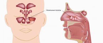
Benign formations formed in the nasal cavity, paranasal sinuses and nasopharynx are divided according to location:
- on soft parts (hemangiomas vascular formations, myxoma, angiofibroma and many other varieties);
- on hard parts;
- mixed (teratoma, meningioma, etc.);
- tumor-like formations (polyps, cavities with fluids, hyperplasia, papillomas).
More often than others, hemangioma develops. It can appear at any stage of life with equal frequency in men and women. Hemangioma usually forms in the anterior part of the nasal septum - where the bones and cartilage meet. It looks like a bright red polyp, often bleeding due to the passage of many capillaries through it.
Malignant tumors of the nose and paranasal sinuses account for approximately 3% of all cancers. If we consider ENT tumors, the first place in frequency will be neoplasms of the larynx and laryngopharynx, and in second place will be the nose and adjacent areas. The maxillary sinus and nasal cavity are most often affected. Men suffer from this disease twice as often as women. Representatives of the stronger sex are at risk.
Cancerous tumors that affect the nose develop quickly, reaching the orbit and eye, the base of the skull, and sometimes its cavity. Squamous cell oncology leads in frequency (more than half). The growth in this case arises from the epithelium. In addition, the nose can become a place for the development of melanoma (a neoplasm developing from pigment), sarcoma (from connective tissue), and adenocarcinoma.
Inverted papilloma occupies a special place. This is a benign neoplasm of the paranasal region with a high risk of becoming malignant, which happens with poor quality treatment.
Inflammation of the sinuses: diagnosis and treatment
Depending on the location, sinusitis can be:
- maxillary;
- ethmoiditis (inflammation of the mucous membrane of the cells of the ethmoid labyrinth);
- frontal sinusitis (inflammation of the frontal sinus);
- sphenoiditis (inflammation of the sphenoid sinus)
If the inflammatory process affects everything, then it is diagnosed as acute pansinusitis. If inflammation affects one half of the head (right or left frontal and maxillary sinuses), then this condition is called hemisinusitis. If more than one, but not all, paranasal sinuses are inflamed, then this process is called polysinusitis (poly - from the word many).
Clinical picture
Symptoms of acute sinusitis may include weakness, headache, and malaise. A febrile state may occur, and there are inflammatory changes in the general blood test. Local manifestations of inflammation are of greatest importance in the diagnosis of this disease.
Friends! Timely and correct treatment will ensure you a speedy recovery!
In all forms the following symptoms appear:
- Difficulty in nasal breathing. It can be constant or periodic, one or two-sided, and may occur due to obstruction (blocking of the lumen) of the nasal openings by swelling.
- Nasal discharge. They can be permanent or temporary, on two or one side. Due to swelling of the mucous membrane of the nasal cavity or anastomosis, there may be no discharge. Often there is a flow of secretion into the nasopharynx.
- Headache.
In severe cases of the disease, swelling of the soft tissues of the face in the projection of the maxillary and frontal sinuses is possible. In some cases, periostitis (inflammation of the periosteum) occurs.
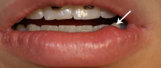
Herpes on the nose: causes of manifestation and exacerbation
As with herpes on the lips, manifestations in the nasal area are caused by viruses of types 1 and 2. The infection may go unnoticed and appear weeks or even months later.
Typically, an exacerbation of infection is provoked by the following circumstances:
- overheating (on the beach in summer) or hypothermia;
- viral or bacterial diseases;
- unbalanced diet;
- stress, destructive situations;
- alcohol abuse, smoking;
- lack of vitamins;
- exacerbation of chronic diseases;
- long course of antibiotics;
- Women may experience infection during menstruation.
Types of nasal cancer
Cancer of the nasal mucosa can be of several types:
- squamous. It arises from epithelial cells, which are flat. This type of disease is diagnosed in most cases (up to 70%);
- adenocarcinoma. Its development begins with glandular cells; similar sinus cancer is noted in 10-20% of cases;
- lymphoma. Cells of the immune system take part in the development of the disease. If we talk about tumors of the nasal cavity and paranasal sinuses, then it takes about 5%. It is important to study the symptoms of nasal cancer and understand when you need to see a specialist;
- melanoma. This is a tumor that is malignant and is formed from pigment cells. It is diagnosed in 3% of patients;
- esthesioneuroblastoma. This cancer of the paranasal sinuses is very rare; it develops from cells of the nervous system.
Diagnostics
Diagnosis of diseases that are accompanied by swelling of the nasal cavity is carried out by otolaryngologists. The examination program includes a survey, during which the specialist determines the time and circumstances of the appearance of swelling and other symptoms, their relationship with various external and internal factors. The doctor conducts an external examination and prescribes the following diagnostic procedures:
- Rhinoscopy.
Allows you to confirm the presence of inflammation, deformities, tumors, polyps, foreign bodies and traumatic injuries. - Lab tests
. A microbiological examination of nasal discharge is performed to detect bacterial infections, and RIF to detect viral diseases. For tumors, histological or cytological examination is prescribed. - Radiography
. In case of injuries, photographs of the nose are taken, and if sinusitis is suspected, X-rays of the paranasal sinuses are taken.
The listed methods can be supplemented by allergy tests, studying the condition of the pharynx and larynx during pharyngoscopy and laryngoscopy, and other studies.
Rhinoendoscopy
Prevention
There are certain precautions that will prevent cancer of the nose and sinuses:
- treat chronic inflammation, as well as benign neoplasms that have arisen in the nose;
- when working in industries that can cause harm to health, use means that provide personal protection;
- give up all habits that are harmful;
- Consult a doctor promptly if you notice symptoms of sinus cancer.