What does a sinus x-ray show?
An x-ray shows the human skull with bone structures, cavities and septa. The method is used at the preparatory stage in surgery.
What can be seen on a sinus x-ray:
- Foreign object in the nasal passages.
- Inflammatory process, thickening of the mucous membrane of the infected area.
- Consequences of injuries to the face and head.
- The presence of exudate (mucous, blood, purulent) in the paranasal and frontal cavities.
- "Airiness" of the sinuses.
- Neoplasms: polyps, tumors, cysts.
- Anomalies in the structure of the facial skeleton.
X-rays help to correct the diagnosis in case of fever or headache of unknown etiology.
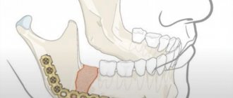
Maxillary sinus cancer
The symptoms of tumors of the maxillary sinus are largely determined by their localization in one or another segment of the upper jaw. It has been established that tumors of the anterioinferior location have a less malignant course than tumors of the superoposterior location. The clinical picture and course of tumors located in the internal or external segments of the maxillary sinus are equally different. The spread of a maxillary sinus tumor into the nasal cavity most often occurs through the natural opening in the middle meatus. During rhinoscopic examination in such cases, a pinkish protrusion with an uneven surface is identified, which bleeds easily when examined with a probe. The next stage is the ethmoidal labyrinth, from where the tumor can penetrate the cranial cavity through the cribriform plate, causing the corresponding symptoms. When the posterior cells of the ethmoidal labyrinth are affected, the tumor spreads into the nasopharynx or into the main sinuses.
Tumor growth into the orbit through the upper wall of the maxillary sinus or from the ethmoidal labyrinth is accompanied by various ophthalmological and neurological symptoms such as oculomotor nerve palsy, complete ophthalmoplegia and optic nerve atrophy. Along with this, protrusion of the eyeball is often observed.
Pain, which is one of the relatively early signs of malignant tumors , is especially pronounced when the maxillary sinuses are affected. Many patients whose tumor is expected to spread towards the orbit complain of painful evening and night headaches.
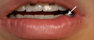
If the tumor is localized at the bottom of the maxillary sinus, it may grow into the hard palate and alveolar process. In such cases, sometimes palpation reveals the pliability of the hard palate and softening of the alveolar process, as well as loosening of the teeth of the upper jaw. As a result of destruction of the facial wall of the maxillary sinus, the tumor process spreads to the soft tissues of the cheek.
A tumor of the maxillary sinus can penetrate through the posterior wall into the pterygopalatine fossa, affecting the maxillary nerve located in this area. This can cause the development of severe neuralgic pain along the branches of the maxillary nerve. Along with excruciating pain, sometimes there is a sensitivity disorder in the skin of the cheek and upper lip.
Malignant tumors of the maxillary sinus are asymptomatic for a long time or are masked by the clinical picture of chronic sinusitis. With further development of the tumor, clinical manifestations are determined by its initial localization (anteroinferior internal, superoposterior internal, superoposterior external, anterioinferior external segments of the sinus) and the direction of growth.
Neoplasms located in the anterioinferior internal segment are characterized by unilateral obstruction of nasal breathing, mucous, mucopurulent or bloody discharge. The tumor spreads into the nasal cavity, onto the alveolar process, hard palate, and when the anterior wall of the sinus is destroyed, it infiltrates the soft tissues of the cheek.
The clinical course of the tumor emanating from the superoposterior internal segment is characterized by difficulty in nasal breathing, mucopurulent and bloody nasal discharge. Further growth of the tumor leads to deformations in the medial corner of the eye and its displacement upward (when the lower wall of the orbit is destroyed) and outward (when the inner wall of the orbit is destroyed and spreads into the ethmoid labyrinth).
A tumor emanating from the superoposterior outer segment causes severe pain in the region of the second branch of the trigeminal nerve. When it grows into the masticatory muscles and the pterygopalatine fossa, the phenomena of contracture of the masticatory muscles develop. Neoplasms of the anterioinferior outer segment cause pain, loosening of teeth, and deformation of the alveolar process of the upper jaw in the posterior regions. When the posterior wall is destroyed, the tumor grows into the temporomandibular joint, masticatory muscles, and pterygopalatine fossa and causes severe contraction of the jaws.
The following stages of maxillary sinus cancer are distinguished : Cancer in situ (sometimes called stage 0 cancer) - in this case, cancer cells are found only in a limited area of the mucous membrane, without penetrating deeper than the lamina propria. Stage I cancer . The tumor is found in the area of only the mucous membrane of the nasal cavity or paranasal sinuses. The cancer does not spread to other parts. Stage II cancer . In this case, the tumor spreads to the bones surrounding the paranasal sinuses or to the bones of the nose and palate, but does not affect the bones of the posterior wall of the sinus or the base of the skull. Stage III cancer . With cancer at this stage, the tumor is found in any of the following places: • Posterior bony wall of the sinus • Tissues under the skin • Orbit • Base of the skull • Ethmoid sinuses. In this case, on the affected side there may be an enlarged lymph node in the neck area up to 3 cm. Stage IV cancer .
This stage of cancer is in turn divided into 3 substages: • Stage A. There is an enlarged lymph node in the neck from 3 to 6 cm, or several lymph nodes in the neck are affected in any area. In this case, the tumor is found in the following areas: o Posterior bony wall of the sinus o Tissue under the skin o Orbit o Base of the skull o Ethmoid sinuses. • Stage B: The tumor has grown into the following areas: o Behind the eye o Into the brain o In the middle of the skull o Into the nerves coming out of the skull o The upper part of the pharynx behind the nose o At the base of the skull. o Or there is a lymph node larger than 6 cm in the neck. • Stage C. The tumor can be in any area of the sinus or near it, and there are metastases in distant organs, for example, in the lungs.
How is a sinus x-ray performed?
The procedure is not painful and does not require preparation. Receive a referral at an appointment with an otolaryngologist, infectious disease specialist or surgeon.
How to do a sinus x-ray:
- The patient removes metal jewelry, glasses, and dentures.
- To protect against radiation, he wears a lead apron or vest with a collar.
- The laboratory assistant indicates how to position yourself correctly relative to the apparatus. Depending on the required projections, the position is changed at the command of a specialist.
- While taking the photo, you should not move and hold your breath.
- After development and decoding, the film with the description is given to the patient.
The conclusion is not considered a final diagnosis, but provides clarifying information for the treating specialist.
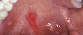
Angiofibroma
Benign vascular space-occupying lesion with slowly aggressive growth; localized in the nasal cavity; grows around the posterior wall of the nasal cavity, along the edges of the pterygopalatine foramen; in the early stages penetrates into the pterygopalatine fossa (arrows), grows into the medial pterygoid plate
a rare, but benign tumor characteristic of the nasal cavity and its sinuses. More common in men. As a rule, it occurs on the side wall of the nasal cavity, as well as in the paranasal sinuses. From the nasal cavity, a tumor can grow into the paranasal sinus and vice versa. Patients usually complain of nasal congestion, discharge, nosebleeds, and pain in the facial nerve area. Sometimes bone tissue is destroyed in the area of tumor growth.
Transitional cell papilloma with bone wall remodeling
A formation in the center of the middle nasal meatus, accumulating a contrast agent, spreads into the maxillary sinus and/or cells of the ethmoid bone labyrinth
Darkening of the sinuses on x-ray
The method is based on the different permeability of hard and soft tissues by electromagnetic waves. Bones block radiation and appear white in the picture. Specialists are interested in blackouts, which determine the nature of the disorder.
Darkening of the sinuses on an x-ray indicates fluid accumulation. This is a sign of inflammation with the release of mucous or purulent secretion. Sometimes, the picture shows thickening of the walls of the mucous membrane lining the sinuses.
New growths stand out as clear dark spots with shadows. Polyps look like peas on a “pedicle”, and cysts have a cavity filled with fluid inside.
Chronic rhinosinusitis
characterized by parietal thickenings caused by hyperplasia of the mucosa and partial fibrous changes in it. The thickness of the mucous membrane ranges from 4-5 mm.
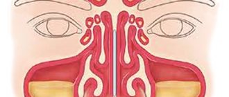
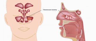
Sinonasal polyposis, hypertrophic sinonasal rhinosinusitis. Non-tumor inflammatory swelling of the mucous membrane.
Recently, there has been an increase in the number of fungal sinusitis. Chronic forms occur under the guise of polypous recurrent sinusitis, the MRI picture is nonspecific, and laboratory diagnosis is difficult. There may be a change in the bony walls of the sinuses due to hyperostosis or destruction of the sinus wall as a result of prolonged pressure from the fungal body.
X-ray of the sinuses during pregnancy
The radiation dose during the study is 20 μSv and is considered safe even when performed repeatedly for an adult. The fetus is susceptible to ionizing radiation, which causes intrauterine developmental defects. Carrying a child is a contraindication to the procedure.
X-rays of the nasal sinuses during pregnancy and breastfeeding are done exclusively for health reasons. The potential benefit of the research must outweigh the harm to the child. After the procedure, a pregnant woman needs an ultrasound of the fetus, and a nursing woman needs to transfer the baby to artificial nutrition for a day.
The most common (58-90%) is squamous cell carcinoma.
1. are asymptomatic for a long time, under the guise of inflammatory changes, especially in the absence of destruction of the walls 2. quickly spread to neighboring structures and by the time of recognition, infiltrate several areas 3. it is difficult or impossible to establish the original site of tumor origin 4. extremely rarely metastasizes to distant organs and tissue 5. it is not possible to clearly define the boundaries of the lesion 6. MR semiotics: tissue formation, spread to surrounding tissues, bone destruction
If bone structures are damaged - the hard palate and the alveolar process of the upper jaw, it is necessary to undergo an additional radiological examination - X-ray CT, which clarifies the presence or absence of bone destruction.
Detection of tumor tissue against the background of soft tissue structures - the pterygopalatine and infratemporal fossa, masticatory muscles, soft tissues of the cheek, as well as the spread of the tumor to the frontal and sphenoid sinuses, the ethmoidal labyrinth intracranially requires MRI (with contrast enhancement). In addition, MR imaging is indispensable in the differential diagnosis of postoperative or post-radiation changes with relapse or continued growth.
Thus, in order to exclude a pathological process and begin treatment on time, it is necessary to undergo a complete radiation examination.
X-ray of the sinuses for children
Pediatricians try to protect preschool children from the harm caused by X-ray radiation to the fragile skeletal system. The procedure is allowed to be performed on patients over 7 years of age.
Before this period, a clear justification for the importance of intervention is necessary - for example, severe facial trauma or severe sinusitis with a risk of inflammation of the meninges.
It is difficult for a young child to sit or stand still during an x-ray. To distract him, they use toys, sedatives, and in emergency cases, anesthesia. At an older age, you can captivate your child with a game in which you should freeze for a short time.
Sign up for the study
Paranasal sinuses and their role.
The sinuses are cavities filled with air and connected to the nose by special openings - anastomoses.
The following types of sinuses are distinguished: maxillary (maxillary), frontal, sphenoid and ethmoidal labyrinth.
All sinuses play a major role in the body:
- they soften the consequences of injury when hit in the face;
- thanks to these cavities, our body reacts correctly to changes in pressure in the environment;
- sinuses are involved in the formation of voice;
- participate in the thermoregulation of air during breathing, heat the inhaled air and moisturize it;
- Due to the air filling the sinuses, the weight of the skull bones decreases.
As we see, the sinuses play a big role, so their condition must be treated with special care.
Interpretation of results
The interpretation of the results of an x-ray examination may be as follows:
- normal (taking into account the patient’s age);
- the presence of dark spots in the paranasal sinuses;
- thickening;
- visible trauma or the presence of a foreign body.
Norm
An X-ray of the sinuses of a healthy person looks like this:
- The nasal septum divides the nasal cavity into symmetrical sides of a triangle.
- The white stripes running to the right and left of the divided area are the nasal passages.
- The triangular cavities on the sides of the nose are the maxillary sinuses.
- Between the eye sockets there is an ethmoid sinus with thin walls, the cells of which should be clearly visible.
- Above the orbits, the frontal sinuses are defined, which can have different shapes. Their separation by bone partitions is allowed.
- There must be air in the sinuses. Their edges, like the contours of the bones, should be clear and even.
Photo of a healthy person
Darkening, cavities and thickening in the image
We can talk about diagnosing a pathology if the images show the following:
- darkening the area;
- thickening;
- the presence of a cavity of different shapes;
- deformation of bone tissue.
Acute inflammation is characterized by thickening of the mucosa and deformation of its borders (with purulent contents). The presence and level of fluid are determined by horizontal demarcation in the sinuses. An x-ray will not tell you about the composition of the contents; for this you will need to make a puncture (puncture). If the inflammation is chronic, the mucous membrane thickens and the lumen in the sinus becomes smaller.
Sinusitis
Sinusitis on an x-ray looks like a darkening, which has characteristic differences in various pathologies.
The pictures should be decrypted like this:
- With hyperplastic sinusitis, there is thickening of the mucosa directly next to the bone. In this case, the internal contour becomes wavy and blurry.
- With catarrhal sinusitis, thickening of the mucosal walls with complete or partial darkening is noted. The presence of a light cavity in the center of the sinus indicates a chronic process.
- With exudative sinusitis, darkening of the paranasal sinuses occurs with a horizontal delimitation, reflecting the level of fluid filling.
- With vasomotor and allergic sinusitis, pronounced swelling of the mucous membrane is noted.
Sinusitis
Sinusitis is an inflammation of the lining of the maxillary sinuses.
The following types of sinusitis are diagnosed:
- Exudative. The presence of fluid in the upper sinuses on one or both sides.
- Parietal. Localization of inflammation near the bone walls. The edges of the mucous membrane are deformed and directed inside the sinus.
- Polypous. There is bulging of areas of the mucous membrane, which can be either single or multiple.
Neoplasms and cysts in the sinuses
If photographs of the paranasal sinuses reveal a cavity with dense contents, this indicates the presence of a benign or malignant formation. In most cases, neoplasms are found by chance during the diagnosis of other pathologies.
- Sinusitis - what is it and how to treat it at home Symptoms of sinusitis and medications for children and adults
A sinus cyst is defined as a light, rounded area located outside the sinus mucosa. Its edges are clear and even.
Bone injuries
If a bone is broken or displaced, it will be visible on an x-ray. Bone injuries may appear on x-rays as dense fragments in the sinuses. The image helps to establish the location of the fracture and the displacement of bone fragments, if present. Severe fractures are accompanied by bleeding, which will appear as fluid in the sinuses. The doctor may also detect an old injury that appears as a callus on the image.
Foreign bodies
The foreign body in the image has contours in accordance with what exactly got into the nasal cavity. Typically, these are beads or other round objects that children place in their noses. On x-rays they appear as areas of varying darkness.
Photo gallery
Right-sided sinusitis Cyst in the left maxillary sinus Sinusitis. Frontal and maxillary sinuses Trauma to the nasal cavity
Preparation for x-ray examination.
The advantages of this type of research include its accessibility, speed of implementation, speed of result processing, simplicity and painlessness. This diagnostic method is also affordable.
Friends! Timely and correct treatment will ensure you a speedy recovery!
No special preparation is required to conduct the study. The procedure is performed on any day, regardless of meal time. Even if the patient is not feeling well, such a diagnosis is easy to carry out without waiting for his condition to improve.
Make an appointment right now!
Call us by phone or use the feedback form
Sign up
Before the examination, you need to remove jewelry and metal objects, including glasses and dentures. Since all these items can affect the accuracy of the study and prevent the ENT doctor from making the correct diagnosis.
During the x-ray, you must fix your head in one position and not move it, otherwise the x-ray will be blurry and you will have to repeat the study.
Why you should entrust your treatment to the ENT Department of Dentistry
ENT dentistry combines two areas of medical services and treatment options - otolaryngology and dentistry . This is a modern format for the clinic’s work on comprehensive rehabilitation in the treatment of traumatic and inflammatory processes in the upper jaw, penetrating into the maxillary sinus or passing along its borders.
We carefully approach statistics and regret to report that the main provider of patients are our colleagues who are stuck in an unsuccessful treatment plan; there are not many initial visits to the clinic. Fortunately, correct and timely suspended non-ideal treatment can always be reversed from an unsuccessful outcome. I am very grateful to our colleagues who do not hesitate to send us such complex patients, despite some reputational damage.
According to our statistics, 70% of patients turned to our Center after unsuccessful conservative treatment of cysts by ENT doctors, an endless number of traumatic “punctures” of the maxillary sinus, or attempts to find and persuade dentists not to remove the dental cyst, but to treat the cyst without removal.
In response to this request, many years ago, ENT dentistry was identified by us as a separate important area of clinical work.
The Center for Private Dentistry “Doctor Levin” has been specializing in providing care to patients with combined ENT and dental pathology for many years. Medical and surgical treatment programs are carried out by candidates of medical sciences, maxillofacial surgeons with ENT training.