Interpretation of results
The interpretation of the results of an x-ray examination may be as follows:
- normal (taking into account the patient’s age);
- the presence of dark spots in the paranasal sinuses;
- thickening;
- visible trauma or the presence of a foreign body.
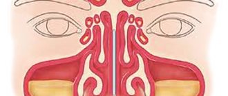
Norm
An X-ray of the sinuses of a healthy person looks like this:
- The nasal septum divides the nasal cavity into symmetrical sides of a triangle.
- The white stripes running to the right and left of the divided area are the nasal passages.
- The triangular cavities on the sides of the nose are the maxillary sinuses.
- Between the eye sockets there is an ethmoid sinus with thin walls, the cells of which should be clearly visible.
- Above the orbits, the frontal sinuses are defined, which can have different shapes. Their separation by bone partitions is allowed.
- There must be air in the sinuses. Their edges, like the contours of the bones, should be clear and even.
Photo of a healthy person
Darkening, cavities and thickening in the image
We can talk about diagnosing a pathology if the images show the following:
- darkening the area;
- thickening;
- the presence of a cavity of different shapes;
- deformation of bone tissue.
Acute inflammation is characterized by thickening of the mucosa and deformation of its borders (with purulent contents). The presence and level of fluid are determined by horizontal demarcation in the sinuses. An x-ray will not tell you about the composition of the contents; for this you will need to make a puncture (puncture). If the inflammation is chronic, the mucous membrane thickens and the lumen in the sinus becomes smaller.
Sinusitis
Sinusitis on an x-ray looks like a darkening, which has characteristic differences in various pathologies.
The pictures should be decrypted like this:
- With hyperplastic sinusitis, there is thickening of the mucosa directly next to the bone. In this case, the internal contour becomes wavy and blurry.
- With catarrhal sinusitis, thickening of the mucosal walls with complete or partial darkening is noted. The presence of a light cavity in the center of the sinus indicates a chronic process.
- With exudative sinusitis, darkening of the paranasal sinuses occurs with a horizontal delimitation, reflecting the level of fluid filling.
- With vasomotor and allergic sinusitis, pronounced swelling of the mucous membrane is noted.
Sinusitis
Sinusitis is an inflammation of the lining of the maxillary sinuses.
The following types of sinusitis are diagnosed:
- Exudative. The presence of fluid in the upper sinuses on one or both sides.
- Parietal. Localization of inflammation near the bone walls. The edges of the mucous membrane are deformed and directed inside the sinus.
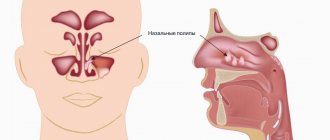
- Polypous. There is bulging of areas of the mucous membrane, which can be either single or multiple.
Neoplasms and cysts in the sinuses
If photographs of the paranasal sinuses reveal a cavity with dense contents, this indicates the presence of a benign or malignant formation. In most cases, neoplasms are found by chance during the diagnosis of other pathologies.
- Sinusitis - what is it and how to treat it at home Symptoms of sinusitis and medications for children and adults
A sinus cyst is defined as a light, rounded area located outside the sinus mucosa. Its edges are clear and even.
Bone injuries
If a bone is broken or displaced, it will be visible on an x-ray. Bone injuries may appear on x-rays as dense fragments in the sinuses. The image helps to establish the location of the fracture and the displacement of bone fragments, if present. Severe fractures are accompanied by bleeding, which will appear as fluid in the sinuses. The doctor may also detect an old injury that appears as a callus on the image.
Foreign bodies
The foreign body in the image has contours in accordance with what exactly got into the nasal cavity. Typically, these are beads or other round objects that children place in their noses. On x-rays they appear as areas of varying darkness.
Photo gallery
Right-sided sinusitis Cyst in the left maxillary sinus Sinusitis. Frontal and maxillary sinuses Trauma to the nasal cavity
Folk remedies
Subtotal darkening of the maxillary sinuses can be treated using traditional methods. Medicines based on medicinal plants are also used in the treatment of inflammatory diseases of the upper respiratory tract. For this purpose, saline solutions, drops, and compresses are used.
The only contraindication is an allergic reaction to the components of the drug.
For young children under 1 year of age, traditional treatment methods should be used with caution so as not to provoke the development of an allergic reaction.
Mustard oil
Rhinitis and sinusitis can be treated with mustard oil. To do this, the oil must be slightly heated, but the temperature must be comfortable so that when the oil is instilled into the nose, the mucous membrane does not burn.
Place 3-4 drops of oil into each nasal passage 2 times a day. The oil has anti-inflammatory and antiviral effects, and also reduces swelling of the nasal mucosa.
Garlic
Garlic has antibacterial properties and can be used to make drops for the common cold. To do this, you need to take 2 cloves of garlic, pass through a press, place in gauze (folded in several layers) and squeeze out the resulting juice.
Then add 3-4 tbsp to the garlic juice. l. water or 2 tbsp. l. olive oil. This must be done so that garlic juice does not lead to a burn of the mucous membrane.
For the first use, 1 drop is instilled into each nasal passage. If no allergic reaction subsequently occurs, then you can instill 2-3 drops into each nasal passage for 7 days.
Such drops are prohibited for use by children. Since the mucous membrane in children has a more immature structure compared to adults. Instillation of such drops may cause burns.
In addition to drops, garlic can be used as an air disinfectant. To do this, the garlic is first peeled and cut into small pieces. Garlic is laid out on plates and placed at different ends of the room. Since garlic is a natural antiseptic, its action will deactivate viruses and bacteria that are released by a sick person.
Propolis solution
To treat sinusitis, use a propolis solution. To do this, take a 10% alcohol solution of propolis. Before use, dilute a small amount with water so as not to irritate the mucous membrane. You need to instill 1-2 drops into each nasal passage for 7 days.
Due to the alcohol content, this method of treatment is contraindicated for children.
Aloe juice
Aloe is used not only to treat inflammatory diseases, but also to prevent viral infections.
For this purpose, drops can be prepared from aloe juice. To do this, take aloe juice and mix it with water in a 3:1 ratio. If such drops are indicated for a small child, then the proportion is 5:1. You need to instill 2-3 drops into each nasal passage for 10 days.
Black radish
Any compresses are prohibited in the phase of acute inflammation, as well as at elevated body temperature. A black radish compress is effective in reducing swelling of the mucous membrane.
To prepare such a compress, you need to take one radish, peel it and grate it. Then you need to mix with vegetable oil and heat slightly. Apply the prepared pulp to the sinus area for 10 minutes. You can use thin cloth or gauze for this purpose. The course of treatment with this remedy is no more than 7 days.
Sea salt
A hypertonic sea salt solution can be purchased at a pharmacy, or you can prepare it yourself at home.
For this purpose, you need to take ½ teaspoon of sea salt and dissolve its contents in a glass of warm water. Then you need to draw the required amount of solution into a sterile syringe and rinse each nasal passage one by one. It is recommended to rinse your nose with saline solution up to 5 times a day.
Children need to rinse their nasal passages by instillation. Until the age of 3, the nasal passages should not be washed with sprays or a syringe. Since a strong stream of solution can promote the advancement of microorganisms to the auditory tube. Since the auditory tube in children is short, this can cause otitis media.
If a subtotal darkening is observed on the x-ray of the maxillary sinuses, this means that a pathological process has begun in them. In some cases, a number of additional diagnostic procedures may be required to clarify the diagnosis.
How does the procedure work and how long does it last?
The duration of the study is several minutes.
X-rays are taken depending on the indications as follows:
- The patient enters a special room. Children are accompanied by adults.
- In a standing or sitting position, the radiologist fixes the head in the position required to take the image. This can be a straight or lateral plane, a projection of the chin, the occipito-frontal part or the occipito-mental part. One of the parents holds the child's head.
- You need to take a deep breath and hold your breath for a few seconds.
- The doctor will inform you that the procedure is complete.
- The picture develops within 15-20 minutes.
Is X-ray dangerous? This is explained in the video from the Live Healthy channel.
Purpose of radiography of the maxillary sinuses
An X-ray of the nose is performed to determine the clinical picture of the disease after taking a blood test for sinusitis.
When assessing the results, the doctor answers the following questions:
- whether the patient has a disease;
- whether there are complications;
- does the treatment give results;
- assessment of the patient's condition after illness.
The treatment tactics depend on the answers received, so it is absolutely impossible to neglect diagnostic procedures for sinusitis.
Symptoms of sinusitis in adults without fever are described here.
Contraindications to X-ray examination
X-ray of the paranasal sinuses has several contraindications. This method is prohibited from doing:
- Pregnant women. Despite the fact that the study has low radiation exposure, it is contraindicated for expectant mothers due to the strong sensitivity of the fetus to radiation exposure. The use of X-ray machines during pregnancy can cause birth defects in the child.
- Children under 15 years old. For patients younger than this age, sinus x-rays may be rarely ordered because gamma rays can negatively affect the child's bone growth.
- People suffering from mental illness. In such cases, patients are unable to fix their head and not move for several minutes.
- A month before IVF. The cells of the reproductive system are considered the most vulnerable to radiation. Irradiated sperm in men and damaged eggs in women are unable to conceive. Therefore, gynecologists recommend undergoing an X-ray examination at least a month before in vitro fertilization.
In very rare cases, a doctor may issue a referral for x-rays for children under 15 years of age and pregnant women. But this happens if the research justifies the risk of probable harm that the diagnosed disease can lead to.
How often can an x-ray be taken for sinusitis, when can it be repeated?
The standard for diagnosis and treatment of sinusitis requires two x-ray examinations: before and after therapy. In certain cases, a second study is carried out two weeks after the start of treatment.
Radiography involves the patient receiving a certain dose of radiation, so it is not recommended to carry it out frequently. The optimal number of examinations is three times a year.
If there is a need for several diagnostic procedures, it is better to replace them with ultrasound or magnetic resonance imaging of the sinuses. It is better to refuse repeated computed tomography because it is a high-tech type of x-ray examination.
Preparing for an X-ray
Preparation for the procedure comes down to two rules:
- What does pneumatization of the sinuses indicate?
- remove jewelry and other metal objects located in the head area (chain, earrings, hairpins, hoop);
- wear a special apron (lead), which is issued immediately before the x-ray.
There are no restrictions related to food or liquid intake.
It is necessary to clarify when and how to take an x-ray when observing the disease over time. For example, if a patient is having a cuckoo sinus irrigate, x-rays are taken before the sinuses are cleared of fluid.
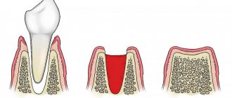
Physiological and pathological mobility of teeth
The movement of the dentition is almost imperceptible; its existence can only be evidenced by the polished areas between the adjacent incisors and molars. All dental units are mobile during the chewing process. It is this reflex that allows them to be kept in good condition. The absence of this reflex can lead to the destruction of tooth enamel and bone tissue.
In the bones of the upper and lower jaws of a person there are special holes, and the dental roots are located strictly in individual cells, while they are firmly connected to the jaw bone by a strong and rigid ligamentous structure called the periodontium.
What can a doctor detect on x-rays?
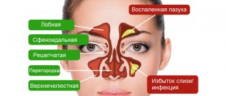
This procedure provides ample opportunities for diagnosing and confirming various diseases of the paranasal sinuses. The resulting images (x-rays) help doctors visualize and examine the following anatomical parts: frontal, maxillary (maxillary) sinuses, ethmoidal labyrinth, orbits and facial bones.
Inflammation of the maxillary and frontal sinuses
An x-ray of the paranasal sinuses can reveal:
- defects in the structure of the organ and adjacent soft tissues;
- the presence of certain inflammatory diseases: acute or chronic sinusitis, sinusitis, sinusitis, pansinusitis;
- traumatic injury to the facial skeleton,
- foreign objects;
- tumor formations (benign and malignant), cysts of the nasal cavity;
- deviated nasal septum;
- developmental defects, congenital and acquired anomalies of PPN.
The method is used everywhere in the practical work of otolaryngologists and is the basis for making a diagnosis.
With the help of radiography, diseases of the ENT organs, injuries, and the presence of foreign objects are well diagnosed. This technique allows you to visualize pathological contents in the paranasal sinuses, changes in the mucous membrane lining the sinuses (swelling, polyps, etc.). Normally, the paranasal sinuses are filled with air and do not differ from the eye sockets. If they contain liquid (pus, blood, etc.), darkenings are formed (reduced pneumatization) with a horizontal upper border.
By taking a timely picture of the paranasal sinuses, it is possible to avoid complications fraught with irreversible consequences, for example, osteomyelitis, meningitis.
- Consultation with a specialist: how to remove pus from the maxillary sinuses?
Total blackout
Total darkening is characteristic of inflammatory diseases of the sinuses, in which exudate of a different nature is produced: mucus, pus, serous fluid. In this case, the sinus is not filled completely, but only half or 2/3 of the total area of the cavity.
If the patient's fluid production is a consequence of a hypersensitivity reaction, then the picture shows formations of the mucous membrane in the form of pads. In radiology, this sign is called “plus shadow”, in which darkening of varying degrees of intensity is observed.
In the image, fluid, pus and changes in the structure of the epithelium may appear in the same color. In this case, to clarify the diagnosis, additional examination methods are prescribed - laboratory, endoscopic, instrumental (CT, ultrasound).
If homogeneous darkening appears more on the right, one should assume edema, on the left - accumulation of pus.
Conclusion
The outstanding ancient physician Hippocrates taught: “Eliminate the cause and the disease will go away.” The use of radiography for the diagnosis of sinusitis fully meets this requirement.
- It is effective, accessible and simple.
- The method has established itself as the main one in the diagnosis of this disease.
- The study makes it possible to establish not only the presence of the disease, but also to evaluate the effectiveness of the treatment. For a more detailed examination, the doctor may also prescribe a CT scan of the maxillary sinuses.
- Reading the image is easy.
- X-rays can be repeated throughout the year.