Briefly about sinusitis
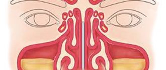
Sinusitis is inflammation of the mucous membrane in the maxillary sinuses. Nathaniel Gaymore was the first to describe these sinuses: the name came from his name. And Leonardo da Vinci was the first to draw the maxillary sinuses.
Essentially, sinusitis is sinusitis caused by various reasons, which are associated with infection in the sinuses and the accumulation of pus in them. It can be rhinogenic, odontogenic, traumatic, allergic.
Rhinogenic occurs due to untreated rhinitis, when the infection penetrates from the nasal cavity into the sinuses. Odontogenic sinusitis develops as a result of infection through a diseased tooth and inflamed tissue around it.
Traumatic rhinitis is a consequence of damage to the nasal septum, maxillary sinus or jaw.
Finally, allergic rhinitis occurs due to allergies to pollen, animal dander, dust mites, medications and other allergens.
Acute sinusitis lasts less than 3 months, recurrent acute sinusitis occurs up to 4 times a year, chronic sinusitis lasts from 3 months or more. An exacerbation of chronic sinusitis is also possible, when new symptoms are added to existing ones.
What will a CT scan of the maxillary sinuses show?
Based on the results of a CT scan of the upper jaw and maxillary sinuses, the doctor determines:
- the location of the nasal septum and bones forming the maxillary cavities;
- sinus drainage pathways;
- symmetry of the left and right sides of the nose and sinuses;
- degree of pneumatization of the sinuses;
- condition of the tear ducts.
CT scan of the upper jaw reveals:
- inflammatory processes, including the presence of exudate in the sinuses;
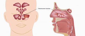
- tumors of the mucous membranes;
- polyps;
- granulomas and cysts;
- abscesses;
- foreign objects in the nasal sinuses (filling materials, root fragments after tooth extraction, dental implants);
- damage or deformation of the nasal bones;
- additional voids (ostia);
- pathological communication of the maxillary sinus with the oral cavity (oroantral fistulas).
Thanks to the high resolution of the computed tomography apparatus, it is possible to detect the smallest pathological formations, assess their size, location and relationship with surrounding tissues. Based on the interpretation of CT data and comparison with the results of other types of examination, the dentist or ENT makes a diagnosis.
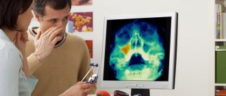
How is a CT scan of the upper jaw performed?
CT scan of the upper jaw and maxillary sinuses looks like this:
- the patient lies down on a retractable couch, the neck is fixed using a special stand;
- the head end of the couch smoothly slides into a ring-shaped device where sensors and an X-ray emitter are located;
- moving in a spiral, the scanning complex takes a series of layer-by-layer images of the studied area;
- if necessary, a contrast agent is injected into the vein and the examination continues;
- Upon completion of the diagnostics, the device turns off.
Any movement of the patient during a CT scan of the upper jaw can degrade the quality of the images, so it is necessary to maintain a static position throughout the entire scanning period.
Do you need an x-ray?
If a survey of complaints, anamnesis, and identification of clinical symptoms is not enough to make a correct diagnosis, then doctors prescribe an x-ray.
X-rays have their own contraindications: pregnancy, breastfeeding, young age of the patient, repeated procedure.
An X-ray of the children's nasopharynx is performed only when other diagnostic methods have failed. The procedure itself is not dangerous for the child. Maxillary sinusoids up to 16 years of age are similar to light spots, have a dense structure, and do not appear hollow. Using the image, you can get an idea of the accumulation of masses of pus, excessive enlargement of the pharyngeal tonsils, and the development of sinusitis in the acute stage.
X-ray of the sinuses during pregnancy
The radiation dose during the study is 20 μSv and is considered safe even when performed repeatedly for an adult. The fetus is susceptible to ionizing radiation, which causes intrauterine developmental defects. Carrying a child is a contraindication to the procedure.
X-rays of the nasal sinuses during pregnancy and breastfeeding are done exclusively for health reasons. The potential benefit of the research must outweigh the harm to the child. After the procedure, a pregnant woman needs an ultrasound of the fetus, and a nursing woman needs to transfer the baby to artificial nutrition for a day.
Preparation for the procedure
Before starting the procedure, you must remove your outer clothing, metal and other jewelry, earrings, piercings, and remove dentures. Next, you need to tell the doctor whether similar procedures have been performed previously, whether there are dental implants, fractures of the nose or facial bone.
During the diagnosis, the patient must stand still and not move. He should rest his nose and chin on the stand of the X-ray machine, which has been previously adjusted to the patient’s height. After this, the doctor will take several pictures from another room and give further instructions to the patient. During the procedure, the patient will sometimes need to hold their breath for a few seconds (no more than 10 seconds). The radiologist will take pictures in two projections, and sometimes while lying down.
The whole procedure will take about 10 minutes.
Maxillary sinus cyst (MSC) is a term that combines pseudocyst and retention cyst [1, 2]. Pseudocyst is an accumulation of inflammatory exudate, often of odontogenic etiology, lifting the mucous membrane of the upper jaw from the bone bottom. A retention (true) cyst is a cyst lined with sinus epithelium and filled with mucus as a result of obstruction of the goblet cell excretory tract. Retention cyst and pseudocyst cannot be differentiated either clinically or radiographically, and therefore are combined with one term used in most studies [3–9].
ICP cysts are a common finding in screening studies. They are detected incidentally in 12.4–22% of people during CT scans of the head and are often asymptomatic [10–15]. There is still no consensus on the symptomatology of cervical cysts. Various sources list a fairly wide range of clinical manifestations of this pathology: headache, dull pain in the projection of the sinus and numbness of the cheek, swelling of the cheek, rhinorrhea, mucus drainage along the back wall of the pharynx, nasal obstruction [1-9]. The International Classification of Headache Disorders [16] clearly states that headache associated with the nose and paranasal sinuses (SNS) is pain associated with acute sinusitis or exacerbation of chronic sinusitis. Currently, there is no convincing evidence linking headaches to SNP cysts. There is an opinion that cysts manifest themselves with inflammatory symptoms and proceed as rhinosinusitis, which is confirmed by a higher value of the Lund-Mackay coefficient when assessing CT scans of the SNP than in healthy individuals [10, 11]. Some authors, not finding pathology in the ostiomeatal complex and on CT scans of the SNP, believe that ICP cysts are not always a variant of rhinosinusitis [14]. JH Wang [15] in his study reports that most often cysts manifest as nasal obstruction (52.5%). T. Hadar [12] notes the presence of complaints of headache in 63% of 38 (63%) patients in a group of 60 people with cervical cysts.
The purpose of the study is to identify symptoms associated with cervical cysts.
Research methods
The study was conducted at the State Clinical Hospital No. 3 (ENT Center) from January 2010 to September 2010.
Criteria for inclusion of patients in the study group:
1. Patients with non-odontogenic cysts of the maxillary sinuses (absence of the bone shell on CT scan of the sinus).
2. Absence of clinical and CT images of sinusitis, deviated nasal septum, neoplasm and other pathology of the nose and SNP.
Criteria for excluding patients from the study group:
1. Suspicion of an odontogenic cyst (periapical dental pathology and the presence of a bone shell on a CT scan of the SNP).
2. Mucocele (characteristic CT picture).
3. Diseases that can cause postnasal drip syndrome, difficulty in nasal breathing and headaches (deviated nasal septum, chronic rhinitis, sinusitis, nasopharyngeal tumors, etc.).
Taking into account the above criteria, 67 patients were selected who were admitted to the hospital for surgical treatment of cervical cysts. All these patients were asked to fill out a questionnaire with a list of symptoms characteristic of this disease. The questionnaire included the most frequently mentioned symptoms in the literature: headache, feeling of pressure and/or heaviness in the sinus area, mucous rhinorrhea or mucus running down the back of the throat, difficulty in nasal breathing, cough in the morning.
If there was a complaint of headache, its location, frequency and intensity were indicated. The intensity of the headache was determined using a visual analogue scale: minor (does not force the patient to lie down or take a painkiller) - 1-3 points, moderate (usually the patient is forced to lie down or take a painkiller) - 4-7 points, severe (tolerable from difficulty, forces you to take a painkiller in a higher dose) - 8-10 points [1].
4-6 months after surgery, patients were recommended to undergo a CT scan of the SNP to exclude cyst recurrence, after which the questionnaires were filled out again.
The study is level 4 (without internal control group) with class C evidence category.
For statistical data processing, we applied the Z-sign test according to Van der Waerden for nonparametric tests [17]. The disappearance of a symptom after surgery was regarded as “+”, its persistence as “-”. The smaller number with the same sign was taken as Z. The p value was determined from the table. As for all biomedical studies, a p value of <0.05 indicated the presence of a statistically significant difference between the compared results with a probability of >95%.
Results and discussion
The nature of the complaints of patients with intracranial cysts, which we identified before and after surgery, is presented in Table. 1.
The table shows that there is a significant connection between HF cysts and the following symptoms: headache in the frontal region, a feeling of heaviness in the projection of the affected sinus, rhinorrhea, mucus flowing down the back wall of the pharynx and difficulty in nasal breathing. Morning cough as part of postnasal drip syndrome was more common preoperatively than postoperatively; however, the insufficient number of patients with this symptom did not allow us to identify the statistical significance of this sign.
The complaint of papular exanthema of the chest and back was the only one in a patient with a cervical cyst; removal of the latter was carried out on the recommendation of a dermatovenerologist. Another patient with recurrent fainting conditions and non-systemic dizziness was admitted to the clinic for removal of a cyst of the upper quadrant at the insistence of neurologists.
Characteristics of headaches in patients with HF cysts are presented in Table. 2.
The table shows that the most common location of headaches caused by a cyst of the upper jaw is the frontal region (67%). It should be noted that after surgical treatment, the frequency and intensity of pain in the forehead area significantly decreased (the persistence of symptoms was noted in 2 cases out of 42). Pain sensations in other localizations did not change significantly after removal of cervical cysts in our patients. Thus, a statistically significant connection between the presence of a cervical cyst and pain in other parts of the head has not been proven.
In 61 (91%) patients, according to CT data, the cysts had a diameter of more than 15 mm. Of the 6 cases of cysts with a diameter of less than 15 mm (9%), they were independent in only 2 patients, the rest were accompanied by large cysts, which suggests that only large cysts have clinical manifestations. Most often, cysts were located in the alveolar bay on the lower wall of the upper quadrant (n=59; 88%), which is consistent with data obtained in other studies. The remaining cases included cysts in the area of the lateral edge of the posterior wall (n=4; 6%), anterior wall (n=3; 4%) and medial wall of the upper quadrant (n=1; 2%).
The study confirms that the most common location of maxillary sinus cysts is its lower wall. Clinical manifestations of the disease are typical for large cysts (15 mm in diameter or more). With a high degree of probability, the following symptoms can be associated with cysts of the maxillary sinus: headache in the frontal region, a feeling of heaviness in the projection of the maxillary sinus, rhinorrhea, mucus flowing down the back wall of the pharynx and difficulty in nasal breathing.
What does sinusitis look like in the picture?
Healthy paranasal sinuses in the picture are similar in shade to the eye sockets. If inflammation develops, they acquire a darker shade with whitish, “milky” contents. The usually smooth edges of the sinus may be curved, and thickening may be observed locally or along the entire edge of the sinus. This picture indicates that pathology has developed in the sinuses.
An otolaryngologist and a radiologist interpret the X-ray. Photos of the sinuses highlight potential abnormalities and darkened areas. The analysis can determine: the presence of potentially affected areas - darkened or sub-darkened areas, whether there is sinusitis - lightened areas of the sinuses.
After treating a runny nose and congestion, the doctor can also determine the nature of the disease - sluggish, stopped, chronic, acute, the presence of tumors or cysts.
Decoding the testimony
The radiologist deciphers what an x-ray of the paranasal sinuses shows. First of all, the condition and location of bone structures and cartilage is assessed. The liquid on the x-ray image appears as an intensely darkened horizontal boundary. Thickening of the mucous membrane indicates swelling in the paranasal sinus. You will receive a transcript of the research results in the form of a conclusion, but the final diagnosis is made by the doctor. Usually, when a pathology is identified that requires a more detailed study, another diagnostic method is prescribed.
How often should x-rays be taken?
Despite the fact that radiation exposure from X-rays using modern equipment is minimal, it should not exceed certain levels per year. The procedure can be done 3-4 times in a row. Before each new x-ray, the health worker must look at the patient’s card, and if he finds an exceeded radiation dosage there, he will refuse the procedure.
During treatment, it is recommended to conduct two x-ray examinations: before and after therapy or 2 weeks after the start of treatment. If the disease is serious, examinations can be done 3 times a year.
CT scan of the upper jaw with contrast
Native CT examination of the upper jaw accurately diagnoses pathologies in the sinuses, lacrimal ducts and anastomoses with the nasal cavity. With the addition of contrast to the procedure, adjacent soft tissues are clearly visualized.
Most often, CT with contrast is prescribed when malignant processes are suspected. Intravenous administration of iodine-based drugs improves the visibility of the circulatory system: tumors have their own network of vessels, which is clearly visible after contrast. By identifying additional foci of blood supply, computed tomography can detect tumors at an early stage.
Examination of sinuses without enhancement does not require special preparation. It is enough to bring with you a referral from your attending physician, an extract from the outpatient card and the results of previous diagnostic measures. Before a CT scan of the upper jaw and maxillary sinuses with contrast, it is necessary to do a blood test for creatinine. If creatinine levels increase, the use of contrast is prohibited, since there is a high risk of problems with removing the medication from the body.
Other contraindications:
- Individual intolerance to the components of the drug.
The patient may develop reactions to iodine - dermatological (skin redness, rash, itching) or systemic (facial swelling, difficulty breathing, bronchospasm, Quincke's edema, anaphylactic shock). Similar phenomena during CT scanning of the upper jaw with contrast cannot be excluded in people with any serious allergies.
- Taking metformin in patients with diabetes mellitus.
This medication reduces the glomerular filtration rate of the kidneys, which increases the damaging effect of the contrast agent. In combination with iodine-containing drugs, metformin can provoke the accumulation of lactic acid in the body (lactic acidosis). This condition is life-threatening. For patients taking metformin, it is important that two days before a CT scan of the upper jaw with contrast, the endocrinologist either discontinues the drug or replaces it with another medicine.
- Hyperfunction of the thyroid gland.
Iodine-based substances accumulate in the tissues of the thyroid gland, which can significantly worsen the patient’s condition. In this situation, the possibility of examination with contrast appears only after preventive preparatory treatment. In any case, CT scanning of the upper jaw and maxillary sinuses using iodine-containing preparations should not be done without consultation with an endocrinologist.
- Age up to 12 years.
It is difficult for young children to get into a vein, and it is also difficult for children to explain the need and sequence of the manipulations performed.
Lactation is not a contraindication to CT scanning of the maxilla with contrast. But since the dye used partially penetrates into breast milk, a woman should take care of the baby’s nutrition in advance. After the procedure, you must skip two feedings in a row, expressing and pouring out the milk.