X-rays allow you to make the correct diagnosis, prescribe appropriate treatment, and also monitor the results of treatment.
Myth about the dangers of X-rays in dentistry
It is important to note that x-rays are an absolutely painless procedure that does not require patient preparation and takes only a few minutes. Many people believe that x-rays are harmful to health. Actually this is not true. Of course, if you take an x-ray every day, there may be unpleasant consequences for the body, but x-rays in dentistry rarely require repeated examination, and therefore are completely harmless to the body.
The X-ray rooms of our dental clinic are equipped with the most modern equipment, which allows you to take an image of the oral cavity without harm to the patient.
How do our dentists conduct a visual examination?
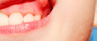
First, you will sit in the dental chair, and the doctor will examine your head and face: there are no changes in the color of the skin, the size of the lymph nodes, or swelling.
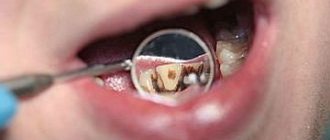
He will then ask you to tilt your head back and open your mouth as wide as possible. This is necessary to assess the condition of the upper and lower palate, tongue, inner cheeks and lips. No tools are required here, just a source of bright light. Even if you are concerned about a specific tooth, all teeth must be examined. After all, it often turns out that the problem lies in a completely different place. To do this, the doctor uses a dental mirror and a special probe.
- A small mirror allows you to carefully examine hard-to-reach areas and illuminate those places that the lamp rays do not reach. Carious cavities, tartar deposits, suppuration, chips, dental growth disorders - all pathologies appear in full view.
- Using a pointed probe (explorer), the dentist checks all suspicious crevices, depressions, cracks and darkened areas. If everything is in order, then the probe moves freely. If the enamel is damaged somewhere, then most likely there is a hidden carious cavity.
- The condition of the gums can be assessed with a periodontal probe. At its tip there is a millimeter scale and a small ball to reduce possible discomfort. With its help, the dentist receives information about the presence of inflammation, bleeding, the depth of periodontal pockets, etc.
A visual examination helps an experienced dentist immediately make a diagnosis in the following cases: caries, gingivitis, periodontitis, malocclusion. However, some diseases that occur latently, for example, periodontal disease and severe forms of caries, can only be detected using hardware diagnostics.
Main stages
Today, dentists use different methods, the choice of which depends on the condition of the oral cavity, the patient’s complaints and other factors. Basic measures include:
- Anamnesis is collected, including an oral interview and examination;
- fillings, crowns and other structures are checked;
- analysis of occlusion, identification of defects in the dentofacial apparatus;
- undergoing an X-ray examination;
- computed tomography (performed if necessary, before implantation);
- making a diagnosis, drawing up a treatment plan.
Diagnostics is an important condition for a full assessment of the clinical picture, identifying problems, and selecting effective therapeutic methods.
What diagnostic methods do we use?
Dr. Granov’s clinic has all the necessary modern equipment for early and accurate diagnosis of various diseases of the teeth and gums. Indeed, often at the initial stage, pathology develops completely unnoticed, it cannot be detected visually, and the person does not feel any symptoms.
- Radiovisiograph/ X-ray visiograph
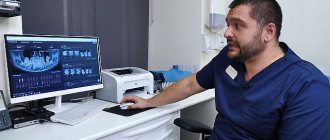
Using this device, you can obtain a targeted X-ray image of a specific area. During the procedure, the patient receives a minimal dose of radiation, so it can be repeated as often as required during the treatment process. Due to the fact that the radiovisiograph displays the image directly on a computer monitor, the dentist can enlarge it, examine it, and evaluate the exact size and location of tumors and carious cavities.
Radiovisiography (dental X-ray) is successfully used to identify pathologies such as dental cysts, tumors, periodontitis, periodontal disease, pulpitis, etc., as well as to determine the quality of treatment of the root system of the tooth.
- Orthopantomograph
An orthopantomogram is a panoramic digital x-ray of the entire maxillofacial system. It clearly shows both teeth, sinuses, bones and joints. The device also initially displays the image on the computer screen. Using an orthopantomograph, you can quickly and accurately determine the condition of all teeth and their roots, the severity of malocclusion, the position of impacted teeth, and the condition of gingival and periodontal tissues. The procedure is absolutely painless and safe, so it is prescribed even to small children and pregnant women.
International magazine
X-ray research methods in dentistry have been and remain a priority compared to other currently existing clinical and laboratory diagnostic methods due to their informativeness, ease of use, relative cheapness and quick receipt of high-quality results. This research method is widely used in absolutely all areas of dentistry and maxillofacial surgery, especially for the diagnosis of diseases of periodontal tissue, periapical tissues, traumatic lesions, cysts and tumor processes in the bone tissue of the maxillofacial area.
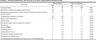
One of the main problems in dentistry today remains acute odontogenic infection. The problem lies in the need for timely and error-free diagnosis of the inflammatory process, as evidenced by the high percentage of errors that occurred during the examination. Of all the currently existing diagnostic tests, almost all of them only reflect the presence of inflammation, and it is not possible to judge from them the depth and nature of the lesion. Therefore, the most rational choice in this matter remains the integrated use of radiation diagnostic methods, which are based on modern technologies.
For each area of dentistry, as well as for maxillofacial surgery, the preferred methods of x-ray diagnostics are different research methods. For example, of all the currently known radiological methods used in periodontology, 5 main groups can be distinguished, arranging them in order of decreasing importance for the dentist and the quality of the resulting image in the following order: computed tomography; orthopantomography; panoramic radiography; targeted radiography.
Three-dimensional dental computed tomography (3D CT) has become an innovative method of x-ray diagnostics in the last decade. The following devices are currently used: 3 DX Accuitomo (Morita, Japan); GALILEOS (Sirona, Germany); Picasso Pro (Vatech, E-WOO, South Korea), Scanora 3D, (Finland). All of them have found wide application in identifying numerous diseases of the dental system, maxillofacial area, maxillary sinuses and TMJ [5].
The above CT scanners are universal diagnostic devices, powerful tools of the 21st century generation, opening an era in the diagnosis of maxillofacial pathologies and dental diseases, with enormous potential for use. 3D CT has clear advantages for both the patient and doctors of various specialties, including dentists and maxillofacial surgeons. The existing experience in using 3D tomographs indicates the high accuracy of the resulting images and, accordingly, the quality of this study, which is important for practical application in all areas of dentistry and maxillofacial surgery, as well as for increasing the level of subsequent treatment for patients based on the obtained images. 3D tomographs are of great practical importance in various fields of medicine, but the following clinical situations are of interest to the dentist: detection of unerupted (impacted and dystopic) teeth in pediatric dentistry; endodontic treatment; traumatic injuries of teeth and maxillofacial area; bone tissue examination before implantation; identification of pathology in periodontal tissues and foci of chronic inflammation in periapecal tissues; arthritis, arthrosis and other pathologies of the TMJ; tumors and tumor-like processes detected in oncostomatology; differential diagnosis of pathological processes in the Himer’s sinus of various etiological origins [2, 3].
In orthodontics, in the last decade, two main CT examination techniques, such as cone beam computed tomography (CBCT) and multislice computed tomography (MSCT), have become promising methods of radiological diagnosis. CBCT is a higher priority for orthodontists due to the higher-quality images it produces combined with a low dose of radiation, which is of particular importance due to the fact that the main patients seen by orthodontists are children. CBCT improves the efficiency of diagnosis and treatment of patients with dentofacial anomalies [1].
Innovation in the field of radiology was the appearance of devices that operate not only from an X-ray tube, but also based on the following phenomena: optical, electrical, laser. For example, a combination of computed tomography and positron emission tomography, which improved the quality of the resulting image of the object under study not only in the field of dentistry, but also in neurology, cardiology and oncology, including in a multidisciplinary clinic [4]. Another such “combined” device is the X-ray laser. In 2009, physicists at Stanford developed the most powerful X-ray laser currently available: the Linac Coherent Light Source (LCLS). Currently, active work is underway to improve it in order to be able to study the substance at the molecular level. Its ideological difference from the old generation X-ray machines is its wavelength of up to 0.15 nanometers. To date, the daytime device is the shortest wavelength. Previously, only synchrotron devices could cope with the task of studying matter at the molecular level, but unlike an X-ray laser, they required an ideal crystal to work, while LCLS can work with individual molecules. Its beam is so precise that it does not deviate more than five micrometers at a distance of five meters. The operating principle of this device is to scatter a coherent X-ray beam to visualize atoms and molecules at the time of examination. Examination using an X-ray laser differs from traditional x-ray examination, being more reminiscent of microscopy, because For a more detailed examination, sampling of material is necessary. Scientists claim that in the future, the operating principle of such devices will form the basis for other, more powerful tomographs and will make it possible to study the body in the clinic as a whole, rather than individual organs and tissues in the laboratory [6].
Improving the quality of existing X-ray examination methods, as well as the development of new diagnostic methods in the future, will allow dentists, maxillofacial surgeons and doctors of related specialties to improve the quality of diagnostic and treatment services provided in hospitals and outpatient clinics, and reduce the time spent on diagnosing each patient. To obtain more highly accurate examination results, an integrated approach is required in diagnosing the patient’s pathology, using all the resources available in the clinic and relying on the results of clinical, laboratory and radiological studies, especially those obtained with radiovisiographs and 3D computed tomographs, in order to evaluate the results obtained, diagnose and differentiate diseases. different etiologies among themselves, drawing up a more accurate treatment plan and subsequent assessment of the quality of its implementation. Thus, the use of modern high-quality devices and methods of X-ray examination, such as 3D computed tomographs in outpatient and hospital settings, increases the effectiveness of treatment of diseases of the hard tissues of teeth, periodontal disease, jaw bones and other tissues of the maxillofacial area, and therefore reduces the likelihood of relapses in patients and possible complications. As a result, the time spent on examining each patient is reduced and the total radiation exposure is reduced, which makes it possible to use these techniques not only in periodontics, therapeutic and orthopedic dentistry, but also in other areas, such as orthodontics and pediatric dentistry.
What additional diagnostics can the dentist prescribe?
- Apex locator
With its help, the doctor can determine the length of the dental root canal as accurately as possible. Due to the complex anatomy, it is difficult to do this in other ways. Selecting the right tools is essential to effectively treating root infections.
- Biopsy
This procedure is carried out if a dental cyst or other neoplasm has been diagnosed. A biopsy is necessary to ensure that the suspicious object is benign. The dentist takes a sample of fluid or tissue using a biopsy needle and sends the finished sample to a laboratory for analysis.
The diagnostic methods used in our clinic are safe and do not cause any discomfort. Thanks to them, the doctor will be able to start treatment on time and keep your smile healthy and beautiful!
Advantages of X-rays in dentistry
One of the most common diseases that occurs in almost every patient is caries. If the disease is detected at an early stage and treated, it is not dangerous. But in the absence of timely intervention, caries causes serious complications (pulpitis, periodontitis and others), which are accompanied by severe toothache and the treatment process in such cases takes a lot of time and effort. This is why it is so important to visit the dentist regularly for preventive examinations. It is recommended to undergo an examination every six months, which may include x-rays.
If a root canal filling is planned, the dentist needs to see its structure and possible individual characteristics. After filling, the specialist prescribes an x-ray for control. This is very important, because if a filling defect is not detected in time, inflammation will develop after some time, which can lead to tooth loss.
More information about caries treatment can be found here