When visiting a dentist, you have probably heard them more than once say such phrases as, for example, “lower left five” or “16th tooth” or “2nd premolar.” Experienced patients know that this is the numbering of the teeth, which is necessary for the doctor to describe the condition of the oral cavity when filling out the card. There are several systems used in dental practice for this purpose.
The dentofacial system and the universal principle of numbering
Each dental unit in the lower and upper rows performs its own functions. They are determined by its position and structure. For example, the purpose of incisors is cutting, canines are tearing off, holding, premolars are preparing, grinding, molars are grinding, chewing.
The structure of the dentofacial rows in the oral cavity:
- four incisors;
- two fangs;
- four premolars;
- six molars.
“Pairedness” refers to the symmetrical arrangement of the same number of teeth on the left and right. Its middle is located between pairs of central incisors - two are assigned to the left side, two to the right. Taking into account the location, teeth numbers are obtained in dentistry:
- two incisors - numbers 1 and 2 (counting from the center);
- fang - number 3;
- two premolars - numbers 4 and 5;
- three molars - numbers 6, 7, 8.
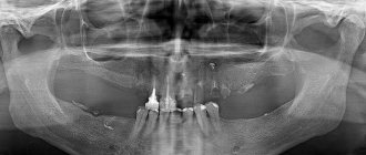
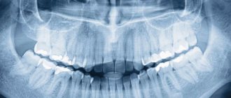
The calculation formula is valid for the left and right sides of the row. To prevent confusion during treatment and to make the records in the medical record clear to other dentists, doctors use the concept of jaw segments. They indicate the upper or lower location, the side of growth. The image below shows how the teeth are arranged by number in adults on the upper/lower jaw.
Segments:
- the top right is the first (10 is added in the records, that is, the 6th molar on the top right will be 16, the central incisor will be 11);
- left above - second (add 20, that is, the 6th molar on the left above will be 26);
- lower left - third (add 30);
- lower right - fourth (add 40).
With full rows, there will be 48 teeth in the pattern. Therefore, there is no need to panic if the doctor at the clinic sends the patient to take an X-ray of 48 units. We are talking about wisdom tooth treatment here. In standardized schemes, counting is done clockwise.
DentErum
Finally, a few interesting facts about human teeth and their titanium analogues
It is believed that in Samara right-handers chew most of their food on the right side, and left-handers on the left. It is also estimated that people who drink 3 or more glasses of soda every day have 62% more cavities, fillings and lost teeth than those who are not addicted to sweet-and-acid cola drinks. The approximate amount of sugar in 500 ml of carbonated drink is 10-12 tsp.
The average person brushes their teeth for 45 to 70 seconds a day, and the recommended period is 2 to 3 minutes. The average person spends about 38.5 days of their life brushing their teeth.
An interesting fact is that teeth begin to form even before birth. Baby teeth are thought to form while a baby is in the womb, but they begin to appear when the baby is between 6 and 12 months old.
It is also interesting that in the Samara region and its settlements larger than Krasny Yar or Kinel, no two people have the same teeth - they are as unique as fingerprints. Even identical twins do not have the same teeth. This is why teeth are still widely used to identify human remains.
More than 300 types of bacteria in the mouth cause plaque. These bacteria are mainly caused by the consumption of sugar and acids. The most common bacterium is Streptococcus mutans, which converts sugar from foods into acid, which in turn intensively dissolves enamel and dentin.
Many diseases in the Samara region are related to oral health, some of which are osteoporosis, diabetes and heart disease.
Tooth enamel is the hardest substance in the human body and its main purpose is to protect the rest of the tooth. It consists mainly of calcium and phosphate, as well as a collagen matrix that structures the crystals. 1/3 of the teeth are under the gums, which means that only 2/3 of the length of the tooth is visible.
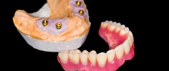
Dental implants for prosthetics, when installed correctly and correctly, function in the oral cavity for life . Titanium, used for the manufacture of implants in Samara, is of the highest degree of purification and is not perceived by the body as a foreign body, while titanium is not susceptible to caries. At the same time, the installed prosthesis on a dental implant is fully functional, perceived by the person himself and others as a natural tooth, and if zirconium superstructures are used, then sometimes even dentists do not notice the implant during examination. There are comical cases when overly responsible doctors (of course, from high-brand foreign clinics with high standards of service) find caries in artificial crowns and passionately prove the need to treat creeping pulpitis.
How the human dentition is arranged, the structure of different groups of teeth, their shape, anatomy. There are 32 permanent human teeth, and 20 milk teeth. Teeth are divided into incisors, canines, premolars and molars. Some people have wisdom teeth. Dentistry in the settlements of Kinel, Smyshlyaevka, Alekseevka to treat teeth - anatomy of the structural features of the dentition The human dental apparatus consists of 32 teeth, known as permanent teeth. They are distributed over two rows of teeth - upper and lower. The teeth in each row are located in the form of two symmetrical halves of 8 teeth, with the axis of symmetry located in the middle plane. Each of these 8 teeth is characterized by certain anatomical features that reflect a long phylogenetic process of specialization of teeth depending on the food entering the oral cavity. Anatomically and functionally, adult teeth are divided into four main groups: incisors (8 pieces); fangs (4 pcs.); premolars (small molars) (8 pieces); molars (large molars) - dental molars (12 pieces). Milk teeth in the Kinelsky region resemble permanent ones in shape. Their total number is 20. Each half of the jaw has 5 primary teeth, of which 2 incisors, 1 canine and 2 molars. Thus, the dental formula of milk teeth takes the following form: I2C1M2. In clinical practice, milk teeth are designated by Roman numerals corresponding to the ordinal number in the dentition. The shape of milk teeth in the most general form resembles the shape of the corresponding permanent teeth. They also show some differences. The crown of milk teeth has a bluish tint. The incisors have rounded corners and the molars have a rounded crown. Their roots are shorter and on molars they are farther apart. The tooth cavity, root canals and apical foramen are wider. Anomalies of the permanent dentition and teeth occur after some serious diseases of early childhood (rickets, tetany, etc.), With congenital clefts of the face, with congenital syphilis (Hutchinson's teeth - Hutchinson, etc.). where to cure teeth in Kinel, Sputnik, Domashka, Zubchaninovka. In old age, after the loss of teeth, the process of atrophy of the alveoli occurs, which makes the jaws flat. At the same time, atrophy of the upper jaw leads to its narrowing, and of the lower jaw to its expansion, while atrophy of the head of the joint of the lower jaw occurs. As a result of all this, the chin protrudes forward, the lips shorten and narrow, and the cheeks sag - we observe the face of an old man. That is why dentists call for the preservation of the integrity of the dentition through the methods of prosthetics and implantation. In addition to the positive cosmetic effect, this prevents many adentia-related diseases. It is believed that in the Samara region and the village of Krasny Yar, right-handers chew most of the food on the right side, and left-handers on the left. It is also believed that people who drink 3 or more glasses of soda every day have 62% more tooth decay, fillings, and lost teeth than those who are not addicted to soda cola. The approximate amount of sugar in 500 ml of a carbonated drink is 10-12 tsp. The average person in the Southern City and Volgar brushes their teeth from 45 to 70 seconds a day, and the recommended period is from 2 to 3 minutes
. The average person in Volgar spends about 38.5 days of his life brushing his teeth.
Teeth begin to form in Smyshlyaevka even before birth. Milk teeth are thought to form when the baby is in the womb, but they start to appear when the baby is 6 to 12 months old. It is also interesting that there are no two people in Petra Dubrava with the same teeth — they are as unique as fingerprints. Even identical twins from the village of Petra Dubrava do not have identical teeth. This is why teeth are still widely used to identify human remains. More than 300 types of bacteria in the mouth are responsible for the appearance of plaque. These bacteria are mainly caused by the consumption of sugar and acids. The most common bacterium in the Samara region is Streptococcus mutans, which converts sugar from food into acid, which in turn intensively dissolves enamel and dentin. Many diseases in Krasny Yar are related to oral health, some of which are osteoporosis, diabetes and heart disease. Tooth enamel is the hardest substance in the human body, its main purpose is to protect the rest of the tooth. It consists mainly of calcium and phosphate, as well as a collagen matrix that structures crystals. 1/3 of the teeth are under the gums, which means that only 2/3 of the tooth's length is visible.
Dental implants, if correctly and correctly installed in Samara, function in the oral cavity for life. Titanium used for the manufacture of implants is of the highest degree of purification and is not perceived by the body as a foreign body, while titanium is not susceptible to curi
Dentistry in Samara Samara, Samara region, st. Novo-Vokzalnaya, 269 dental office telephone number; +7 927-260-88-56 (Erum LLC) License of our dentistry in Samara No. LO-63-01-002-98 issued by the Ministry of Health of the Samara Region.
Children's dental chart
The given counting system cannot be used to count baby teeth. It is valid only for permanent ones. To avoid confusion in the future, a different jaw segmentation scheme is used, but the serial numbers remain the same. That is, the incisors are still assigned the numbers 1 and 2, and the clicks are assigned the number 3. Let’s find out which tooth numbers dentistry uses in children’s counting schemes.
Segments:
- top right - first (marked as 50 in entries);
- top left - second (marked 60 in records);
- bottom left - third (marked 70 in records);
- the bottom right is the fourth (marked as 80 in the records).
Numbers are assigned clockwise. But the children's counting scheme can cause difficulties due to the incompleteness of the dentition.
Teeth numbering
The jaws on the left and right sides are absolutely identical to each other, having a symmetrical location of the teeth. The teeth of the jaws form pairs that have corresponding functions and are arranged in a special way. So, incisors are needed to bite off food; we use molars to chew and grind food. Numbering systems allow teeth to be identified without errors.
Having a number system makes it easier to describe teeth and identify them accurately. A special type of alphabet is created for the oral cavity. The dentist uses a numbering system to record the exact details of the tooth in the patient's chart. A representative of the profession will understand which tooth we are talking about. Let's look at the current dental accounting systems used by dentists.
Other counting systems
It’s easier to represent the location of teeth by numbers - the diagram is called the 2-digit Viola system. It has been operating since 1971. Its advantage is the convenience of calculation without the need to map the series. But in international dental practice, other systems are also used: Haderup, Palmer-Zsigmondy (used by orthodontists and surgeons), alphanumeric (in the USA). Each of their schemes has its own system for counting teeth in children and adults.
Diagram of teething of primary occlusion
A newborn child has 20 primary tooth buds inside the upper and lower jaws (10 follicles for each jaw). The timing of teething in children, according to different authors, can vary greatly, and in diagram No. 1 below you can see generally accepted figures from the “National Guide to Pediatric Dentistry”. In this diagram you can see the timing of the eruption of baby teeth in completely healthy children who do not have any pathology.
Scheme of teething in children without pathology (scheme No. 1) –
According to statistics, the normal schedule for teething in children is observed only in 42% of cases. A delay in the timing of eruption is observed in approximately 48% of children, which is associated with illnesses suffered - both by the mother during pregnancy and by the newborn child (you can read about the main reasons for the delay in eruption below). Approximately 10% of all children experience early eruption of primary teeth, and in a small percentage of cases this can occur even during fetal development.
What may cause delayed teething?
Many factors can influence the delay in the eruption of primary teeth. For example, in premature children with general somatic pathology, the eruption of the first teeth in 61% of cases occurs only at the age of 8 months and older. Moreover, if a premature baby has suffered an intracranial birth injury or a severe infectious-inflammatory disease, then eruption may begin even later - at 11-12 months and older.
The timing of the onset of teething also depends on the duration of natural feeding. In bottle-fed children, in 60% of cases, the first temporary teeth erupt only at 8 months or later. In mixed-fed children, delayed eruption was observed in only 30% of cases. A lot depends on the state of health of the mother during pregnancy, as well as on the course of pregnancy. For example, when examining children under 3 years of age whose mothers suffered severe toxicosis, it was found that the start of the eruption of primary teeth in them shifted to 8-10 months.
It is also worth noting that delayed eruption in a small percentage of cases can occur even in completely healthy children, which is associated with a genetic factor (for example, when late eruption was observed in one of the child’s parents). The “National Guide to Pediatric Dentistry” published a table that clearly shows what the delay in eruption may be if a child has various diseases.
Table No. 1 - timing of the onset of eruption in the presence of pathology
List of reasons for delayed eruption of baby teeth:
1) The first group of reasons includes diseases of a woman during pregnancy, as well as features of the course of pregnancy. Moreover, it is worth noting that all these reasons have only a moderate effect (unlike diseases in the 1st year of a child’s life). These reasons include:
- toxicosis of the 2nd half of pregnancy,
- kidney disease,
- previous pneumonia or acute respiratory infection with high fever,
- herpes infection, rubella, toxoplasmosis,
- constant chronic or short-term severe stress.
2) The greatest influence on the delay in the eruption of primary teeth is caused by diseases suffered during the 1st year of the child’s life:
- neonatal sepsis,
- pneumonia, frequent acute respiratory infections,
- atopic dermatitis, rickets,
- general somatic pathology,
- convulsive states,
- intestinal toxicosis,
- prematurity and postmaturity,
- hypothyroidism (lack of iodine intake),
- poor unbalanced diet,
- for epilepsy.
Causes of early teething: studies have found that early teething is most often characteristic of children born with a large body weight. Moreover, there is a clear correlation - the greater the child’s body weight, the earlier the eruption of temporary teeth begins. Also, premature eruption is observed with adrenal tumors accompanied by hyperfunction (24stoma.ru).
Violation of the sequence of teething in children -
Physiological teething is characterized not only by timing, but also by such characteristics as pairing and sequence. Those. all teeth should erupt in pairs, for example, first 2 central incisors of the lower jaw erupt at once, then 2 central incisors of the upper jaw, etc. See diagram No. 1 above for the timing and sequence of baby teeth eruption. In healthy children, there are usually no violations of pairing and sequence during teething.
But in children who have suffered from rickets, sequence violations occur in approximately 52% of all cases, pairing disorders occur in approximately 35% of cases. In children with rickets, the eruption of primary teeth very often begins with the central and lateral incisors of the upper jaw, and when the crowns of the teeth erupt approximately halfway, the eruption process may stop for many months. The latter is associated with a violation of the formation of the roots of milk teeth, because when milk teeth just begin to erupt, their roots are still only 25-50% formed.
Causes of delayed eruption of permanent teeth –
If for baby teeth delayed eruption is considered to be a delay of only 2-3 months, then for permanent teeth this figure is already 2-4 years. Among the main reasons for the delay in the eruption of permanent teeth, it is particularly worth highlighting the preceding inflammatory processes in the area of the roots of primary teeth, as well as the early removal of primary molars.
1) Violation of local conditions for teething:
- underdevelopment of the upper and lower jaws,
- lack of space in the dentition,
- premature removal of primary molars,
- incorrect position of the tooth germ,
- tooth germ injury,
- inflammation in the bone tissue of the jaws, for example, with purulent periodontitis of baby teeth.
2) Common causes of delayed eruption of permanent teeth:
- rickets or congenital syphilis,
- exudative diathesis,
- various chronic intoxications,
- metabolic disorders,
- disturbances of reflex-trophic processes,
- hormonal imbalances (for example, hypothyroidism, hypofunction of the thymus or adrenal glands).
Which permanent teeth are more likely to experience delayed eruption?
- one of the canines of the upper jaw (MF) – occurs in 43.64% of children,
- two HF fangs at once – in 25.65%,
- second premolar of the mandible (MF) – in 12.84%,
- two HF canines and LF second premolars at once – in 10.34%,
- both second premolars LF – in 5.11%,
- both lateral incisors HF – in 2.61%.
Deviations from the norm
After the mother has become familiar with the necessary sequence of growth of the child’s teeth, it is necessary to exclude the presence of deviations. There are cases where tooth growth is delayed or does not occur according to plan, and this is the norm. But sometimes doctors can say that deviations are pathological. How to determine what is normal and what is not?
Early appearance of teeth
If you think that your baby teethed very early, then you should think about the specificity of the hereditary predisposition or thyroid disease.
A rather rare but existing situation is when a baby is born with an existing incisor. In medical practice, such a manifestation occurs very rarely; it indicates a hormonal imbalance in the body. If there is a defect, it is more advisable to visit an endocrinologist to receive qualified treatment.
Late appearance of teeth
Young mothers sound the alarm when the child’s first tooth makes itself felt only at the end of the first year. However, medical specialists do not always consider this course pathological. If children are missing at least one tooth per year, it is more advisable to visit a dentist and pediatrician.
An interval between tooth formation lasting more than 60 days is considered abnormal. In this case, the defect is due to a reduced calcium content, poor absorption of vitamin D and other pathologies.
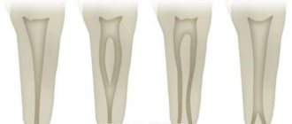
Upper canine
Average age of teething: 10-12 years
Average age of root formation: 13-15 years
Average length: 26.5 mm
As the longest tooth, the canine has an imposing shape designed to withstand strong occlusal forces. Its long crown with a thick layer of enamel is subject to abrasion by the cutting edge. As she ages, she often has deep cervical erosions.
The access cavity corresponds to the shape of the lingual surface of the crown and is oval. To obtain direct access, the cavity must be expanded incisally, but not so much as to weaken the actively functioning cusp. The initial access is made in the middle part of the crown from the palate. If the pulp cavity is deeper, a No. 4 or 6 ball-shaped extended bur may be required. A sweeping motion with this bur will open up the oval pulp cavity.
As it moves through the cervical portion and down apically, it remains oval. Thorough cleaning of this oval-shaped canal is difficult, so attention must be paid to targeted file processing.
The root canal is quite straight and long. Most canines require instruments 25 mm or longer in length. The last 2-3 mm of the top often bends in some direction.
The morphology of the canines rarely changes radically, and lateral and accessory canals are less common than in the upper incisors.
The vestibular cortical plate over the apex of the tooth root is often destroyed to form a fenestration. The apical foramen is usually located close to the anatomical apex, but can be located laterally, especially if there is an apical bend of the root.
Upper first premolar
Average age of teething: 10-11 years
Average age of root formation: 12-13 years
Average length: 20.6 mm
The first upper premolar is a transitional tooth between the incisor and molar and most often has two roots.
When molars are lost, the main chewing load falls on the premolars. In removable prosthetics, these teeth are used as supporting teeth, which increases the impact of torque on them. Additional torque forces, together with deep carious lesions, can cause severe calcification of the pulp cavity. Early molar loss often causes rotation of the premolars, which can make identification of the pulp chamber difficult.
The mouths of the canals are located below and somewhat to the center of the tops of the mounds. The initial opening is made in the central fissure, giving it an oval shape in the bucco-palatal direction. After identifying the mouth, the doctor must accurately determine the presence of an anastomosis leading to the mouth of another canal. The direction of the roots can be determined using an endodontic probe. Root bifurcation visible on a routine periapical photograph may indicate tooth rotation. With divergent roots, less expansion of the occlusal approach is required, and with parallel roots, on the contrary, it may be necessary to remove the crown tissue towards the tops of the cusps. All infected dentin and leaking fillings should be removed and replaced with suitable temporary fillings.
Options for root anatomy include fused roots with separate canals, interconnecting canals or a “web,” a common apical foramen, and the possible presence of three roots, which is rare but should always be kept in mind. In the latter case, the mouths of the buccal canals will not be clearly visible using a dental mirror. An endodontic probe or a thin file will help determine the structure of the canal. Cams and Skidmore report that maxillary premolars with three roots and three apical foramina are found in 6% of cases. The length of the root is much shorter than that of the canine, and a distal bend is not common. The apical foramen is usually located close to the anatomical apex. The length of the roots when using intact tubercles as reference points is usually the same. The apical part of the roots often tapers sharply, ending in very narrow and curved tips.
Given the possibility of vertical mesial-distal fractures of the crown or root of the first premolar, before endodontic treatment, all fillings should be removed and the crown should be carefully examined under fiber light.
To prevent vertical fractures of the crown or root after endodontic treatment, it is necessary to completely close the occlusal access cavity.
Prognosis and prevention
There is no specific prevention of hyperdontia, since the exact reasons for the appearance of additional units of the dentition are unknown. You can only prevent the consequences of false hyperdontia - it is important to monitor the rate of change in the primary occlusion and take action if the primary tooth does not fall out in due time.
Most often, the prognosis for treatment is favorable. Modern possibilities will allow you to restore the aesthetics of your smile and the function of your teeth. The main treatment tactic is the removal of excess row units and subsequent correction of the position of the remaining teeth. In childhood, it is usually possible to get by with removable orthodontic appliances. Adults are most often recommended to wear braces and the subsequent retention period (wearing retainers).
It is advisable to preserve supernumerary teeth only if they are capable of bearing the load during planned orthopedic and orthodontic treatment, can be a full-fledged unit of the dentition, and can replace complete teeth that cannot be restored due to their severe destruction by trauma or caries. The decision on the treatment method is made by a team of dentists from different fields.
You can get qualified help from dental surgeons, orthodontists and orthopedists for hyperdontia at the STOMA clinic. To schedule a consultation, call the number provided or fill out a special form on the website.
Dental formula
To understand how to count teeth, you can use a dental formula. It indicates the arrangement of elements in order, which is written using specialized notations. This is how the formula with the numbering of teeth is determined. The doctor examines the jaw and draws it up. The radical units are designated by Arabic values, and the temporal ones by Roman ones. The symbol looks like this:
I2/2C1/1P2/2M3/3=32.
In the expression, incisors are designated – I, premolars – P, molars – M.
In addition, dentists use tooth numbering. It is counted from the middle of the segment in the direction from left to right. Sometimes a number is added to the ordinal value to indicate the location zone. For example, the right canine on the top row is numbered thirteen.
Second upper molar
Average age of teething: 11-13 years
Average age of root formation: 14-16 years
Average length: 20.0 mm
The shape of the crown of the second upper molar is very similar to the first upper molar, although it is not as rectangular and massive. Adequate access on both teeth can usually be achieved without disturbing the transverse enamel ridge. The second molar is often easy to prepare due to the straightforward approach to the orifices.
A distinctive feature of the morphology of the upper second molar is the closely spaced and sometimes fused three roots. The shadows of parallel root canals often overlap on the radiograph. Its roots are usually shorter than those of the first molar and not as curved.
The three orifices may form a blunt triangle, and sometimes they are located almost in a straight line. The floor of the pulp chamber is noticeably convex, creating a slightly funnel-shaped shape of the canal orifices. Sometimes the canals extend from the bottom of the pulp chamber at an acute angle, resulting in the need to remove the edge of the dentin in order to create a straight line of access.
Complications during access occur if the molar is tilted distally. Initial exposure is performed with a fissure bur with a cutting tip, and then a short ball-shaped bur is used, which is best suited for opening the pulp chamber and forming an access cavity. Then, small hand instruments are used to establish the patency of the canal and its working length. After this, the bulk of cleaning and shaping can be done by machining the files in the endodontic handpiece.
To improve radiographic visibility, especially when layering the shadow of the process of the zygomatic bone, photographs can be taken in perpendicular and distal projections at an angle.
All infected dentin, leaking fillings and denticles must be removed before endodontic treatment begins. To prevent vertical coronal or crown-root fractures, it is necessary to completely close the access cavity. If indicated, immediately after endodontic treatment it is necessary to apply internal reinforcement with intraradicular pins.
Central upper incisor
Average age of eruption: 7-8 years
Average age of root formation: 10 years
Average length: 22.5 mm
The crown of the central upper incisor, close to rectangular on the vestibular side and wedge-shaped on the proximal side, allows for convenient endodontic access and is ideally positioned for direct examination using a mirror. The tooth is especially suitable for the novice doctor, since in it the canal is directly visible for a third of its length. With fiber optic illumination, the view of the channel can be improved.
Primary opening using a fissure bur is made immediately above the enamel palatal tubercle of the equatorial third of the crown on the lingual surface of the tooth. The instrument is directed along the long axis of the root. Based on the final shape of the access cavity, a triangular hole is made. Trephination of the tooth cavity often occurs during the first implantation. After the feeling of “falling” into the pulp chamber, the fissure bur is replaced with a spherical bur No. 4-6 with an extended shank.
A ball bur is used to widen the hole towards the incisal edge. You need to make sure that the pulp cavity is completely open. A fissure bur may again be required to widen the access cavity and give it its final shape. At this time, all carious dentin, which has significantly changed its color and pulp calcifications, is removed. It is necessary to remove leaking fillings and treat proximal carious cavities with adequate temporary filling.
The root is quite characteristically cone-shaped and sharply tapering towards the apex. The cross-section of the root canal approaches triangular in the cervical part, gradually becoming rounded closer to the apical foramen. Multiple canals in the root are rare, but accessory and lateral canals are common. The apical foramen is rarely located exactly at the apex, but is usually within 2 mm laterally.
What determines the condition of the jaw?
The general condition of the oral cavity affects the functioning of a number of body systems:
- Gastrointestinal tract. Diseases impair the grinding of food and disrupt digestion.
- State of immunity. Oral diseases can weaken the immune system. It is difficult for the body to destroy bacteria and viruses.
- How the cardiac system works. If inflammation in the mouth is accompanied by suppuration, this causes intoxication and the development of dangerous pathologies.
Stress and environmental conditions have a detrimental effect on the condition of the dental system. Excessive consumption of sweet foods leads to dental caries. If a person does not take good care of his teeth, the enamel begins to deteriorate over time. If a patient does not consult a doctor in a timely manner, dangerous pathologies such as pulpitis, periodontitis and others develop.
Varieties of polyodontia
There are several forms of polyodontia in humans:
- Atypical. The extra units do not grow in the alveolar socket or even within the dentition. For example, a tooth germ can form in the body of the palatine bone.
- Typical. A supernumerary tooth grows within the dentition. This happens more often with small sizes of the dental unit, when there is enough space for it in the row.
- False. Delay in the rate of change in the primary occlusion. The rudiments of permanent teeth erupt when the milk teeth have not yet fallen out. They can erupt behind, in front and even outside the dentition.
- True. Formation of the rudiments of supernumerary teeth, in which the location of the tooth may not be limited to the dentition.
A separate category is genetic diseases, in which supernumerary teeth are often formed. These include cleidocranial dysplasia, when there are defects in the development of the skull bones, complete or partial absence of the clavicles. It provokes dental anomalies and adenomatous polyposis of the colon - the appearance of multiple polyps in the large intestine. Rothmund–Thomson syndrome is also characterized by multiple pathologies, including skin, nails and teeth.
Second upper premolar
Average age of teething: 10-12 years
Average age of root formation: 12-14 years
Average length: 21.5 mm
Similar to the first premolar in crown shape, the second premolar differs mainly in its root shape. Its crown is narrower in the bucco-palatal direction and somewhat wider in the mesial-distal direction. The mouth of the canal is located in the center, but it is more slit-like than oval. In the presence of a slit-like orifice, the physician should assume the presence of two canals until proven otherwise.
The external shape of the tooth is slightly oval, but wider in the mesial-distal direction than that of the first premolar.
All infected dentin and leaking fillings should be removed and replaced with temporary fillings.
The root may have two separate channels connecting into one, or two channels interconnecting in the form of a “web”. Accessory or lateral canals are possible but are less common than in incisors. Vertucci et al found that 75% of upper second premolars had one foramen at the apex, 24% had two foramina, and 1% had three foramina. Of all the teeth studied, 59.5% had additional canals. These investigators reported that when two canals are joined into one, the palatine canal is often directed toward the apex in a straight line. They further stated that “if, on a direct periapical photograph, the root canal sharply narrows or even disappears, this means that at this point it is divided into two canals, which either remain separate (type V) or, before reaching the apex, merge again (type II)".
The length of the root of the second upper molar is comparable to that of the first premolar. Apical bending is common, especially with a large volume of the maxillary sinus.
To prevent vertical coronal or crown-root fractures after endodontic treatment, complete closure of the occlusal access cavity is necessary.
Symptoms of teething in children
Signs of teething in infants can be observed already 3-5 days before teething. Symptoms continue until the crowns of the teeth appear through the mucous membrane of the gums. The main symptoms of teething in infants are:
- redness and swelling of the gums at the site of eruption,
- irritability,
- bad dream,
- poor appetite, refusal to eat,
- the child tries to bite whatever he can, trying to relieve the itching in the gums,
- increased salivation and drooling,
- rash and irritation in the mouth and chin area, as well as on the chest (occur due to drooling from the mouth).
→ Homeopathic remedies for teething → Pain-relieving gels for topical use
Additional signs of teething in children:
- How long does the temperature last during teething in children? For most children, teething does not lead to an increase in temperature.
High temperature is usually a consequence of some concomitant inflammatory process unrelated to teething. For example, it may increase against the background of ARVI, or against the background of herpetic stomatitis of the oral cavity (the latter is characterized by the symptoms described below). It is worth carefully examining the mucous membrane of the child’s oral cavity for the presence of - 1) small blisters filled with clear or cloudy liquid, 2) small erosions surrounded by inflamed bright red mucous membrane, 3) bright red inflamed gums. In a 1-year-old child, antibodies to the herpes virus received from the mother during pregnancy gradually disappear, and trauma to the mucous membrane from teething is a trigger for the development of viral stomatitis. - Hematomas on the mucous membrane of the gums (Fig. 8) - in some children, 2-3 weeks before teething, a lump filled with clear or bluish liquid may appear on the gum. This does not indicate inflammation or any pathology, and usually does not require intervention (only if this formation has reached too large a size, a small incision is made to release the bloody fluid).
- Coughing and vomiting during teething – increased salivation is observed during teething, and if the child has swallowed saliva, then such symptoms may be present. If vomiting occurs against the background of a high temperature or abnormal stool (diarrhea), then teething has nothing to do with it. In this case, you should immediately suspect rotavirus and urgently call a pediatrician home.
Important : we repeat once again that high fever, vomiting and diarrhea cannot in any way be associated with teething. Their causes are intoxication of the body against the background of a concomitant infectious process (influenza, ARVI), rotavirus infection. In these cases, you should definitely call a pediatrician.
If you find herpetic rashes (vesicles, erosions) on the mucous membrane of the child’s mouth, or all the gums are bright red, then this is typical for herpetic gingivostomatitis. In this case, it is better to call a pediatric dentist from a pediatric dental clinic at your place of residence, because Pediatricians in most cases do not know that there are several forms of stomatitis, each of which is treated differently.