Development of teeth in the embryonic period and after birth
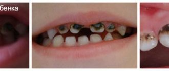
At the 6-7th week of embryogenesis, the first signs of teeth appear. At the time of birth, the crowns of the central incisors are formed in the child, the crowns of the lateral incisors are not fully formed, half of the crown of the canines is present, as well as the chewing surfaces of the remaining teeth. Many surfaces have incomplete mineralization.
In each jaw of a newborn there are 18 dental follicles, including 8 permanent and 10 temporary. They all have different degrees of mineralization. By about five years of age, the growth of the rudiments of permanent teeth is activated and the process of physiological resorption of the roots of baby teeth begins. Against the background of these changes, a change in bite begins - temporary to permanent.
Methodology
There are several ways to take an X-ray of a child's jaw or lower skull.
Intraoral radiography
Intraoral x-rays are carried out using special dental devices, which help take targeted images of one or more adjacent teeth (one to four). Such an examination allows us to assess the condition of the hard tissues of primary and permanent teeth, periodontal and periodontal tissues, jaw bones for the diagnosis of retention and follicular cysts, oncological tumors, anomalies in the location and number of erupted teeth and rudiments, etc.
When performing a study, a blank of x-ray film is placed in a special paper envelope, which is inserted into the oral cavity and placed in the area of the desired tooth. The X-ray machine is brought to the face from the outside and placed as close as possible to the tooth/teeth being examined.
In pediatric dentistry, intraoral radiography is often prescribed to diagnose pathologies of the palatal suture. Although, it must be said that if we are talking about a very young patient (2-3 years), it is quite difficult to carry out such an x-ray, because it can be difficult to explain to a child why it is needed and that it will not hurt.
Extraoral methods
The most common method of performing extraoral x-rays is a panoramic photograph, or orthopantomogram. In such photographs, you can see in a direct projection the entire upper and lower dentition, as well as the structure of the patient’s jaw. Orthopantomogram images allow you to make a detailed interpretation and description of the condition of teeth and gums, to recognize even hidden pathologies or those that have just begun to develop (for example, periodontal disease, caries).
You can see what a panoramic x-ray of a child’s jaw looks like in a photo on the Internet.
Features of the shadow picture of temporary teeth
On x-rays, the roots of primary teeth are shorter and the bifurcation angle of molars is greater than that of permanent teeth. The root canals are wider, and the dental cavity is larger in volume. The size of the periodontal gap in a child may vary, and this is considered a normal variant.
The dental follicle appears on an x-ray as a rounded clearing and has a clear rim of compaction. In its cavity there is a rudiment, which changes at different stages. First, points are visible along the cutting edge, which gradually form a single contour of the crown.
Content:
- Indications for orthopantomography
- How does the procedure work?
- Preparation
- Contraindications
- What can be seen when interpreting an x-ray
- Negative effects of X-rays: dispelling myths
- 3D tomogram
This X-ray diagnostic examination provides the dentist with the most complete information about the condition of the teeth, gums and periosteum. This procedure (also called orthopantomography) allows you to accurately determine the cause of pain, identify an asymptomatic disease, and determine treatment tactics and orthodontic correction. Our medical center is equipped with modern equipment that provides a clear image of the tooth and minimal radiation exposure.
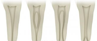
What unformed root tips look like on an x-ray
In unformed tips, the length of the root is almost normal, the walls are parallel to each other. The wide root canal ends in a funnel-shaped extension. In the area of the apex, the periodontal fissure merges with the growth zone, which incompetent specialists sometimes identify as a pathology.
Throughout the entire length of the root and at the apex, a compact plate of the socket wall is differentiated. This picture is typical for the upper central and lateral incisors in 8-year-old children, for the lower central incisors in 6-year-old children, as well as for the lower lateral incisors and first lower molars in 7-8 year old children.
The issue of X-ray safety for children
Any X-ray radiation, including in the dental industry, carries a certain degree of danger to the patient’s health. Research in the field of medicine confirms the possibility of developing acute radiation sickness when exposed to increased radiation. However, X-ray equipment is not enough to provoke it.
X-raying of a child’s primary teeth can be safe if the examination schedule and radiation safety standards are followed.
The permissible annual exposure rate, according to regulatory documentation, is 5 mSV (millisievert). In the case of small patients, as well as pregnant and lactating women, this figure is halved.
Dental radiography is characterized by minimal mZV indicators. With digital X-rays, the amount of single exposure is 0.01-0.03 mSv. In this case, the permissible single dose is 0.1 mSV. The development of radiation sickness is discussed only when this figure increases by (0.7 ZV).
X-rays are often prescribed periodically, for example, as part of a long therapeutic course. How often can dental x-rays be taken? According to WHO, if 5-6 such procedures are carried out during the year, the radiation background of a small patient will not be disturbed. However, one should not lose sight of the factor of the presence of its own background radiation in the area in which the child lives. For example, for Moscow this figure is 20 µSV.
There is no official evidence that chronic doses of irradiation to the oral cavity (for example, long-term therapy) result in the development of cancer.
What can be seen when interpreting an x-ray
In the picture you can see:
- Borders, cavity of the maxillary sinuses.
- Position of the jaw joints.
- Condition of periodontal tissues, canals, periosteum
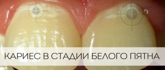
- Carious lesions: their exact localization, the size of the focus of dentin destruction.
- Cysts, granulomas, their size and location.
- Foci of the inflammatory process.
- Previously placed fillings (how tightly they fit to the dentin, are there any secondary carious cavities under them, etc.), dental implants.
- Unerupted permanent molars, incisors and canines.
Contraindications
The picture is not taken for children until they reach the age of 5. Also, the use of this diagnostic method is limited during pregnancy. According to strict indications and with the permission of the gynecologist, only digital radiography is allowed using all possible means of protecting the chest and abdominal area.
But even taking into account the minimum x-ray load, it is not recommended to take a panoramic photograph of the teeth in the first trimester of pregnancy. It is for this reason that you should visit the dentist and perform all the required treatment measures at the stage of planning conception.
Operating principle
The technique is based on X-ray radiation, which is combined with special programs and equipment. X-rays are a special type of radiation that can pass through the tissues of our body. These rays pass through some tissues (for example, abdominal organs) very easily, but through others (for example, muscles, ligaments or bone tissue) - worse, since they are partially or completely absorbed.
When taking a panoramic photo, the body receives a certain amount of radiation. Some of the rays are retained by tissues, while the rest passes through them. All information is sent to a special computer, which analyzes the received data and forms a contour image of the organs located in the path of radiation. As a result, the most accurate image of the oral cavity is obtained, on which the roots of all teeth, periodontal tissue, etc. are clearly visible.
Indications for orthopantomography
A panoramic photograph of teeth is required by a specialist in the following cases:
- Comprehensive diagnostics before implantation. Before installing a dental implant, the doctor needs to evaluate the density and structure of the bone tissue, the distance to the maxillary sinuses (in case of maxillary prosthetics) and to the mandibular nerves. The wrong choice of implant type can impair the sensitivity of the facial nerves.
- Treatment of complex caries. As a rule, in doubtful cases, when it is important to assess the depth of the lesion and find out whether the canal is affected, an x-ray of a separate area is taken. But in a situation where caries is detected on several dental units, it is better to do an orthopantomography to determine the condition of the jaw as a whole.
- Control of the eruption of permanent molars, canines and incisors. Based on the results of the image, the correct formation of the dentition in children and the growth characteristics of “eights” are determined. Their incorrect location can negatively affect the bite, which often requires surgical correction.
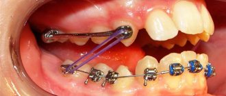
Monitoring of treatment and implantation. This approach allows you to promptly identify possible complications and, if necessary, make changes to the treatment plan.- Preparation for correcting the bite before installing braces and plates.
- Additional diagnostics. Sometimes the patient of the dental clinic himself cannot clearly indicate the localization of the pain syndrome, and on the very surface of the enamel there are no obvious signs of carious processes, and the mucous membrane of the oral cavity is not changed.
- Assessing the severity of damage in trauma.
- Examination before surgery according to a dental or ENT profile.
- Inspection of the roots before prosthetics and installation of a “bridge”.
As practice shows, not everyone follows recommendations regarding regular visits to a specialist (at least once every 6-7 months if there are no complaints). As a result, many end up in the dentist's chair in already extremely advanced cases, for example, when the upper part of several dental units is missing, all the symptoms of long-term progressive periodontal diseases are present. In such a situation, the doctor always suggests an x-ray examination, after which he will clearly determine a step-by-step treatment strategy.
This diagnostic method is also used in otolaryngology to examine the nasal passages and nasal septum to identify the connection between the infectious process in the maxillary sinuses and damage to the upper teeth. The examination is sometimes recommended by pediatric dentists when deciding whether to treat primary teeth. It is a rare child who will be able to sit quietly in the dentist’s chair for a long time, and even more so when it comes to filling a root canal. The image will help you decide whether it is worth engaging in complex therapy or whether it is better to immediately remove the tooth painlessly if this does not affect the formation of the bite in the future.
3D tomogram
An alternative to radiography is a more informative computed tomography. The essence of the method is to take several pictures of layer-by-layer sections of a certain area at once. Three-dimensional orthopantomography is often used in preliminary computer modeling of upcoming dental implantation or prosthetics.
It is this diagnostic technique that is insisted upon when it is necessary to differentiate neoplasms found in the periodontium and tissues of the palate, and to correct complex forms of malocclusion (especially at a later age).
Whether a panoramic photograph of the tooth or a tomography is needed is determined by the attending physician. At the subsequent consultation, the dentist talks in detail about all the problems found, explains what therapy is ahead, and answers all questions.