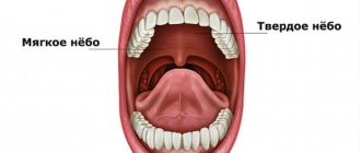
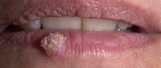
Problem: a 55-year-old woman came to the Family Dentistry with a complaint about a round, protruding formation in the oral cavity, on the cheek, which did not hurt, but created discomfort. The preliminary diagnosis made by the surgeon was fibroma of the oral mucosa.
Solution: surgical removal of fibroids from the oral mucosa using a laser was performed. The diagnosis of fibroma was confirmed by histology.
Surgeon V.P. Alaverdov explained to the patient during the consultation what kind of formation was in her mouth (preliminary diagnosis - fibroma), and how to eliminate it. Since the operation does not take long and is performed under local anesthesia, cheek fibroids can be removed on the day of the consultation.
Fibroma
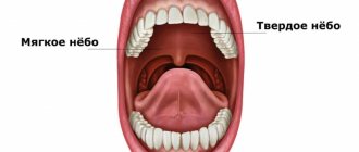
is a benign neoplasm of connective tissue. In appearance, oral fibroma is a raised, round formation with a wide base or stalk. Fibroma can be located on the mucous membrane of the cheeks, lips, tongue or soft palate. The surface of the fibroma is smooth, without growths or other changes in the mucosa.
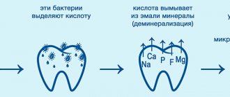
The appearance of fibroids is provoked by frequent injuries to the oral mucosa, for example, with sharp edges of teeth or dentures, frequent cheek biting, as well as chronic inflammatory processes (stomatitis, gingivitis, periodontitis, etc.). Clarifying the cause of fibroma is important in order to prevent its secondary formation, eliminate the traumatic factor, or compensate for chronic diseases. In the case under consideration, the cause was injury to the cheek by the sharp cusps of the distant teeth. It is recommended to consult a dentist about smoothing out the bumps.
Fibroids usually grow slowly in size, and foreign body discomfort is the reason to see a doctor when the fibroids become large enough.
Fibroma is not dangerous in itself, but frequent trauma to the fibroma can lead to its malignant degeneration.
In this case, there are two formations that are located on the buccal mucosa. By all characteristics, these neoplasms correspond to fibroma (an exact diagnosis is established only after histology) - they have a dense consistency, rise above the surface of the mucosa, are located on a stalk, there are no changes on the surface.
The treatment for oral fibroids is surgical removal. The operation is performed under local anesthesia and does not require any preparation.
The surgeon offered to remove the fibroma with a laser on the same visit, to which the patient agreed.
Reasons for appearance
Fibroids on the gums most often appear in children under 10 years of age and adolescents. In adults, the pathology is less common. The growth of a tumor can be provoked by the following factors:
- permanent tissue injury from fillings, artificial crowns, dentures, solid food,
- stomatitis, gingivitis, periodontitis,
- taking certain medications - estrogens, anticonvulsants, cyclosporine.
In children and adolescents, the growth of fibroids is usually due to a hereditary predisposition; in adults, it is caused by injuries to the mucous membranes.
Types of oral fibroids
- Dense (hard) fibroma . The formation consists of coarse connective tissue fibers containing a small number of nuclei, tightly adjacent to each other. This type of fibroma is most often located on the gums or hard palate.
- Soft fibroma . The neoplasm has a softer structure due to the formation of thin and loose fibers, the structure of which contains a large number of nuclei. This tumor is localized on the tongue and inside the oral cavity on the cheeks. In some cases, mixed neoplasms such as fibrohemangiomas or fibrolipomas may occur.
- Fibroma from irritation . This neoplasm is not a tumor and is quite common. It develops as a result of damage by mechanical or chemical means. This fibroma is located on the mucous membrane of the oral cavity and looks like a pink papule with clear boundaries. As it grows, a dense, rounded nodule appears. With constant trauma to the fibroma, lumpiness and ulceration may appear on its surface.
- Symmetrical bean-shaped fibromas with a dense consistency are usually located near three molars on the surface of the gums of the upper jaw. Such a tumor is not a true fibroma, but is an overgrowth of the gums and is accompanied by tissue scarring.
- Lobular fibroma . This neoplasm is distinguished by a lumpy surface that occurs as a result of reactive hyperplasia of the gum tissue when it is regularly injured, for example, by a removable denture.
- Fibrous epulis . This neoplasm of dense consistency is located on the gums and has slow growth.
Symptoms
A benign tumor forms slowly and does not cause discomfort, so it may remain undetected for some time. If the fibroma reaches a large size and is constantly injured, the following may occur:
- swelling and redness of the gum tissue,
- discomfort and pain of varying intensity.
Constant trauma to the tumor can lead to the beginning of active growth and transition to a malignant form, infection, loosening and tooth loss. Therefore, at the first signs of fibroma, it is important to consult a doctor.
Signs of oral fibroma
Fibroma looks like a pink hemispherical formation, rising above the general surface of the mucous membrane and having a wide, strong base or thin stalk. Fibroids do not cause pain. Its surface is smooth and does not have any growths, unlike papilloma. As a rule, no changes in tissues and mucous membranes are observed in the area of the fibroma, but in some cases ulcerations may appear over the neoplasm. In this case, an infection develops followed by inflammation, expressed in redness, swelling and pain in the area where the fibroma is located.
A standard fibroma in the oral cavity grows slowly, almost unnoticeably. And if it is constantly exposed to trauma, then the growth of the tumor may slow down, and the tumor itself will be within the initial stage of development. It is worth considering that constant injuries lead to complications: the tumor degenerates into a malignant one.
Treatment methods
Fibroids on the gums are usually removed with a laser or radio wave method. The operation lasts up to 40 minutes. If the neoplasm is large, to prevent deformation of the mucosa, the wound surface is covered with a flap formed from adjacent tissues.
If fibroma is formed due to tissue injury from a filling, crown or prosthesis, you can do without surgery. The tumor may shrink or disappear after the provoking factor is eliminated. In this case, treatment includes refilling or replacing the prosthesis.
Advantages of laser in removing oral fibroids
Using a laser to remove fibroids in the mouth has its advantages:
- the wound is sterile, since there is no contact with the instrument (tissues are evaporated by a high-temperature laser beam);
- there is no bleeding, since the vessels are immediately sealed with a laser;
- postoperative swelling is reduced.
The Family Dentistry uses the PICASSO diode laser (USA). All specialists working with lasers have undergone certified training.
The choice of method of operation remains with the surgeon, but in clinics where both a scalpel and a laser are used, there is more opportunity to choose the most optimal method for removing fibroids.
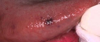
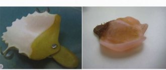
Under local anesthesia, both fragments of fibroma of the oral mucosa were removed completely, including the base. The duration of the operation is 10 minutes.
The photo shows the view immediately after removing the fibroma with a laser - the wound is small, there is no bleeding:
As in the case of all other neoplasms, the removed tissues are sent for histological examination to exclude malignancy of the formation and make an accurate diagnosis.
In this case, the mucous membrane will heal on its own; no stitches or tissue replanting are required.
Signs of the disease
Fibroma in the oral cavity has the appearance of a spherical formation, painless to the touch. The neoplasm rests on a low stalk, so it protrudes above the surface. The tumor has a smooth surface and is completely free of growths, erosions or any visible changes in the mucous membrane. Fibroids, as a rule, grow and increase in size quite slowly and relatively imperceptibly for the host. If the tumor has not been injured, it may remain “quiet” for some time, maintaining its normal size. If the traumatic factor has not been eliminated, over time a malignant tumor may form in its place.
Forms of tongue fibroids
As we described in the article on fibroids photo, tumors of the tongue have 2 main forms:
- Soft
- Solid
And 3 more additional forms:
- Symmetrical fibroma
- Lobular
- Fibrous epulis
Although fibroids are more often a skin disease, tongue fibroids should be diagnosed and treated by a dentist. You may also need to consult an oncologist, and if the patient has dentures, also an orthopedic dentist.
Fibroma is a benign tumor consisting of mature connective tissue. Fibroma of the oral mucosa is a common reason for visiting a dental surgeon [1]. However, true fibroma in the oral cavity is rare. According to N.I. Kriheli et al. [2] basically this term describes various tumor-like formations that are a response to chronic mechanical irritation and trauma, can develop as a result of biting, in the presence of sharp edges of teeth, fillings, dentures and, as a rule, more often occur in the elderly . Knowledge of medical tactics and surgical methods is necessary for the successful treatment of patients with this type of pathology and the prevention of the development of malignant neoplasms.
Purpose of the study
— apply the tactics of traditional surgical treatment of patients with benign neoplasms of the oral mucosa using the example of fibroid removal.
Material and methods.
A patient contacted the Department of Hospital Surgical Dentistry and Maxillofacial Surgery of the Moscow State Medical University with complaints of a formation under the edge of the body of a complete removable denture on the lower jaw, which arose about 1 year ago. Over time, the formation gradually increased in size, and minor pain in its area periodically appeared when wearing a prosthesis. I had not seen a doctor before.
Status locales: on the inner surface of the lower lip and along the transitional fold in the area of missing teeth 4.1, 4.2, 4.3 against the background of an unchanged mucous membrane, a new formation of soft elastic consistency is determined, consisting of two lobes, measuring 1.5×1×1 cm, with clear boundaries, on palpation it is inactive and slightly painful, the mucous membrane over the formation is slightly hyperemic.
Diagnosis: lobular fibroma of the mucous membrane of the lower lip in the area of missing teeth 4.1, 4.2, 4.3. Fibrous growth of the mucous membrane of the lower lip in the area of missing teeth 4.1, 4.2, 4.3 - D10.0 (ICD-10).
The patient is indicated for surgical treatment. The method of choice is traditional surgical treatment - excision of the tumor with a metal scalpel within healthy tissue. An alternative option for surgical intervention using a laser scalpel is possible. Conservative treatment is necessary at the stage of relieving local inflammatory phenomena (hyperemia, swelling, pain) if present. Progress of the operation: under infiltration anesthesia Sol. Ultracaini DS forte (4%, 1:100,000, 1.7 ml) the neoplasm is excised within healthy tissue. Hemostasis. Iodoform turunda was sutured into the wound. The material was sent for pathohistological examination. Assignments and recommendations are given.
Results.
A fibroma of the lower lip mucosa was removed. Postoperative management of the patient was carried out using general anti-inflammatory therapy, local antiseptic treatment and application of epithelializing agents to the wound area. Within a month, complete epithelization of the mucous membrane occurred, and the anatomical and functional integrity of the vestibule of the oral cavity was restored. The patient was referred for the manufacture of a new complete removable denture for the lower jaw.
Conclusion.
The stages of the operation to remove benign neoplasms of the oral mucosa are shown using the example of removing fibroids from the mucous membrane of the lower lip. The tactics of postoperative management of the patient in the conditions of a daily outpatient consultation with a dental surgeon are shown. It should be noted that the choice of treatment tactics in each specific case is individual and depends on the stage of the disease.