Caries is a progressive pathology that does not go away on its own without treatment. It gradually develops and moves from one stage to another, covering more and more tooth tissues. The second degree of the disease is called superficial caries. In this article we will tell you why it occurs, how it manifests itself and how it is treated.
In this article
- Causes of caries
- Reasons for the transition of caries from the first stage to the superficial
- Classification of early stages of caries
- How is caries diagnosed?
- Treatment of superficial caries
- What happens if superficial caries is not treated?
- Prevention of superficial caries
Superficial caries is the second stage of carious lesions, in which the cavity formed in the enamel does not affect the dentin. This form of the disease occurs due to the progression of the first degree of caries. At the second stage, subjective signs of the disease appear, that is, the patient’s complaints. But there is still a chance to cure it without installing a filling. Let us describe in more detail the process of development of superficial caries.
Causes of caries
The carious process occurs as a result of the influence of various factors on the human body - external and internal. But the main cause is cariogenic microorganisms, the waste products of which destroy the solid components of the enamel. The acidic environment created by these bacteria corrodes the outer coating of the tooth, which does not have time to recover naturally. There are fewer minerals in the enamel, it softens and cannot fully protect the internal dental tissues from external factors. The area of demineralization gradually increases and forms a carious cavity.
However, microbes alone are not enough to cause caries. There must be factors predisposing to the disease. The main ones are:
- Poor oral hygiene. Food debris stuck in the interdental spaces is a favorable environment for the active proliferation of bacteria. Also, irregular tooth brushing causes plaque formation, and one gram of dental plaque contains over 250 billion microbes. In this regard, caries often affects people who neglect basic hygiene rules.
- Abuse of sweet foods. Sweets disrupt the balance of minerals in the oral fluid. Because of this, trace elements are washed out from the enamel. It becomes vulnerable to bacteria that feed primarily on carbohydrates. For this reason, children who eat sweets in large quantities are more likely to suffer from caries.
- Abnormal structure of teeth: crowding, malocclusion, deep narrow fissures, etc. This complicates the process of cleaning the mouth from food debris and plaque.
- Genetically determined weakness of enamel, congenital pathologies affecting the condition of teeth and bones.
- Decreased salivation. Saliva contains substances that neutralize acids produced by bacteria. With insufficient salivary fluid production, microbes multiply faster.
- Lack of phosphorus, fluorine, calcium and magnesium in the body, which ensure the resistance of enamel to external factors.
Now let's look at the main reasons why superficial caries occurs.
Reasons for the transition of caries from the first stage to the superficial
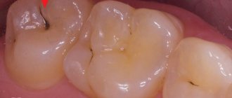
The initial stage of caries is called the white or chalky spot stage, when an area of demineralization forms on the enamel. It does not have time to recover and gradually collapses. Externally, the white spot differs from the healthy part of the tooth: it is not shiny and has a rough structure. However, at this stage of caries development there are no complaints from the patient. He does not feel discomfort while eating or brushing his teeth; There is no increased tissue sensitivity or other symptoms. The disease can only be detected during an examination by a dentist.
Lack of treatment leads to further demineralization of the enamel. Bacteria continue to multiply in the oral cavity. Bad habits, consumption of sweets and other factors can aggravate the situation. Superficial caries affects only the enamel; the internal tissues of the tooth are not yet involved in the pathological process. In this case, the patient begins to complain of pain after eating sweets. The pain goes away immediately after the irritant is removed. There may also be discomfort when the tooth is exposed to hot or cold drinks.
At the second stage, it is still possible to stop the pathology through remineralization, that is, without installing a filling. Let's look at the types of superficial caries.
Installation of a filling for initial caries
Treatment of initial dental caries in most situations does not require the use of a drill, but in some cases filling is performed. This usually occurs at the dark spot stage, when the doctor is not confident in the success of remineralizing therapy, and for some reason treatment using Icon technology cannot be carried out. Light filling for initial caries is considered the most effective, since it has high functional and aesthetic indicators.
Classification of early stages of caries
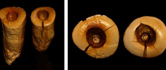
At the first and second stages, hydroxyapatite, the main substance of enamel, is destroyed. It forms 5 pathological zones, which differ from each other in the depth of penetration into the tooth tissue. Each of these zones has its own characteristics:
- Superficial: stable and dense zone with small microporosity in the area of demineralization.
- Subsurface: in the affected area of the tooth, the calcium content decreases by 15%. Because of this, the permeability of the enamel increases.
- Central: calcium concentration drops by another 25-30%, and the number of micropores increases by 20%.
- Intermediate: mineralization is at a very low level - 2-4%, but the natural process of enamel restoration has not yet stopped.
- Internal: the carious lesion is located closer to the dentin, although within the enamel. There is a risk that superficial caries will progress to the third stage.
Initial and superficial caries can be cured without preparation and filling. However, treatment should be started as early as possible.
Clinical researches
Asept products are clinically proven effective. For example, clinical studies have proven that regular use of professional toothpaste ASEPTA REMINERALIZATION improved the condition of the enamel by 64% and reduced tooth sensitivity by 66% after just 4 weeks. According to tests, using ASEPTA propolis gum gel for a week can reduce gum inflammation by 31%.
In addition, clinical studies have proven that regular use of preventive toothpaste ASEPTA ACTIVE for a month can reduce bleeding gums by 60%, improve the overall condition of the oral cavity by 44% and reduce inflammation by 33%.
Sources:
- Report on the determination/confirmation of the preventive properties of personal oral hygiene products “ASEPTA PLUS” Remineralization doctor-researcher A.A. Leontyev, head Department of Preventive Dentistry, Doctor of Medical Sciences, Professor S.B. Ulitovsky First St. Petersburg State Medical University named after. acad. I.P. Pavlova, Department of Preventive Dentistry
- The effectiveness of the use of Asept “adhesive balm” and Asept “gel with propolis” in the treatment of chronic generalized periodontitis and gingivitis in the acute stage (Municipal Dental Clinic No. 4, Bryansk, Kaminskaya T. M. Head of the therapeutic department Kaminskaya Tatyana Mikhailovna MUZ City Dental Clinic No. 4, Bryansk
- Clinical and laboratory assessment of the influence of domestic therapeutic and prophylactic toothpaste based on plant extracts on the condition of the oral cavity in patients with simple marginal gingivitis. Doctor of Medical Sciences, Professor Elovikova T.M.1, Candidate of Chemical Sciences, Associate Professor Ermishina E.Yu. 2, Doctor of Technical Sciences Associate Professor Belokonova N.A. 2 Department of Therapeutic Dentistry USMU1, Department of General Chemistry USMU2
How is caries diagnosed?
Caries can be detected during an external examination, but some of its symptoms are similar to those of enamel hypoplasia and fluorosis, so differential diagnosis is necessary. To achieve this, dentists use the following methods:
- Treatment of teeth with hydrogen peroxide. Areas of demineralization become more noticeable.
- Caries indicators. The enamel is coated with a special compound that turns the affected areas blue.
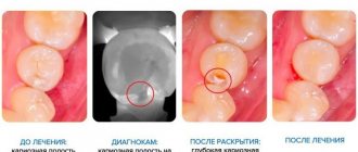
- Transillumination is the transillumination of a tooth with a directed beam of light from a special device.
- Fissurotomy: The dentist widens or grinds down the natural grooves on the chewing surface of the tooth to more closely examine the fissures.
- Laser diagnostics. This is the most modern research method. The dentist directs a laser beam at the tooth and illuminates it. The device shows whether there is a carious lesion and how deep it has spread.
- Radiography. It is used for the development of caries at the root or under the gum. The photographs show the carious lesion, its scale and stage.
After the examination, a method of caries treatment is selected.
Diagnostic methods
Clinical diagnosis, carried out as part of an examination of the oral cavity, involves the use of various techniques to determine the degree of development of pathology. Over time, superficial caries can develop into a more severe form that affects dentin and the pulp chamber, so it is important to promptly select the optimal treatment method.
Current examination methods used in clinics include:
- Visual inspection and percussion - assessment of the condition of the dentition by tapping the elements, allowing to identify the patient’s reaction;
- Probing is a technique that allows you to determine the presence of superficial caries by detecting a rough texture on the surface of the enamel;
- Staining – the use of special dyes that form markers in affected areas characterized by insufficient mineralization;
- Antiseptic drying - applying a special composition to the dentition, which, under the influence of a directed light flux, indicates defective areas;
- Differential diagnosis is a procedure that involves comparing the basic symptoms observed in the patient.
The final cost of dental services consists of many factors - as a rule, the price for caries treatment in St. Petersburg clinics is agreed upon based on the results of a preliminary diagnosis, which determines the structure of the treatment plan.
Treatment of superficial caries
For superficial caries, various treatment methods can be prescribed depending on the extent of the carious process. At the very beginning of the second stage, remineralization can be applied, during which a special composition containing large quantities of fluorine and other minerals is applied to the enamel. They strengthen the tooth surface and prevent bacteria from penetrating into dentin.
For home use, the patient is prescribed anti-caries toothpastes, gels and rinses with antibacterial properties: SPLAT Professional “Whitening Plus”, Stomysens® Desensitizing Repairing Treatment, Biorepair Delicate Gums Mouthwash, etc.
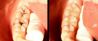
Another method of treating superficial caries is Icon infiltration, which does not involve drilling the tooth with a drill. The procedure is carried out in several stages:
- Removal of affected tissue using hand tools.
- Drying the tooth surface with warm air.
- Treating enamel with alcohol or other antiseptic.
- Application of infiltrate - liquid filling.
- Examining the tooth with a lamp, which helps the composition harden.
The procedure is prescribed mainly to children because they are afraid of the drill, but it is also suitable for treating adult patients.
If superficial caries has almost reached the third stage, filling is required. It is carried out according to the following algorithm:
- removal of dental plaque and tartar removal;
- drilling of a carious cavity, preparation carried out using a drill;
- isolation of the tooth crown using an elastic rubber dam;
- antiseptic treatment with chlorhexidine solution;
- applying an adhesive material - a special glue that improves the fit of the filling;
- layer-by-layer filling of the cavity with a filling compound;
- grinding and polishing the hardened filling.
Both procedures are safe and can be completed in 20-30 minutes. They do not cause complications. Filling can be performed under local anesthesia or without painkillers. Modern fillings usually have an unlimited service life. If it is installed correctly, and the patient after treatment is engaged in the prevention of dental diseases, there will be no need to re-treat the tooth.
How is the treatment carried out?
During treatment, a drill is used and the dental nerve is affected, so it is impossible to do without anesthesia if you want to achieve high-quality treatment. If the nerve was once removed from the tooth, then you do not need to use an anesthetic.
Modern anesthetics are used as anesthesia - Ultracain, Septanest and other drugs with a similar effect.
Removal of infected tissue
Since patients turn to the dentist when caries has already penetrated not only into the enamel, but also into the dentin, it is necessary to remove tissue damaged by putrefactive processes in order to prevent the further spread of bacteria, as well as to form a cavity suitable for filling.
Modern technologies suggest replacing the drill with a laser, but this method is not characterized by reduced pain, and its cost is often unreasonably high, since laser tissue removal is not common in all clinics and is used extremely rarely.
After cleansing the caries-pigmented tissues, it is necessary to rinse the cavity and treat it with an antiseptic.
Treating the cavity with an antiseptic
Chlorhexidine or drugs with a similar effect are used as an antiseptic, which disinfect tooth tissue and reduce the activity of enzymes, which are a breeding ground for bacteria.
Dental nerve extraction
Removal of the dental nerve is indicated only in the case of a complicated form of acute caries, when destructive processes have already reached the nerve, exposing it. In this case, the patient experiences acute pain.
Now the removal of a nerve, both partial and complete, is most often completed in one visit to the dentist; the procedure is assisted by an X-ray machine, which allows you to evaluate the accuracy of the manipulation during the treatment process. Thanks to modern painkillers, there is no pain when the nerve is removed.
Restoration of the anatomical shape of the tooth
The last stage of restoring a tooth that has undergone caries is restoring its original shape. The materials used for this are composites, which are placed into the hole, and then ground and polished so that the finished filling does not cause any inconvenience to the patient, does not rub the cheek, does not injure the gums and tongue, and does not interfere with neighboring teeth.
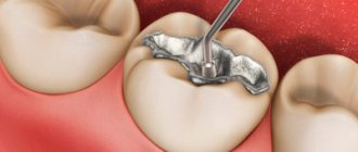
For chewing teeth, which are subject to increased load, composites and ceramic inlays and onlays are used as cement. There are times when a doctor uses several types of material to restore the function and shape of a tooth.
What happens if superficial caries is not treated?
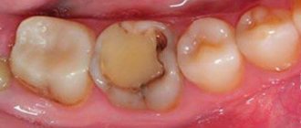
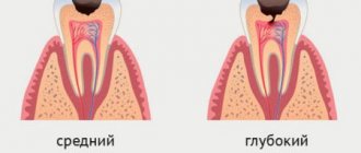
If there is no treatment, the pathology continues to progress, and the third, middle stage of carious lesion begins. The patient's enamel and tooth sensitivity increases; he reacts painfully to sweet and sour foods, temperature changes and a toothbrush. The carious cavity extends beyond the enamel and penetrates into the dentin. It is no longer possible to do without preparation and installation of a filling.
Then caries begins to destroy the internal tissues of the tooth. The pain is almost constant; it can occur even in the absence of irritants. This suggests that the pathological process is located next to the pulp or has already affected part of the nerve. During treatment it has to be removed. The tooth becomes dead and darkens. There is a risk of losing it: it can break at any moment. In addition, caries can spread to adjacent tissues. Complications from periodontal and bone tissue are possible.
Complicated caries can cause diseases that are not related to teeth, for example, chronic tonsillitis, sore throat or systemic infection. This happens infrequently, but it is better to exclude the possibility of such scenarios and engage in prevention. Let's find out what it is.
What complications can there be after treatment?
- Inflammation or even necrosis of the pulp - the tissue that fills the tooth cavity. Occurs after a long time due to inaccurate preparation of rotted tissue and excessive pressure on the pulp;
- secondary caries occurs due to the fact that during preparation some parts of the tissue affected by bacteria were not removed;
- inflammation of the gums due to injury;
- periodontitis, which occurs due to increased pressure on the tooth that occurs after the formation of an anatomically incorrect filling;
- loss of a filling due to insufficient strengthening or improper formation of a carious cavity.
Prevention of superficial caries
Caries prevention should be done from childhood. Dental treatment is expensive, so it is better to make every effort to prevent pathology. To do this you need to follow a number of simple rules:
- Brush your teeth twice a day. For additional care, use flosses, rinses and irrigators.
- Limit your intake of sugary foods, including soda. It is better to eat foods that contain nutrients, vitamins and minerals.
- Visit your dentist once every six months for ultrasonic cleaning. This is especially true for coffee and tea lovers.
- Give up bad habits, especially smoking.
At the first symptoms of caries or other dental diseases, consult a doctor. Don't try to treat them yourself. People with healthy teeth should come for a preventive examination to the dentist at least twice a year, children should be examined even more often - 2-3 times a year.
Icon system
The Ikon system is a progressive technique that allows you to treat caries at the initial stage without drilling, without damaging areas of healthy enamel. The kit consists of an etching gel and a composite infiltration material.
The affected tooth is isolated using a rubber dam. Softened areas of enamel are removed manually with a dental instrument. Using a special applicator, a gel is applied that corrodes demineralized enamel fragments. Healthy tissues are not damaged. After 2 minutes, the gel should be thoroughly washed off and the enamel surface dried.
Icon set.
The next step is to apply an infiltrate to fill the defect and fix it using a photopolymer lamp. Excess material is removed and the surface is polished. This order of the procedure allows you to get excellent results.
The system is used for surface enamel defects. If there is a deep cavity, it is advisable to use standard preparation methods.
The Ikon system is the most progressive treatment method for superficial carious processes. If you visit your dentist in time, the unpleasant drilling procedure can be avoided.
The choice of treatment method is made individually. The doctor examines the problem area, determines the size of the defect, and assesses the condition of the enamel. Based on this data, the doctor chooses the most effective method.
The Ikon system is used in leading clinics in Moscow and is recognized by the best specialists all over the world.