What you will learn in this article:
- What is this anyway? Root caries, definition.
- Its prevalence and relevance of this problem. Epidemiology of root caries.
- What is it like? Classification of root caries.
- What causes this problem? The mechanism of development of tooth root caries.
- How to find this disease in a patient? Diagnosis of root caries.
- What will we see in the mouth? Clinical manifestations of root caries.
- How is this treated? Tooth root caries treatment.
Ready? Then let's get started!
Epidemiology of root caries
The incidence of root caries is constantly increasing in older people. This is due to the fact that:
- There are more people with periodontal diseases, since their prevention is ineffective
- Dental care has improved, and retirees now have many more teeth in their mouths.
- Well, life expectancy has increased, where would we be without it?
Be that as it may, in our country today people suffer from root caries
- 1.3% aged 25-29 years
- and 35.2% (aged 55-64 years)
Clinical researches
Clinical studies have proven that regular use of professional toothpaste ASEPTA REMINERALIZATION improved the condition of the enamel by 64% and reduced tooth sensitivity by 66% after just 4 weeks.
Sources:
- Report on the determination/confirmation of the preventive properties of personal oral hygiene products “ASEPTA PLUS” Remineralization doctor-researcher A.A. Leontyev, head Department of Preventive Dentistry, Doctor of Medical Sciences, Professor S.B. Ulitovsky First St. Petersburg State Medical University named after. acad. I.P. Pavlova, Department of Preventive Dentistry
- Clinical experience in using the Asepta series of products Fuchs Elena Ivanovna Assistant of the Department of Therapeutic and Pediatric Dentistry State Budgetary Educational Institution of Higher Professional Education Ryazan State Medical University named after Academician I.P. Pavlova of the Ministry of Health and Social Development of the Russian Federation (GBOU VPO RyazSMU Ministry of Health and Social Development of Russia)
- Evaluation of the clinical effectiveness of a combined drug of local etiotropic action in the treatment of inflammatory periodontal diseases (E.L. Kalichkina, E.A. Tyo, Z.Z. Abubakarova) E.L. Kalichkina, candidate of medical sciences, assistant of the department of therapeutic dentistry, Kemerovo State Medical Academy of Roszdrav, Kemerovo E.A. Tyo, MD, professor, head of the department of therapeutic dentistry, Kemerovo State Medical Academy of Roszdrav, Kemerovo Abubakarova, assistant of the department of therapeutic dentistry, Kemerovo State Medical Academy of Roszdrav, Kemerovo GOU HPE Kemerovo State Medical Academy of Roszdrav , Kemerovo
Classification of root caries
In the International Classification of Diseases (ICD-10), root caries is located in section K02 dental caries. This is the classification:
K02 Dental caries
- K02.0 Enamel caries
- K02.1 Dentin caries – here too
- K02.2 Cement caries - here it is
- K02.3 Suspended dental caries – here too
- K02.4 Odontoclasia
- K02.8 Other specified dental caries
- K02.9 Dental caries, unspecified
Classification of root caries according to Leus
Leus P.A. , Borisenko L.G.
According to the depth of damage to root tissue:
- No cavity formation
- With the formation of a cavity
According to the course of root caries:
- Active lesion
- Suspended caries
- Secondary caries (active or inactive)
- Unspecified
Measures to prevent dental caries
The following preventive measures will help to avoid damage to the integrity of tooth enamel and damage to dentin:
- Regular (twice a year) visits to the dentist for a preventive examination.
- Thorough regular cleaning of teeth, interdental spaces, tongue using a toothbrush, paste, floss, and, if possible, an irrigator.
- Drinking drinking water rich in fluoride and other beneficial elements.
- Preventive intake of vitamin complexes as recommended by the dentist.
Prevention reduces the risk of dental caries and other dental diseases by more than 70%.
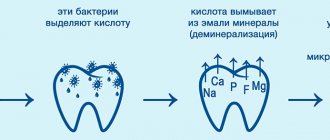
The mechanism of development of tooth root caries
Here we will explain the risk factors leading to root caries. They are divided into 2 groups:
- Factors that influence periodontal disease are:
- Insufficient prevention, incl. hygiene, both personal and professional
- Periodontal disease itself, and associated gum recession (exposure of the root)
- Elderly age
- Anatomical features of the patient’s mouth (small vestibule, short frenulum, bite pathology)
AS A RESULT – disruption of gum attachment and exposure of the root
- Once the root is exposed, the factors that cause caries come into play.
- Poor nutrition (carbohydrates)
- Fluoride deficiency (in food and toothpaste)
- Insufficient quantity or composition of saliva
AS A RESULT – the development of caries on the root surface.
One more question remains: why does caries develop faster on the root than on the crown of the tooth?
Answer: Because root cement is more sensitive to acids than highly mineralized enamel. For comparison: enamel dissolution begins at a pH less than 5.5. And cement - at 6.2 - 6.7. There is a difference?
Prevention of secondary caries
The occurrence of carious lesions indicates the activity of pathogenic microorganisms that destroy the protective layer of enamel and tooth tissue. Predisposition to the disease may be genetic, but to a greater extent this dental disease is associated with hygiene and/or nutrition. Moderate caries is considered a developed stage of the disease: often many patients, having noticed a carious stain, think that it will “resolve” by itself, and consult a doctor only when they feel pain and discomfort. The main goal of prevention is to prevent the development of the disease and its transition to the middle and deep stages, when it is no longer possible to do without a drill. The key to success here is simple: regular and high-quality hygiene, proper nutrition and preventive visits to the doctor. If you follow at least these points, then the chance of developing the disease will be minimal.
Diagnosis of root caries
Root caries is diagnosed like other diseases using basic and additional research methods:
- The main ones are questioning, inspection, probing, percussion, palpation. With their help, you can see the carious cavity on the root surface, determine its topography (on which root surface it is located), depth, edges, etc.
- Additional:
- This is an index assessment of hygiene (OHI-S, PLI), gum condition (GI, gum recession index), periodontal condition (CPI)
- Also, diagnostic tests: (saliva pH test).
- X-ray examination (bite-wing radiography, orthopantomogram)
Using X-rays, you can find hidden carious cavities under the gum, or adjacent to an adjacent tooth.
Prognosis and consequences of the disease
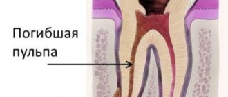
Treatment of this disease, if the dentist has sufficient experience, is successful - all unpleasant sensations in the sealed cavity completely disappear, and the tooth again acquires aesthetic appeal. If an untreated area remains under the filling, the disease will progress further. For this reason, it is necessary to carefully consider the choice of clinic and attending physician.
If average caries is not treated in a timely manner, the disease will enter an advanced stage, which is fraught with severe pain, the development of pulpitis and periodontitis, and then tooth loss.
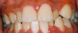
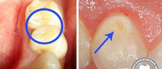
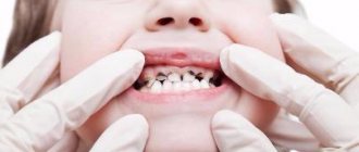
Clinical manifestations of root caries.
The first is complaints. The patient most often complains of:
- An aesthetic defect (not beautiful) if there is caries on the vestibular surface of the front teeth.
- Discomfort while eating, pain when brushing teeth
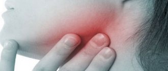
- Symptoms of periodontal disease (bleeding gums, loose teeth, etc.)
- Short-term pain from irritants (teeth hurt from ice cream, hot tea). The pain disappears when the cause is removed.
Next, we look into the oral cavity and see the clinical picture. We will consider it taking into account the Leus classification
Along the way, root caries occurs:
- Active lesion - the edges of the cavity are undermined. The cavity is filled with softened tissues. There is a tendency towards a rapid increase in its size.
- Suspended caries - the edges of the cavity are smooth, dense, flat (not undermined). The bottom of the cavity is dense and shiny. Don't strive to get bigger.
- Secondary caries is the resumption of the process in the area of remission or after treatment (at the site of the filling).
According to the depth of the lesion:
- Without cavity formation - a white spot. Density is less than normal cement.
- With the formation of a cavity - any of those described in paragraph 1. carious defects.
Depending on what surface of the tooth it affects
Caries can affect any surface of the root, or 2 surfaces, or all surfaces in a circle (circular spread)
And lastly, according to ICD-10. There are cement caries and dentin caries.
- Cementum caries affects only the cementum of the tooth root
- Dentin caries also spreads deeper into dentin
The classification is over, and so are the clinical manifestations.
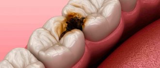
To summarize, root caries is a spot or cavity on the surface of the root of a tooth. Its edges are undermined or have smooth edges.
The cavity may be located above or below the gum.
Symptoms of average caries
In dentistry, there are cases when average caries does not manifest itself at all (especially in chronic form) or the patient simply does not notice the discomfort. However, more often the opposite happens. At this stage, more than one third of the dentin is affected, so the patient begins to feel the manifestations of the disease.
Complaints with average caries
- mild aching pain (may be absent)
- reaction to stimuli (chemical, temperature)
- discomfort when chewing, feeling of teeth on edge
- tooth color change
Treatment of tooth root caries
Treatment of root caries, as well as simple caries, begins with motivation and prevention. Read more about this in our article at the link.
Further, treatment tactics depend on the depth of the carious lesion:
Caries in the spot stage is treated with conservative retherapy. Apply:
- Fluorine preparations – varnishes, gels, solutions (Fluorine in the form of sodium fluoride, amino fluoride, tin fluoride, etc.)
- Calcium preparations (10% Calcium gluconate).
- Antiseptics (Chlorhexidine 1-2%)
Applications of these drugs contribute to the remineralization of tooth tissue, stopping and reversing the development of caries in the stain stage. Root cement (due to its porosity) is easier to remineralize than other dental tissues.
- Desynthesizers – reduce tooth sensitivity.
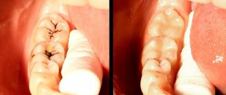
Caries in the stage of hard tissue defect is treated with preparation and filling.
Next, we will describe the differences between each stage of treatment.
- Disclosure. In case of root caries, the entrance to the carious cavity is wide, without overhanging edges. Therefore, no disclosure is made.
- Extension. Preventive expansion of the cavity (preparation of healthy dentin) is carried out only in case of rapidly progressing root caries.
- Necrectomy. Because the distance from the bottom of the cavity to the pulp is small, necrectomy is carried out carefully. Only softened dentin is removed.
- Formation of a carious cavity. The cavity is oval in shape. If it is located on the proximal surface, an additional platform is created on the oral one. If the cavity is located in the enamel area, create a bevel 2-5 mm wide.
- Filling. Due to the high humidity near the gum (gingival fluid), a filling material is chosen that is not afraid of moisture. This is GIC, amalgam or compomer.
And in conclusion, we summarize the main elements of the article:
- Root caries is a cavity in the root of a tooth.
- Prevalence 35.2% in the age group 55-64 years
- WHO classification – cement caries. And clinical classification of prof. Devina
- 2-stage development mechanism: root exposure + cariogenic factors
- Diagnostics - like standard caries + periodontal examination.
- Clinic: Spot caries and Cavity caries. Rapidly progressive with undermined edges and remission with a smooth surface
- Treatment: Rem. Therapy – caries spots or preparation/filling – cavity caries.
That's all. Learn, develop, read other articles. Good luck!
Causes of the disease
There are several factors that lead to the occurrence of this disease:
- Cariogenic microflora of the oral cavity.
- Increased carbohydrate content in the diet.
- Reduced resistance of hard tissues.
- Lack of oral hygiene.
- Malocclusion.
- Genetic predisposition to the disease.
- Plaque or tartar.
- Weakening of the immune system.
- Bleeding gums.
- Changes in the composition of saliva.
- Lack of calcium.
- Fluoride deficiency.
- Poor diet.
- Lack of vitamins.
- Abuse of carbonated drinks.
The main cause of average caries is late diagnosis of the disease at an early stage.
Historical reference
It is believed that the “history” of caries begins with a change in the human diet, that is, from the time when coarse, unprocessed food began to be replaced by food subject to heat, steam and other processing, as well as from the moment when “sweets” appeared. Judging by archaeological finds, for example, by the teeth of a man who lived 13-14 thousand years ago found near the city of Lucca (Italy, Tuscany region), we can conclude that already then caries was encountered and attempts were made to treat it. In the teeth found near Lucca, cavities were found that reached the pulp, and on the walls of the teeth there were traces of drilling with some stone tools. Moreover, the teeth had fillings made of plant resin with plant fragments. This means that ancient healers tried to protect carious cavities from the penetration of food, and the plants added to the resin composition apparently served as an anesthetic.
The healers of the Mayan civilization, judging by other archaeological finds, made fillings from complex plant compositions with the addition of various minerals. In ancient Rus', caries in the initial stages was treated with honey, and in advanced stages - with propolis, which was applied to the tooth cavity, previously cleaned of decay products and affected tissues; to relieve pain, they applied a cloth soaked in a mixture of vodka and table salt. In ancient Rus', caries in the initial stages was treated with honey, and in advanced stages - with propolis, which was applied to the tooth cavity, previously cleaned of decay products and affected tissues; to relieve pain, they applied a cloth soaked in a mixture of vodka and table salt. In medieval Europe, where the development of medicine was hampered by the dogmas of the church, it was believed that caries developed due to the activity of “tooth worms”, and it was treated with poisons and hot metals introduced without anesthesia into the dental cavity affected by the disease. Moreover, these manipulations were carried out not by doctors, but by barbers. The poor people, having no means for “treatment”, received very strange, unscientific recommendations from the “doctors”: sit under the moonlight with your mouth open, tie a frog to your jaw, apply a mixture of oil and bird droppings, “rub” the affected area with the tooth of a deceased person , etc.
In the 15th century, healers began using metals, including gold, to fill teeth. In 1790, John Greenwood, the personal dentist of the first US President George Washington, came up with a primitive device for drilling teeth, where a foot drive from a spinning wheel was used to rotate the drill. More than 80 years later, in 1871, the American doctor James Morrison patented a dental drill he invented, which gave impetus to the development of therapeutic dentistry. Throughout the 20th century, therapeutic dentistry developed rapidly, adding more and more new techniques, materials and tools for the treatment of dental diseases, including caries. Today, caries treatment is the most common dental service, with high treatment effectiveness at any stage.
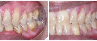
Cleaning in dentistry from stone and plaque
1243900247100
Professional teeth cleaning involves removing the accumulation of all pathogenic microflora around the root.
It involves not only removing plaque, but also stones. This elimination of negative factors protects teeth for a long time.
Antiseptic treatment of affected areas
Treatment of root caries involves drilling out all affected areas with a drill and treating the carious cavity with antiseptics and special preparations. A 2% chlorhexidine solution or a gel based on it is used as an antiseptic for the treatment of carious cavities.
Who is more likely to develop caries?
- People with a sweet tooth and lovers of sweet sodas are most often susceptible to tooth decay. Temporary teeth in children and permanent teeth in adolescents are especially vulnerable. Sugar creates an acidic environment in the mouth, and the acid, in turn, destroys the enamel. Of course, more than one holiday is complete without sweets and lemonade. In addition, carbohydrates are a source of pleasure and joy, an excellent product for stimulating brain function. However, a competent approach is required in organizing the intake of these products.
- Children aged 5-6 years begin the eruption of permanent teeth. There is a peak in carious tooth decay during this period of time, and scientists attribute it to a lack of calcium for new permanent teeth. Temporary teeth have to act as “donors”, and the risk of destruction increases. Sometimes the destruction of the enamel is so small that it does not manifest itself for several months, and is practically not noticeable upon visual inspection. However, in dentin, the deep layer of the tooth, destruction progresses. The destruction of the incisors may not require treatment, because these teeth are already undergoing involution (reverse development, resorption of roots), and within a few days they can be replaced by permanent ones. And canines and chewing teeth - molars - must be restored, otherwise caries will lead to rapid destruction and even early tooth loss.
- Adolescent teeth within 5-6 years after eruption are considered immature, since the maturation of crystals in the enamel and dentin continues. Insufficient dental care and the accumulation of carbohydrate products in areas that are least susceptible to self-cleaning can lead to the development of superficial caries on several teeth at once.
- Pregnant women should be identified as a separate risk group. At this crucial moment, a change in metabolic processes occurs in the body of the expectant mother; and slowly ongoing carious processes can become acute.
Caries, like all diseases, has an initial stage, when the changes are reversible and the inflammatory process can be reversed. It is important to identify it in time.
Caries and its causes, classification and effect on the body
Caries (lat. caries - “rotting”) is a pathological process in the hard tissues of the tooth, which develops under the long-term complex influence of unfavorable external and internal factors and leads to softening of hard tissues and the formation of defects in the form of cavities. Caries, if left untreated, disrupts the chewing process up to the complete loss of this function, therefore, has an extremely negative effect on digestive function and causes diseases of the gastrointestinal tract. In general, caries has a negative impact on the general condition of the body and on a person’s quality of life, being a constant source of infection and intoxication.1
Contraindications
Caries is a disease that has no absolute contraindications to treatment. But if certain factors are present, treatment may be delayed or interrupted until they are eliminated. These factors include: the presence of intolerance to drugs and materials that are planned for use in treatment; concomitant diseases aggravating treatment; inadequate psycho-emotional state of the patient before treatment; acute lesions of the oral mucosa and red border of the lips; acute inflammatory diseases of organs and tissues of the mouth; a life-threatening acute condition/disease or exacerbation of a chronic disease (including myocardial infarction, acute cerebrovascular accident), which developed less than 6 months before seeking this dental care; periodontal tissue diseases in the acute stage; unsatisfactory hygienic condition of the oral cavity.
What to do to protect your gums
If the gum is not treated, inflammation will remain and all treatment will become meaningless. The gums are protected by diathermocoagulation and retraction.
Diathermocoagulation
Diathermocoagulation – excision of excess areas of periodontitis with a coagulation knife. The knife immediately cauterizes the bleeding edges. The procedure is performed when there are severe changes in the gums. In such cases, filling is postponed until the soft tissue is completely regenerated.
Retraction
If the condition of the gums is advanced, treatment of caries becomes impossible - the mucous membrane bleeds and interferes with tooth treatment. This gum is excised or a special device is applied to move the gum back. But more often, retraction is performed - lowering the gingival contour or moving apart the overhanging edges of the gums using special hemostatic threads. A temporary filling is placed until complete healing.