If you want to maintain the beauty and integrity of your teeth until old age, be sure to go to the dentist a couple of times a year. Some pathologies may occur without symptoms, but this does not mean that the process is not dangerous for the oral cavity. These include superficial caries.
Its symptoms may be hidden, and changes may not be visually noticeable due to the location of the tooth at the end of the jaw. However, ignoring superficial caries often leads to irreversible consequences.
What it is?
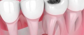
Superficial caries is characterized by the appearance of a yellowish carious spot on the surface of the enamel. A feature of this disease is the absence of damage to the deep layers. This stage is transitional, that is, in the absence of treatment for superficial caries, the infection gradually moves deeper and affects the entire tooth.
Most often, superficial caries is localized in depressions, the so-called fissures. According to statistics, initial carious lesions are observed in 85% of the population, but not everyone knows about it. For the same reason, many patients come to the dentist too late, when the tooth is completely susceptible to deep caries.
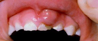
Complicated caries in children
In children and adolescents, complications, like the caries that precede them, develop at lightning speed. This is due to insufficient mineralization of enamel in childhood and the tendency of young patients to infectious diseases caused by streptococci - sore throat, bronchitis, measles and scarlet fever.
Advanced forms of caries in children lead to:
- the development of its circular shape, when, starting from the neck of the tooth, decay spreads around its crown, and over time the tooth breaks off;
- spread of infection along the surface of the crown (planar caries). In this case, the tooth completely darkens, the child complains of aching pain;
- acute and chronic pulpitis;
- infection of the rudiments of permanent teeth, resulting in multiple caries after their eruption.
Causes
Experts have come to the conclusion that the cause of the development of superficial caries is pathogenic microorganisms - streptococci. Note that these bacteria are present on the mucous membrane of every person; in minimal quantities they do not cause harm. Once disposition factors occur in the body, there is a significant increase in “bad” bacteria. These factors include:
- Low body defenses.
- Avitaminosis.
- Lack of useful microelements.
- Poor nutrition.
- Poor quality water.
Secondary reasons include the following:
- Insufficient oral hygiene.
- Pieces of food stuck between teeth that were not removed in a timely manner.
- Accompanying illnesses.
Genetic predisposition should also be noted. If your close relatives have similar problems, most likely you will also experience superficial caries. The sooner treatment is started, the higher the chances of avoiding serious complications.
Classification of disease severity according to ICDAS
To register the status, this system also suggests using 6 codes:
- Initial focal changes in the color of the enamel surface of the tooth, which are visible only after drying.
- Clear, clearly visible changes in the enamel in the form of whitish and dark spots.
- Localized enamel lesions, which manifest themselves in limited areas of destruction.
- Destruction of enamel in a certain place along the entire thickness of its layer, down to dentin.
- The presence of a clearly visible carious cavity in the dentin.
- Severe destruction of dentin, which extends to at least half the volume of the crown.
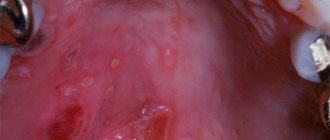
Symptoms and diagnosis
Superficial caries can be recognized by some symptoms. Typically, patients complain of sudden, sharp pain of short duration. Painful sensations occur when eating too cold, hot, sour or spicy food. The pain is especially pronounced when eating sweets.
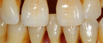
If superficial caries is located on the nearby teeth, the disease can be recognized visually by examining the oral cavity in a mirror. The affected enamel has yellowish spots, it is covered with a coating, and in some cases small depressions are observed.
The initial stage of a superficial disease reacts sharply to mechanical irritants, so if you experience discomfort when brushing your teeth, this is a good reason to see a dentist for an in-person examination. Sometimes a latent course of superficial caries is observed, that is, there are no visible symptoms, and a diagnosis can only be made in the dentist’s chair.
It will not be possible to determine the stage of caries on your own, so if the above signs appear, do not delay your visit to the dentist. Any specialist at Dr. Jacques’ dental center can easily diagnose the disease. It is enough to examine the enamel using special dental instruments. In more complex cases, radiography is prescribed; this will help to examine the enamel in more detail and assess the extent of the damage. After radiography and other diagnostic manipulations, the tactics for further treatment of superficial caries are determined.
Prevention
Proven methods for preventing superficial caries stem from the causes of its occurrence. So, take on board the basic measures to prevent it:
- The main rule
: carry out thorough and systematic hygiene of teeth and oral mucosa. A medium-hard toothbrush and fresh-smelling toothpaste are classics that we all know from childhood. But don’t forget about dental floss: it will be a good protection if you use it after meals. Don’t ignore chewing gum (sugar-free, of course!) – combine business with pleasure. - Try
to avoid unhealthy snacks between meals, especially sweet and carbohydrate ones. Sweets, cookies and cakes are bad for your teeth, because rarely does a person immediately rush to brush their teeth after eating them. It would be worth it! - Take care
of your dental health. Check your mouth regularly. If you see abnormal pigmentation or surface deformation, do not delay in any case, but contact us for help! - Visit
the dentist once, or better yet twice a year, even if you have no complaints. The doctor will detect the slightest violation that cannot be noticed by yourself. This will allow you to begin to take measures at the very first stage, including dealing with caries at the early stage of the stain conservatively - without a drill or filling, as well as promptly treating superficial caries without affecting the dentin. In addition, the specialist will recommend procedures to maintain dental health, such as professional cleaning or treating teeth with special calcium-containing products and fluoride varnish, and will also remove tartar. - Remember
, the sooner you see a doctor at our medical center, the faster and more successfully superficial caries will be cured, which means you won’t have to suffer and be afraid of dire consequences!
In our center, qualified, understanding and most friendly
doctors !
What happens if superficial caries is not treated?
Treatment of initial caries should be prescribed immediately after the problem is discovered. The fact is that if you do not destroy the source of bacteria accumulation, this will lead to the disease moving to the next stage, which in turn will be more difficult and more expensive to cure. Well, if you continue to ignore it, then in this case you will spend a lot on treatment in the future. Even if the disease does not manifest itself in any way and does not cause discomfort, therapy should be carried out immediately after the discovery of superficial caries.
In more detail, in the absence of timely treatment, the growth of pathogenic microorganisms is observed. First, the tooth is completely affected, caries from the surface of the enamel moves into the deeper layers of the tooth and leads to a violation of the integrity of the tooth, the development of inflammation, and putrid breath. The bacteria then spread throughout the oral cavity and cause identical pathology on neighboring teeth.
If you continue to ignore the problems, this can lead to serious gum diseases such as periodontal disease or periodontitis. First, pain occurs in the gum area, then gaps between the teeth and exposed roots are observed. Ultimately, you risk acquiring chronic, incurable periodontal disease, the pathological processes of which will ultimately lead to tooth loss.
Anatomical classification according to Black
This system is used by a huge number of dentists around the world. It identifies six classes of disease:
| Black class | Localization of carious defect |
| First | Fissures of chewing teeth (grooves) and blind pits on incisors |
| Second | Lateral areas of molars in contact with each other |
| Third | Contact areas of incisors and canines with preserved shape of cutting edges |
| Fourth | Lateral surfaces of the anterior teeth with destruction of the cutting edges |
| Fifth | Cervical areas of all units on the anterior or lingual side |
| Sixth | Incisal edges of incisors, cusps of molars |
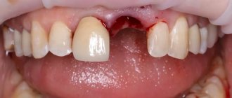
Treatment Options
Only a specialist can determine treatment tactics. If we are talking about the treatment of superficial caries without deepening, then you can get by with professional cleaning and laser without the need for drilling. If caries has deformed the shape and contour of the tooth, then you cannot do without a drill. Treatment of initial caries should be comprehensive:
- First, the doctor performs professional cleaning using a special paste, then polishing is performed.
- Using a drill, the defect is smoothed and the contour is aligned. Since this degree of disease does not penetrate into the deep layers of the tooth, it is absolutely painless.
- Using a rubber dam, saliva is isolated.
- The surface of the affected tooth is coated with a special acid, this is necessary to attach the filling.
- Next, the dentist applies the filling material. At the end of the procedure, grinding and polishing is carried out.
The dental clinic of Dr. Jacques practices the treatment of initial caries without the use of a drill. The contours of the tooth are smoothed using polishing discs and rubber tips, then ground and polished. Laser therapy is also practiced. You can learn more about this treatment of initial caries by scheduling a consultation by phone.
Example of treatment of superficial caries on video
How is caries diagnosed?
Caries can be detected during an external examination, but some of its symptoms are similar to those of enamel hypoplasia and fluorosis, so differential diagnosis is necessary. To achieve this, dentists use the following methods:
- Treatment of teeth with hydrogen peroxide. Areas of demineralization become more noticeable.
- Caries indicators. The enamel is coated with a special compound that turns the affected areas blue.
- Transillumination is the transillumination of a tooth with a directed beam of light from a special device.
- Fissurotomy: The dentist widens or grinds down the natural grooves on the chewing surface of the tooth to more closely examine the fissures.
- Laser diagnostics. This is the most modern research method. The dentist directs a laser beam at the tooth and illuminates it. The device shows whether there is a carious lesion and how deep it has spread.
- Radiography. It is used for the development of caries at the root or under the gum. The photographs show the carious lesion, its scale and stage.
After the examination, a method of caries treatment is selected.
Preventive actions
Prevention of initial caries is aimed at proper oral hygiene. Try to brush your teeth after every meal. Use an antibacterial mouthwash and remember to brush the insides of your cheeks and tongue. Buy dental floss to easily remove food debris between your teeth. If finances allow, purchase an irrigator. This device thoroughly cleans teeth and removes pathogenic microorganisms from the enamel surface.
Let's consider a few more tips:
- Eat a balanced diet so that your body does not experience a deficiency of vitamins and microelements. Strengthen your immune system in every possible way. And, the most important rule, visit the dentist more often and promptly begin treatment for various oral ailments.
- It is important to maintain normal oral microflora. For this reason, you need to start treating colds in a timely manner and check your stomach condition once a year.
- An excellent preventative measure is to coat the enamel with fluoride varnish. This coating prevents bacteria from multiplying and prevents the development of superficial caries. For the highest efficiency, the procedure is carried out only 2 times a year.
- Dose out sweets, especially for children. Such products provoke the growth of bacteria. You should also limit liquids, which wash calcium from the body and make the enamel more vulnerable to the development of superficial caries.
How our teeth work
The crown part of the tooth is the part that protrudes above the gum. It has a surface layer - enamel, and an inner layer - dentin. The thickness of the enamel layer is very pronounced in the area of the cutting edges of the front teeth, the cusps of the canines and chewing teeth - molars and premolars. Enamel is the strongest fabric, however, it can also be vulnerable. The grooves between the cusps of the chewing teeth, the area in the cervical region, and the boundaries of the old filling are places where food debris can most often accumulate and cariogenic bacteria multiply. If you clean these areas regularly, twice a day, you can assume that your teeth are safe. If you allow the soft coating to remain, even in small quantities, disaster cannot be avoided.
Indices used in dental examination
The prevalence of caries is expressed as a percentage. To do this, the number of people who were found to have certain manifestations of dental caries (except for focal demineralization) is divided by the total number of people examined in this group and multiplied by 100.
In order to assess the prevalence of dental caries in a particular region or compare the value of this indicator in different regions, the following assessment criteria for the level of prevalence among 12-year-old children are used:
Intensity level
LOW - 0-30% MEDIUM - 31 - 80% HIGH - 81 - 100%
To assess the intensity of dental caries, the following indices are used:
a) intensity of caries of temporary (baby) teeth: index kp (z) - the sum of teeth affected by untreated caries and filled in one individual;
index kp (n) - the sum of surfaces affected by untreated caries and filled in one individual;
In order to calculate the average value of the indices kp(z) and kp(p) in a group of subjects, one should determine the index for each person examined, add up all the values and divide the resulting amount by the number of people in the group.
b) intensity of caries of permanent teeth:
index KPU(z) - the sum of carious, filled and extracted teeth in one individual;
index KPU (n) - the sum of all surfaces of the teeth on which caries or a filling is diagnosed in one individual. (If a tooth is removed, then in this index it is considered 5 surfaces).
When determining these indices, early forms of dental caries in the form of white and pigmented spots are not taken into account. In order to calculate the average value of indices for a group, you should find the sum of individual indices and divide it by the number of people examined in this group.
c) assessment of the intensity of dental caries among the population. To compare the intensity of dental caries between different regions or countries, the average values of the KPU index are used.
WHO distinguishes 5 levels of intensity of dental caries:
Periodontal indices. CPITN Index
To assess the prevalence and intensity of periodontal diseases, almost all countries use the index of need for the treatment of periodontal diseases - CPITN . This index was proposed by specialists of the WHO working group to assess the condition of periodontal tissues during epidemiological surveys of the population. Currently, the scope of the index has expanded, and it is used to plan and evaluate the effectiveness of prevention programs, as well as calculate the required number of dental personnel. In addition, the CPITN index is currently used in clinical practice to examine and monitor the periodontal condition of individual patients. In this regard, the CPITN index can be considered a screening test at both the population and individual levels. This index registers only those clinical signs that may undergo reverse development: inflammatory changes in the gums, which are judged by bleeding, tartar. The index does not record irreversible changes (gingival recession, tooth mobility, loss of epithelial attachment), does not indicate the activity of the process and cannot be used to plan specific clinical treatment in patients with developed periodontitis. The main advantages of the CPITN index are the simplicity and speed of its determination, information content and the ability to compare results. To determine the CPITN index, the dentition is conventionally divided into 6 parts (sextants), including the following teeth: 17/14 13/23 24/27 34/37 43/33 47/44.
The periodontium is examined in each sextant, and for epidemiological purposes only in the area of the so-called “index” teeth. When using the index for clinical practice, the periodontium is examined in the area of all teeth and the most severe lesion is identified. It should be remembered that a sextant is examined if it contains two or more teeth that cannot be removed. If only one tooth remains in the sextant, it is included in the adjacent sextant, and this sextant is excluded from the examination. In the adult population, starting from 20 years of age and older, 10 index teeth are examined, which are identified as the most informative: 17/16 11 26/27 47/46 31 36/37.
When examining each pair of molars, only one code characterizing the worst condition is taken into account and recorded. For persons under 20 years of age, 6 index teeth are examined during an epidemiological survey: 16, 11, 26, 36, 31, 46
CODE 1 : bleeding observed during or after probing. Note: bleeding may appear immediately or after 10-30 seconds. after probing. CODE 2 : tartar or other plaque-retaining factors (overhanging edges of fillings, etc.) are visible or felt during probing. CODE 3 : pathological pocket 4 or 5 mm (the edge of the gum is in the black area of the probe or the 3.5 mm mark is hidden). CODE 4 : pathological pocket 6 mm deep or more (with the 5.5 mm mark or black area of the probe hidden in the pocket). CODE X : When only one or no teeth are present in the sextant (third molars are excluded unless they are in place of second molars).
To determine the need for periodontal disease treatment, population groups or individual patients can be categorized based on the following criteria. 0: CODE 0 (healthy) or X (excluded) for all 6 sextants means that there is no need for treatment for this patient. 1: A CODE of 1 or higher indicates that this patient needs to improve his oral hygiene status. 2: a) CODE 2 or higher indicates the need for professional hygiene and the elimination of factors that contribute to plaque retention. In addition, the patient needs training in oral hygiene. b) CODE 3 indicates the need for oral hygiene and curettage, which usually reduces inflammation and reduces pocket depth to values equal to or less than 3 mm. 3: Sextant with CODE 4 can sometimes be successfully treated with deep curettage and adequate oral hygiene. In other cases, this treatment does not help, and then complex treatment is required, which includes deep curettage. The prevalence and intensity of periodontal disease in the population is assessed based on the results of a survey of 15-year-old adolescents.
Gingivitis Index (GIA)
To assess the severity of gingivitis (and subsequently record the dynamics of the process), the papillary-marginal-alveolar index (PMA) . Various modifications of this index have been proposed, but in practice the PMA index as modified by Parma (1960) is more often used.
The RMA index is assessed using the following codes and criteria:
0—no inflammation; 1 - inflammation of only the gingival papilla (P); 2 - inflammation of the marginal gum (M); 3 - inflammation of the alveolar gum (A).
The PMA index is calculated using the formula: sum of PMA = ———————- x 100% 3 x number of teeth The number of teeth (while maintaining the integrity of the dentition) is taken into account depending on age: 6 – 11 years – 24 teeth, 12 – 14 years - 28 teeth, 15 years and older - 30 teeth.
Note: if there are missing teeth, then divide by the number of teeth present in the oral cavity. Normally, the PMA index is 0. The higher the digital value of the index, the higher the intensity of gingivitis.
Evaluation criteria for the RMA index:
30% or less - mild gingivitis; 31-60% - moderate severity; 61% and above—severe degree.
Oral hygiene assessment
Fedorov-Volodkina hygienic index (1971)
The index is recommended to be used to assess the hygienic state of the oral cavity in children under 5-6 years of age. To determine the index, the labial surface of six teeth is examined: 43, 42, 41, 31, 32, 33. These teeth are stained using special solutions (Schiller-Pisarev, fuchsin, erythrosine) and the presence of plaque is assessed using the following codes: 1 - plaque not identified; 2 - staining one quarter of the surface of the tooth crown; 3 - staining half the surface of the tooth crown; 4 - staining three quarters of the surface of the tooth crown; 5 - staining the entire surface of the tooth crown. Determination of supra- and subgingival tartar is carried out using a dental probe.
Codes and criteria for assessing dental calculus
O —tartar is not detected; 1 - supragingival tartar, covering no more than 1/3 of the tooth surface; 2 - supragingival tartar, covering more than 1/3, but less than 2/3 of the tooth surface, or the presence of individual deposits of subgingival tartar in the cervical area of the tooth; 3 - supragingival calculus covering more than 2/3 of the tooth surface, or significant deposits of subgingival calculus around the cervical area of the tooth.
The calculation of the index consists of the values obtained for each component of the index, divided by the number of surfaces surveyed, followed by the summation of both values.
Formula for calculation:
IGR-U stone values = ———————————- + ——————————— quantity. number of surfaces surfaces
Evaluation criteria
a) IGR-U values: Level of oral hygiene 0, 0 - 1, 2 good 1, 3 - 3, 0 satisfactory 3, 1 - 6, 0 poor
b) Values of plaque or tartar indicators: 0.0 - 0.6 good 0.7 - 1.8 satisfactory 1.9 - 3.0 bad
Oral Hygiene Performance Index (OHP) Podshadley, Haley, (1968)
To quantitatively assess dental plaque, 6 teeth are stained: 16, 26, 11, 31—vestibular surfaces; 36, 46 - lingual surfaces.
1 - medial 2 - distal 3 - mid-cervical 4 - central 5 - mid-occlusal
Codes and criteria for assessing dental plaque
O - no staining 1 - staining detected
The index is calculated by determining the code for each tooth by adding the codes for each section. Then the codes for all examined teeth are summed up and the resulting sum is divided by the number of teeth:
The index is calculated using the following formula:
sum of codes of all teeth RNR = —————————————————— number of teeth examined
Evaluation criteria
Index value Hygiene level 0 excellent 0.1 - 0.6 good 0.7 - 1.6 satisfactory 1.7 or more unsatisfactory
Dental aesthetic index NC Cons et al., (1986)
A special dental aesthetic index is used to assess the condition of the bite. In the clinic, the index is used at the individual level and when conducting epidemiological surveys of the population. This index determines the position of the teeth and the state of the bite in the sagittal, vertical and transversal directions. Recommended for use from 12 years of age in key age groups. The examination is carried out visually and using a button probe. The index includes the definition of the following components: • lack of teeth; • crowding in the incisal segments; • gap in the incisal segments; • diastema; • deviations in the anterior part of the upper jaw; • deviations in the anterior region of the lower jaw; • anterior maxillary overlap; • anterior mandibular overlap; • vertical front gap; • anteroposterior relationship of molars. Lack of teeth. The number of incisors, canines and premolars on the upper and lower jaws is counted (from 15 to 25 and from 35 to 45) and the number of missing teeth in this group is determined.
Teeth are not considered removed if: 1. with a missing tooth, the space is closed; 2. the baby tooth is in the dentition, but the permanent tooth has not yet erupted; 3. The space is restored with a bridge. Crowding in the incisal segments. Each segment consists of 4 incisors. Crowding is a condition of the dental arches when the space between the right and left canines is not enough to accommodate all 4 incisors in a normal position. The teeth may be rotated or out of line with the arch.
Codes and evaluation criteria
O - no crowding 1 - crowding of one segment 2 - crowding of two segments
Spacing in incisal segments. Gap is a condition where the space located between the right and left canine teeth is greater than the space required to accommodate all 4 incisors in their normal position. If one of the incisors has proximal surfaces without interproximal contact, the segment is considered to have a spacing. If the bite is mixed, you should not consider the space from a recently fallen temporary tooth to be empty if it is obvious that the permanent tooth will erupt soon.
Codes and criteria:
O - no gap in segment 1 - one segment with a gap 2 - two segments with a gap. If in doubt, evaluate to a lower score. Diastema is the space between the two permanent upper central incisors. Measurements are taken with a button probe at any level between the mesial surfaces of the teeth and expressed in mm.
The largest deviation between adjacent teeth is measured. To do this, the tip of a button probe is placed on the labial surface of the tooth most deviated in the lingual direction or rotated around its axis at an angle of 90° to the normal line of the dental arch. Registered in mm. Anterior maxillary overlap. The measurement is carried out in central occlusion. The working part of the periodontal probe is placed parallel to the occlusal plane and the distance (in mm) from the labial-incisal edge of the most protruding upper incisor in relation to the labial surface of the lower incisor projected onto it is assessed. This index component is not taken into account if all the upper incisors are missing and/or in a lingual position (crossbite). If the incisors close to the edge, then you can put code 0. Anterior mandibular overlap. This sign is assessed when either lower incisor is advanced anteriorly or vestibular to the opposing upper incisor. The greatest forward movement of the tooth (in mm) is recorded. Measurements are carried out in the same way as on the upper jaw.
If there is no occlusion on the first molars due to the absence of one or two teeth, incomplete eruption or disruption of their shape due to caries or filling, then the relationship of the canines or premolars is determined.
Codes and criteria:
O - norm 1 - displacement by 1/2 of the tuberosity mesially or distally in relation to the norm 2 - displacement by the size of the tuberosity mesially or distally in relation to the norm.
The dental aesthetic index allows you to analyze each of the components of the index or group them by anomalies of the dentition and bite.
Basic diagnostic methods
Independent determination of dentin caries is possible based on pain and other unpleasant symptoms. However, only a doctor can make an accurate diagnosis after conducting a full dental diagnosis. It includes basic and additional research methods.
The main diagnostic methods for dentin caries include the following:
- Probing - using a sharp probe, the doctor determines the presence and depth of carious cavities. If the bottom of the cavity has a soft structure and a light brown color, this may indicate an acute course of caries. Dense pigmented dentin in the bottom area indicates a chronic form of the disease. Also, with the help of probing, you can detect hanging edges of tooth enamel, chips on it, and by the localization of pain during probing, determine the depth of the carious lesion.
- Percussion is tapping on the teeth with tweezers or the back of a dental probe.
- Palpation - feeling.
These methods are used to determine the viability of the pulp, which is especially important in differential diagnosis.
Additional research methods include:
- Thermometry - measuring the temperature of dental tissues.
- Electroodontometry is the study of pulp using electric current.
And the following methods are used to assess the condition of the pulp:
- X-ray - an X-ray of the tooth helps determine the exact location of the carious cavity and its depth.
- Transillumination - the transillumination method is widely used in the diagnosis of hidden dentin caries, for example, with an isolated, atypical form of progression. When transilluminated, healthy tissues appear transparent, while those affected by caries create a shadow effect.
When the doctor has made a preliminary diagnosis based on the above methods, differential diagnosis of dentin caries with other types of dental diseases - enamel caries, pulpitis, periodontitis may be required.