Seal
Pathological enlargement of the soft tissue of the gums is called hyperplasia or fibromatosis. This disease is not directly related to dental diseases, but, nevertheless, is included in the dental category. In the vast majority of cases, gum hyperplasia is observed in adults. However, in the presence of a genetic factor, tissue proliferation can begin earlier - at the age of 1 to 10 years.
Khashchenko Stanislav Sergeevich is a dental surgeon at the Dentoclass clinic.
What symptoms can be used to identify hypertrophy?
You can see the increased size of the gums visually. Hyperplasia is a non-inflammatory disease that can occur in both adults and children.
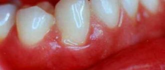
The first and most important symptom is the growth of gums, which seem to be on the tooth. This can be observed in the area of one tooth, between dental units, or throughout the entire jaw. In this case, the mucous membrane may not change its color, but may become bright red, and even turn a little blue. Patients with hypertrophy speak not only about the aesthetic disadvantage of the periodontium, but also about the occurrence of difficulties when chewing food, and pain when pressing on the gums.
Pathology can occur not only in humans, but also in cats and dogs. However, hyperplasia in animals is not contagious and is caused by other reasons.
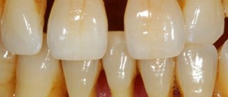
Enamel hyperplasia
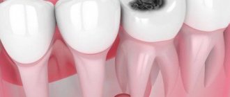
Dental hyperplasia is an increase in dental tissue cells, resulting in the formation of small oval seals on the surface of the tooth enamel. They can be located on the surface of the tooth root, on the neck of the tooth, and also on the surface of the dental crown. Depending on the location of the enamel seal, molar, cervical and coronal hyperplasia of teeth is distinguished.
Typically, tooth enamel hyperplasia does not require treatment, since it does not annoy the patient and does not cause pain. The exception is cervical hyperplasia, in which enamel seals are located close to the gum and constantly injure it. In this case, the doctor will polish off the seal and prescribe the patient phosphate-containing drugs for further treatment.
Reasons for appearance
Gum overgrowth can be a consequence of taking certain medications. There are a number of studies that have proven that periodontal hyperplasia is a consequence of taking antibiotics and other medications. In all the cases studied, either the dosage of the medications was violated, or the patient self-medicated, that is, he prescribed medications for himself, without consulting a specialist.
Other reasons include:
- genetics: in this case, the pathology manifests itself already at a young age, as a rule, at the time of the eruption of milk and then molars;
- hormonal changes in the body during pregnancy;
- blood diseases;
- incorrect structure of the maxillofacial apparatus.
Varieties
Depending on the prevalence, generalized and focal hyperplasia . If overgrown gums cover a large surface of several or all teeth, then this type of disease is called generalized. If it grows only near one tooth, then this type is called focal. It should be noted that generalized hyperplasia is much more common than focal hyperplasia.
Depending on the causes of hyperplasia, it can be medicinal or fibrous.
- Drug-induced hyperplasia occurs as a result of taking medications, the side effect of which is the extensive proliferation of gingival tissue.
- The fibrous form is rare and is a hereditary disease. The first symptoms appear in infancy during teething. In the future, the disease progresses, the gums gradually grow, become thicker, and over time, pockets begin to form, which are similar in appearance to the manifestation of periodontitis.
Forms of the disease and stages of development
There are two forms of pathology: generalized, that is, affecting one or even both jaws, and limited, which appears in a small specific area.
In the generalized form of the disease, there may initially be several lesions in different places in the oral cavity, which later merge into one. A limited form of hyperplasia may not manifest itself at all for a long time.
Stages of disease development:
Stage 1 – the gingival papillae increase in size, the periodontium is slightly enlarged and extends onto the tooth. Stage 2 – the tissue grows over half of the crown part of the tooth. Stage 3 – most of the tooth is hidden under the gum. In this case, the mucous membrane is regularly injured, which causes bleeding, swelling and general inflammation of the oral cavity.
Also, periodontal hypertrophy is divided into fibrous and medicinal forms. Fibrous is extremely rare. Due to similar symptoms - the appearance of periodontal pockets that are filled with plaque, the pathology is often confused with periodontitis. The most common form of the disease is the drug form.
Gum former: what is it and why is it needed?
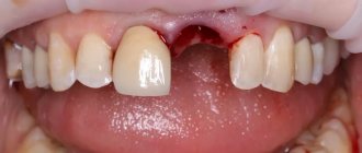
The gum abutment temporarily replaces the abutment that connects the root and crown. And at the same time prepares the space for completion of implantation - installation of the prosthesis. Many patients ask why a gum former is needed? Let's figure it out.
This construction is necessary to restore and maintain the contour of the gum tissue around the artificial tooth. As you know, after a tooth is removed, the gums and jawbone underneath stop receiving chewing load. Therefore, the tissues begin to atrophy. This process develops rapidly - in just 4-6 months, the gums seriously decrease in size - both in height and width. To restore the normal size of periodontal tissues, a gum former is used after implantation. As is already clear from the name of this element, it forms an even level or, one might say, a contour of the mucous membrane around the artificial crown. That is, so that the gums fit nicely, tightly, in a semicircle - exactly like on the adjacent living teeth.
New teeth in 1 day - All-ON-4 - 180,000 rub.
All inclusive!
3D modeling of the structure with a prosthesis, implantation of 4 Osstem implants, installation of a fixed prosthesis on the same day. Free consultation with an implantologist +7 (495) 215-52-31 or write to us
Complications in patients with hypertrophy
In addition to aesthetic problems, the disease leads to other negative consequences.
The patient experiences pain when chewing food, and the mucous membrane becomes sensitive to food irritants.
But the most important complication is the difficulty of maintaining high-quality hygiene. The accumulation of plaque under the gum leads to the formation of tartar, the mucous membrane becomes inflamed, periodontal bleeding and bad breath appear. All this can lead to the development of caries, pulpitis, and periodontitis.
In addition, periodontitis, an inflammatory gum disease, may develop. The consequence of periodontitis can be the destruction of the ligamentous apparatus with which the tooth is held in the socket.
In childhood, periodontal hypertrophy can lead to abnormal tooth growth, malocclusion, and, without proper treatment, problems with the maxillofacial apparatus. Therefore, you cannot delay treatment, and when the first signs of the disease appear, you should immediately contact a specialist.
What it is
Hyperplasia is a random rapid increase in gum tissue cells. Hyperplasia is not a tumor; it is benign changes in cell tissues, in which the cells themselves continue to perform their functions without changes. With hyperplasia, unlike oncological diseases, cells do not grow into surrounding tissues and do not metastasize. Many doctors are inclined to believe that it is not always a pathology, but only an experienced doctor can decide on a treatment method.
Gingival hyperplasia is manifested by excessive growth of tissue that can partially or completely cover the entire crown of the tooth. With this disease, only the gum tissue grows; the cheek and tongue tissues are not affected by changes. Hyperplasia is not an inflammatory process, so there is no drug treatment for it. In addition, it is necessary to correctly carry out differential diagnosis of the disease, being able to distinguish it from cysts and other tumor neoplasms. And only an experienced dentist can do this. Therefore, if you find that your gums are growing by leaps and bounds, consult a doctor immediately.
Gum hyperplasia is also accompanied by increased blood supply to the gums, so when brushing your teeth, the gums often bleed. Often patients mistake this disease for gingivitis or periodontitis and self-medicate, which does not bring any effect. In order not to waste time and not to advance the disease to such a stage when the tooth is completely under the gum, you must immediately consult a doctor.
Treatment
The first thing a doctor does is diagnose the pathology. The disease is easily confused with other gum diseases such as gingivitis and periodontitis.
If drug-induced hyperplasia is diagnosed, they try to eliminate the disease by discontinuing medications. If this is not enough, surgery is performed to remove excess gum.
That is, if therapeutic methods are not enough, the pathology is eliminated using gingivectomy. The essence of the operation is to excise the overhanging edge of the gum. After gingivectomy, when the gums have healed, damaged teeth are treated. Plaque is removed using professional oral hygiene, and the enamel is strengthened with fluoride-containing products.
The sooner treatment begins, the easier it will be to eliminate the pathology and return the gums to a healthy and attractive appearance.
Augmentation of keratinized gums
According to periodontal terminology, the attached gum is that part of the soft tissue that is firmly and tightly connected to the underlying periosteum, teeth and bone. The keratinized part of the gums provides stabilization of the gingival margin, promotes the distribution of forces in the muscle fibers during chewing, and also protects other tissues from any damage and traumatic effects.
Niklaus Lang and Harald concluded that 2 mm of keratinized gums and less than 1 mm of attached gums is sufficient to maintain adequate gum condition. Jan Wennström, in turn, concluded that the attached gum, among other things, also affects the general periodontal status of the patient. According to Mehdi Adibrad, the lack of adequate keratinized mucosa around implants with accompanying superstructures increases the risk of plaque accumulation, gingival inflammation, bleeding, and of course, recession. Ingvar Eriksson believes that adequate width of attached gingiva is that which is sufficient to prevent the risk of recession, based on the individual clinical characteristics of the patient. The role of keratinized gum is still not fully understood, but to preserve it, doctors use free gum grafts, perform apical reposition of flaps and soft tissue augmentation.
The two case reports below describe the effective use of free gingival grafts to increase attached gingival volume.
Clinical case 1
A 26-year-old somatically healthy man sought help with the main complaint of sensitivity and loss of gum level in the area of the lower front teeth. Clinical examination revealed tortoanomalies of teeth 24 and 25, gingival recession, and keratinized mucosal deficiency with abnormal attachment of the lower lip frenulum (Figure 1). A targeted radiograph showed bone loss. The treatment plan included augmentation of the keratinized gingiva using a free gingival graft. After anesthesia was administered, the recipient area was prepared using a scalpel (#15C blade) (Hu-Friedy). The graft was formed from the right area of the hard palate (photo 2).
Photo 1
Photo 2
The wound in the palate was closed using 3-0 silk sutures (Figure 3). The graft was secured to the gum area using 5-0 monofilament sutures (Figure 4).
Photo 3
Photo 4
The recipient area was covered with a periodontal dressing (Figure 5), and after 2 weeks there were signs of adequate wound healing with restoration of connective tissue attachment. To prevent a recurrence of recession, the patient was referred to an orthodontist, but later, unfortunately, refused his help.
Photo 5
Clinical case 2
A 55-year-old woman sought dental care with the main complaint of receding gums in the area of her lower anterior teeth. Clinical examination revealed deficits in gingival attachment, signs of recession, horizontal bone loss, and grade I mobility of the mandibular teeth (Figure 6).
Photo 6
The patient wanted to keep her own teeth. The treatment plan involved splinting the problem area and augmenting the keratinized gums using a free gingival graft. After anesthesia was administered, the recipient area was prepared using a scalpel (#15C blade) (Figure 7).
Photo 7
The area was prepared in such a way as to preserve as much as possible a good periosteal bed for subsequent revascularization of the graft material. A graft was formed from the palate (Figure 8) and the wound was sutured using Surgicel and 3-0 silk sutures (Figure 9). The autograft was secured to the recipient site using 4-0 monofilament sutures (Figure 10).
Photo 8
Photo 9-10
The recipient site was covered with a periodontal dressing, and after 2 weeks, both the donor and recipient sites showed signs of adequate healing (Figures 11-12).
Photo 11-12
Augmentation of keratinized gingival areas helps minimize the risk of recession as reconstructive procedures restore connective tissue attachment. New keratinized tissue can quite adequately withstand the effects of negative factors, provided that surgical interventions to restore it were performed according to a strict surgical algorithm.
Author: Dr. Manthan Desai
How to treat gum hyperplasia?
Drug-induced hyperplasia can occur at any age. Sometimes stopping taking provoking drugs leads to normalization of the gums. In all other cases, surgery is required.
Excess tissue is removed quite easily. Modern anesthetics allow you to completely get rid of pain. But initially it is important to identify the cause of hyperplasia, since if tissue growth was provoked by taking certain medications, then they need to be replaced to avoid relapse. The operation can be performed traditionally using a scalpel, or using modern equipment - the laser allows you to make a more accurate and sterile incision, and it also stops capillaries, which promotes rapid healing of the wound.
Hyperplasia photos before and after treatment
Hyperplasia is not a dangerous disease, but it significantly worsens the quality of life: the aesthetics of a smile is disrupted, which causes psychological problems, and eating becomes very difficult. Therefore, you should not delay contacting a doctor - the surgical operation is performed quickly and without pain. But it is worth remembering that it may have to be done more than once, since gum tissue has the ability to grow again and again.
What it looks like and what it is made of
Almost all patients are interested in what the gum former looks like and whether it will be visible when smiling or talking. The design is somewhat reminiscent of a solid screw with a thick cylindrical “hat”. Less commonly, its upper part is presented in the form of a truncated cone. The lower part looks like a threaded screw that is secured to the root of the implant. The crown part of the structure protrudes above the gum by only 1.5-2 mm, and does not fully replace the tooth. This is necessary so as not to put too much stress on the bone while chewing food while the implant takes root. After all, the main function of the structure is to form a sufficient amount of healthy gum tissue.
Shapers are made from titanium, or more precisely from an alloy of this metal. Titanium components are biologically inert - they do not reject, do not rust and do not release harmful substances into the human body. This metal is also very durable and, at the same time, very light.
Patients sometimes confuse formers with abutments. These are different designs, but in some ways their functions are similar. Shapers are a temporary measure and are installed on the chewing part of the row, i.e. they are not visible during a conversation or when a person smiles. Abutments are already a permanent element; they differ in shape and composition. An abutment serves to attach a crown (temporary or permanent) to an artificial root.
How long does it take to heal and care for the structure?
After installing the component, the question arises: how long will it take for the gums to heal and how to wear the former correctly? After all, the patient wants to quickly get into the usual rhythm of life. In the absence of complications, wound healing and restoration of the gum contour occurs within 1-2 weeks. But sometimes the process can take a month.
The speed of healing depends not only on the individual characteristics of each person, but also on proper care. No matter how much the patient would like to complete the implantation as quickly as possible, he must carefully follow all the instructions prescribed by the dentist.
Post-procedure care includes:
- gentle brushing of teeth: you need to act very carefully so that the seams do not tear and the gums are not injured. A soft bristle brush is required
- oral baths: you can use Chlorhexidine, Miramistin or herbal infusions to relieve inflammation - whatever your implantologist prescribes. Intensive rinsing is excluded, because... the seams may be damaged,
- diet: food should be softened to reduce the chewing load (for faster healing). For a while, you should give up hard and dry foods, too hot or cold foods,
- decreased physical activity, no heavy lifting,
- antibiotics: prescribed to patients who are simultaneously restoring several teeth to reduce the risk of bacterial inflammation.
For what types of implantation is it installed?
Let's figure out when a gum former is installed. Today, dental implantation is carried out using several methods:
- with delayed loading (classical method): first, a metal implant is screwed into the bone, after it has healed, the gum is incised and it is the turn of the gum former. It is installed for about a couple of weeks. However, in a number of situations, the implantologist fixes it almost immediately after implantation. As you can see, with this method, a shaper is definitely needed,
- with immediate loading of the implant: the entire process - from the introduction of an artificial root to the installation of a crown - takes a maximum of 7 days. Here, there is no need for a former, and it is not installed, because temporary abutments are immediately fixed, and a prosthesis is placed on top of them (this is if we are talking about the restoration of 1-2 teeth). That is, the abutment itself and the crown together with it can act as a former.
The construction is not installed during prosthetics on implants if the patient does not have all teeth or there is a large segment of missing ones. There is no need to change the shape of the gums, because... the prosthesis contains its plastic analogue - it is designed to hide the unevenness of the natural mucosa, at least at the first stage, when the implants take root. Afterwards, you can undergo plastic surgery and correct the position of the gums.
Diagnosis of gum fibromatosis
The examination involves examining your mouth in the dentist's office. The mucous membranes are checked for soreness, color shade, tissue density and their level relative to the tooth crown.
Fibromatosis is classified as a tumor disease, so during the examination it is important to differentiate it from other types of neoplasms. To do this, additional diagnostics are prescribed:
- Biopsy and histological analysis of tissues make it possible to separate the disease from similar manifestations of poliomyelitis, periodontitis, and cancer.
- X-ray evaluates the condition of the alveolar processes of bone tissue and the structure of the jaws as a whole.
Problems and complications after installation
Complications after the procedure can arise due to the characteristics of the body, improper care, or due to a poorly installed design. The patient may also notice that the gum former is loose. This sign indicates an incorrect installation. If you miss time, the structure may fall out of the “socket” and the metal implant will become overgrown with soft tissue. In this case, you must urgently contact the clinic so that the dentist can screw the component into place or install a new one.
The patient should be alert to the following symptoms (if they do not go away within 5-7 days): swelling, pain, bleeding, fever. If there are complications, the final stage of implantation is postponed until the tissue has completely healed. The implantologist will determine how long to wear the structure individually.