Classification of dentinogenesis imperfecta
Shields classification distinguishes the following types:
The first type of dentinogenesis imperfecta. The enamel structure remains within acceptable normal limits. The structures of peripulpal dentin are susceptible to destruction. The dentinal tubes narrow and are usually obliterated. Dentin hypomineralization may also occur. Precollagen fibers do not undergo transformation into collagen fibers.
Stanton-Capdepont syndrome (or type II). This subtype is characterized by disturbances in the structure of tooth enamel, a decrease in its thickness, heterogeneity of the dentin structure, developing calcification of the pulp chambers, and changes in the degenerative type of cement. The number of tubes in the dentin decreases, and the level of minerals is also greatly reduced.
The third type of dentinogenesis imperfecta. This form of pathology is characterized by fibrous changes in the dentinal matrix.
Causes
An anomaly appears when there is a disturbance in the development of hard dental tissues. The patient develops amorphous, disorganized, atubular dentin with a high content of organic microelements. The first category of disease is characterized by a lack of collagen. In the second type of disease, the main provoking factor is a defect in the DSPP gene. The provocateur of the third type of pathology is a violation of the phosphoprotein matrix, which is part of the hard tissues of the masticatory organs. In newborns (1 case per 17.5 thousand), the disease has a hereditary factor.
The mechanism of infection can be autosomal dominant, recessive or non-mutational. The genetic risk of the lesion is 50%. To identify the cause of the anomaly and develop the correct treatment plan, it is better to contact a private dental clinic. Only qualified specialists will be able to understand this issue. Self-medication is unacceptable. After all, such heterogeneous genetic damage can combine several diseases with different etiologies, which have the same clinical picture.
Diagnostics
One of the most important stages in diagnosing this pathology is collecting an anamnesis coupled with identifying unfavorable hereditary factors. The clinical manifestations of the disease directly depend on its form.
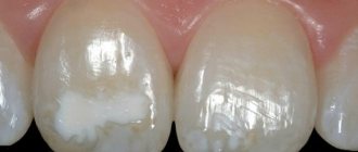
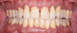
On physical examination, a change in the shade of tooth enamel is observed. The crown takes on a spherical shape and noticeably narrows in the neck area. Additionally, severe abrasion is detected in combination with a “decrease in the lower third of the face.”
It is necessary to differentiate dentinogenesis imperfecta not only from amelogenesis imperfecta, dentin dysplasia, acid necrosis, anomalies of tooth development, but also from other pathological changes in dentin associated with a number of other, more rare diseases. In particular, it is necessary to mention brachioskeletogenital syndrome, marble disease, hypophosphatasia, and Ehlers-Danlos syndrome.
In order to identify genetic factors in the occurrence of pathology, a medical genetic consultation may be indicated.
Disorders of enamel maturation with hypoplasia and taurodontism
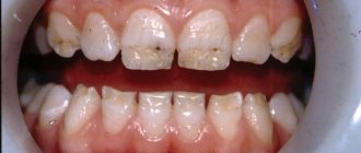
Some experts combine it with hypomaturation, others separate it separately. The formation of enamel tissue is disrupted during tissue differentiation. The pathology may be accompanied by grooved or thin hypoplasia.
The enamel turns into various colors - from white and yellow to unpleasant brown, becomes covered with opaque specks and acquires a pathological tendency to abrasion. The photographs show large cavities in the incisors and symptoms of taurodontism. In multi-rooted teeth, the pulp cavity is expanded and the crown is lengthened. The root is short, and the distance from the occlusal surface to the bifurcation zone is increased.
Treatment of dentinogenesis imperfecta
The treatment process for this pathology is a complex of measures. The main goal is to preserve the tooth through gentle treatment. It is for this reason that the use of remineralizing therapy is recommended. Among the necessary medications to restore normal mineral composition, calcium and fluorine-containing drugs are used.
In case of pathological changes in the hard tissues of the tooth root, endodontic treatment and covering of the tooth with a crown are carried out. If a tooth had to be removed, prosthetics or implantation are performed.
In order to mask a defect in the dentition, and also to prevent dentoalveolar deformation, removable dentures are used. To correct the reduction of the lower third of the face, wearing a special mouthguard is prescribed.
Anomalies of dental development - symptoms and treatment
Treatment depends on the type of anomaly and may include tooth extraction, prosthetics with crowns and veneers, surgical and therapeutic intervention.
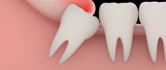
Supernumerary teeth must be removed. If the crown or root part of adjacent teeth is abnormal or destroyed due to a pathological process, then after a thorough examination the corresponding complete tooth is removed, and the supernumerary one is moved into the dentition. Supernumerary teeth most often have irregular shape, size or structure, so treatment is often completed with restoration with composite material or the creation of an artificial crown or veneer.
for patients with hypodentia and edentia . Partial absence of teeth, as a rule, is combined with a violation of the position of adjacent teeth and antagonist teeth, as well as with anomalies in the shape and size of the dental arches. Therapy will consist of orthodontic correction followed by prosthetics and implantation [7]. Patients with true primary edentia are very rare; they should wear complete removable dentures from an early age. Children often respond well to prosthetics and quickly adapt to them. Due to dynamic age-related changes occurring in the dental system, all removable dentures are replaced with new ones every 1.5-2 years. When making dentures, the doctor and technician take into account the morphological and functional characteristics of the dental system.
Treatment of anomalies in tooth size (macro- and microdentia) is usually orthopedic. If macrodentia and microdentia are combined with anomalies in the shape and size of the dentition, malocclusion, then preliminary orthodontic preparation will be required. It consists in the correct placement of teeth and allows you to subsequently qualitatively restore the desired tooth anatomy with crowns or veneers. In case of mild pathology with a small difference in the height or width of the teeth and their normal relationship with the size of the jaws and the type of face, orthodontic treatment can be limited.
Treatment of patients with abnormal tooth shapes involves performing composite restorations and making crowns or veneers that restore the anatomical shape. For concomitant pathologies that require correction of the bite, shape and size of the dental arches, orthodontic preparation will be required.
Treatment of anomalies in the structure of hard dental tissues (hypoplasia, hyperplasia, hypercementosis, amelogenesis imperfecta, medicinal and toxic effects on dental tissues, etc.) is complex and includes therapeutic, orthopedic and orthodontic care.
Treatment of teething anomalies . Early eruption of primary teeth usually does not require orthodontic intervention. If there is a violation, it may be necessary to switch to artificial nutrition instead of breastfeeding. Early eruption of permanent teeth is often caused by untimely loss of baby teeth. It often requires correction of the position of adjacent teeth and antagonist teeth to prevent complications. If the permanent tooth has already erupted, but the baby tooth has not yet fallen out, then the latter is removed, and the direction of eruption of the permanent tooth is corrected, if necessary.
For late eruption of primary teeth, gum massage will be helpful. Delayed eruption of a permanent tooth is usually corrected by an orthodontist. To do this, orthodontic preparation is carried out on the brace system: a place is created for the impacted tooth and a support is prepared for its traction. If it is necessary to open or remove an impacted tooth, the treatment is carried out by a surgeon [1].
Anomalies in the position of teeth are corrected using prosthetics with veneers and crowns, therapeutic restoration and orthodontic treatment with removable (in childhood) and non-removable devices: aligners and brace systems.
Symptoms
Symptoms largely depend on the category of pathology:
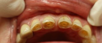
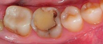
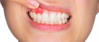
- If the patient suffers from the first form, his units erupt in time and are of normal size. But the patient complains of bleeding gums, as well as mobility of the chewing organs. Its dentin has a pronounced gray-watery color. Sometimes crowns resemble the shape of onions. A high degree of enamel abrasion and root fractures are often diagnosed.
- The second form of pathology has an isolated character. Dental tissues are stained amber or have a gray-watery tint. Less commonly, chewing organs are purple in color. Sometimes the crowns become short and have an onion-like shape. A distinctive feature is the progressive obliteration of the pulp chambers, which forms before the units erupt. If a chip appears on the crown, the dentin quickly becomes exposed, the height of the bite decreases, and the head in the temporomandibular joint shifts. Gradually, the teeth acquire a brown tint. At the same time, the roots become thinner and curved. Additional signs: high degree of electrical excitability of the pulp, delay in temporary organs of root resorption. But, in some patients, the change of milk units occurs ahead of schedule. In such a situation, caries rarely occurs.
- The third type is distinguished by the fact that the permanent masticatory organs are bell-shaped. Their color is brownish-yellowish or bluish-gray. Typically, in this situation, the pulp chambers are characterized by multiple openings.
It is useful to remineralize teeth. During fluoridation, the enamel is saturated with mineral elements. This technology restores the structure of the crown. The procedure can be artificial or natural. In the latter case, the enamel is restored due to the patient’s nutritious diet and taking vitamin and mineral complexes. With the artificial technique, the enamel is covered with protective compounds that include fluoride or mixtures without fluorides.
Provoking factors
Since dentinogenesis imperfecta is a hereditary disease, gene mutations that manifest themselves at the stage of bone formation, blood vessels and dentin formation in the fetus act as provoking factors.
The role of other risk factors for the development of pathology is unclear. However, it is obvious that the general state of health, immune status, quality of nutrition and oral care, injuries, and lack of planned sanitation of the oral cavity have a negative impact on the development and severity of the disease.
Therefore, the role of preventive measures against any dental diseases remains important in this case.
Prevention
The percentage of the disease is higher among patients with acute osteogenesis, when there is a qualitative and quantitative collagen deficiency. Specific methods for preventing pathology have not yet been developed. For prevention purposes, you should carefully observe oral hygiene rules and undergo routine dental examinations. It is necessary to carry out sanitation of the oral cavity in a timely manner. It is necessary to visit the dentist at least once every six months in order to identify the disease in the early stages, as well as to begin treatment of the pathology in a timely manner. If necessary, consult a geneticist.
Diagnostic measures
Diagnosis of dentinogenesis imperfecta involves both standard diagnostic dental procedures (examination of the oral cavity and radiography) and special ones, such as clinical and genealogical research aimed at identifying hereditary dental diseases and their characteristics.
With the help of an examination, the color and shape of the crown part, the presence of stains on it, and the degree of abrasion of the teeth are determined. In particular, the spherical shape of the crown may indicate imperfect dentinogenesis.
Based on radiographs, the condition of the root part of the tooth is determined (presence of canal obliteration, bone resorption) and the thickness of the walls of the pulp chamber.
When conducting a hereditary genealogical examination, a pedigree of family members is compiled (preferably no less than 3-4 generations) and the presence of genetically determined diseases of the jaw apparatus is determined.
The data obtained serve to clarify the diagnosis and make a prognosis for the offspring.
Characteristics of Pulposeptin paste and instructions for use in dentistry in the treatment of gangrenous pulpitis.
In this publication we will talk about the pathology called “Turner’s teeth”.
Here https://www.vash-dentist.ru/lechenie/zubyi/ultrazvuka-v-endodontii.html all about the benefits of ultrasonic expansion of the root canal of a tooth.
Reviews
Dentinogenesis imperfecta is a relatively rare, but in some of its forms, a serious disease that must be identified and treated without delay.
Moreover, in many families, due to the hereditary nature of the disease, it is not a surprise. If you or your children have been treated for dentinogenesis incomplete, please share your personal experience about this rather rare disease. You can leave a comment at the bottom of this page.
If you find an error, please select a piece of text and press Ctrl+Enter.
Tags: enamel destruction
Did you like the article? stay tuned
Previous article
The Korkhaus apparatus as the best solution in orthodontics for eliminating diastemas
Next article
Suturing the tooth socket is a simple and safe way to restore the alveolar process after extraction