Caries is a common dental pathology affecting hard tissues. During the pathological process, a vital element, calcium, is washed out of the enamel coating. This leads to the destruction of enamel, the formation of pathogenic cavities, and then inflammation of the pulp and connective tissue that holds the tooth. Lack of treatment threatens not only complete tooth loss, but also the development of concomitant life-threatening diseases.
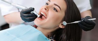
To quickly eliminate the pathology and prevent its progression, it is important to consult a doctor in a timely manner. Specialists at the Levoberezhnaya dental clinic diagnose and treat caries of any degree. To make an appointment with a doctor, call us and a consultant will select the most convenient appointment time for you.
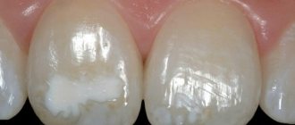
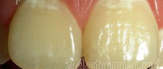
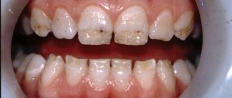
Enamel caries
Light and dark spots on teeth are one of the signs of enamel caries. This is a superficial carious lesion that does not affect the internal tissues - dentin and pulp. There is no pain yet, but the smile is already ruined.
Then a carious cavity forms, the tooth begins to react to cold and hot, sour and sweet. A good reason to rush to the dentist is the opportunity to eliminate caries in the spot stage without drilling and filling.
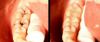
Silk thread method
Despite the fact that modern dentistry has high-precision computer diagnostic methods in its arsenal, the silk thread method is still used by doctors during the initial examination, mainly to identify approximal caries. To do this, floss is passed between the teeth using sawing movements (it used to be a silk thread, hence the name).
If there are carious lesions between the teeth, their edges are uneven, and the thread begins to cling. This method is used after cleaning the enamel and between the teeth that do not border the filled ones. The thread may touch tartar or filling, which reduces the reliability of the method.
Causes
Among the versions of the origin of caries, Miller’s theory, presented in 1898, is generally accepted. Miller even then stated that the disease is caused by cariogenic microorganisms that live in the microflora of the oral cavity of every person - Streptococcus mutans and Lactobacillus. They begin to harm only under certain conditions. By consuming the remains of carbohydrate foods, bacteria produce organic acids.
Together with four dozen other microelements, apatites make up 99% of enamel. Without them, it becomes porous, vulnerable, fragile, loses its shine and gradually collapses. This happens so quickly that it does not have time to replenish mineral losses from food entering the human body.
The two most important causes of enamel caries
- consuming excessive amounts of carbohydrate foods – in particular sweets and starchy foods;
- poor oral hygiene: if you don’t brush your teeth every time after eating, leftover carbohydrates will become food for microbes.
An indirect factor leading to the appearance of enamel caries is the buffer capacity and the volume of saliva secreted. Buffer capacity is the ability of saliva to neutralize acids and alkalis, thereby increasing the pH level and creating unfavorable conditions for bacteria. If there is not enough saliva, then the buffer capacity will not be enough to cope with this task.
Read more about theories of caries occurrence.
Etiology
The main reason at the local level is the presence in the mouth of:
- Cariogenic bacteria - these can be streptococci, lactobacilli and some other species.
- Residues of carbohydrate foods, mainly sugars, where these microorganisms multiply well and comfortably.
As a result of their “violent activity,” the acid-base balance of the oral cavity decreases to a critical level of pH 5.5 and below. Normally, the indicator should be at a pH level of 6.8–7.4. That is, the environment becomes too acidic.
Next, the acids dissolve crystals of hydroxyapatite, a substance that makes up 75% of all enamel minerals. The destruction occurs so quickly that the teeth do not have time to “absorb” all the necessary elements supplied with food.
There is also a second reason - drinking water with a low fluoride content (less than 0.5 mg/l). If the body systematically lacks fluoride, thinning of the enamel will occur regardless of the acidity of the oral fluid.
Demineralized enamel also lacks phosphorus, magnesium, sodium, chlorine, and potassium.
Factors that provoke enamel demineralization
- failure to comply with hygiene rules;
- insufficient secretion of the salivary glands;
- eating soft foods high in simple carbohydrates;
- inflammation of the gums;
- wearing dentures or orthodontic appliances, which prevents the thorough removal of hard deposits;
- crowded teeth - if they overlap each other, it becomes difficult to clean the interdental spaces;
- diseases such as rickets, diabetes, pathology of the adrenal glands;
- hormonal changes.
Classification and clinical picture
Taking into account the stage of development, there are 2 types of enamel caries:
- At the spot stage – the integrity of the coronal part is not broken, there is no cavity;
- Superficial caries - the lesion has spread into the thickness of the enamel, but the cavity does not reach the dentin.
The earliest sign is dull white spots on the enamel. Initially, they are visible only on the dried surface of the tooth. Such defects are single, located in the cervical zone (at the gingival edge), on chewing surfaces - fissures or between crowns.
Over time, the surface of the spots becomes rough, they change color - from white to yellowish, light brown.
With superficial caries, a cavity no more than 3 mm deep is formed in the enamel. The thickness of the enamel itself ranges from 2.8-3.0 mm. With the appearance of a “hole,” a pathological reaction to chemical, thermal and mechanical stimuli occurs. It becomes unpleasant from tooth contact with sour and sweet, hot and cold. Unpleasant sensations often develop into pain. The same reactions appear when pressing and biting on a tooth.
Probing
The doctor uses a periodontal or button probe to determine the condition of the teeth. By examining different areas of the tooth with a probe, he finds out how sensitive the reaction of the dental tissues is.
Probing also makes it possible to identify an inflamed pulp - in this case, the patient feels a sharp pain from the touch of the probe. At an advanced stage of the disease or an acute course, the enamel may be loose and even break off when touched by a probe.
Examination and diagnosis
To identify the problem, methods of extraoral and intraoral examination of the oral cavity are used - extraoral and intraoral. Diagnosis is painless, but there may be discomfort when teeth come into contact with the probe.
Matte chalky and brown spots, to the surface of which the probe “does not cling” - a sign of an inactive form of caries in the stain stage. Softened areas of light brown color, in which the probe “gets stuck,” indicate a pathological process.
Vital coloring
The most commonly used is methylene blue (blue). A swab soaked in it is applied to the surface of the teeth, dried and thoroughly cleaned of plaque, for 3 minutes. Damaged areas are painted. After 20-40 minutes the paint is washed off.
Laser diagnostics
Using the DiangoDENT installation, you can also diagnose enamel caries. Demineralized tissues reflect laser waves with a length of 680 nm or more. When this happens, the device notifies you with a signal.
Ultraviolet diagnostics
The affected areas become dark in ultraviolet rays, they contrast with healthy tissues, which are illuminated in blue. The Pluraflex device is used to conduct the study.
Differential diagnosis
When diagnosing caries at the spot stage, it is necessary to exclude non-carious lesions with similar manifestations - hypoplasia and early stage fluorosis, as well as age-related pigmentation.
With hypoplasia, spots appear on the outside of the incisors and canines closer to the cutting edge of the crown. They remain as shiny as healthy enamel. There are many fluorescent spots, they are located chaotically and do not become pigmented from contact with methylene blue. Age-related pigmentation (an organic film on teeth) is easily removed with soft plaque removal instruments.
It is important to differentiate superficial carious lesions from erosion of hard dental tissues and wedge-shaped defects. In both cases, there are clearly defined defects on the crowns. Their walls are dense, the probe does not cling to them.
Types of caries
Depending on the area of damage, caries of enamel, dentin or the surface of the tooth root is distinguished. Based on the location of the pathogenic focus, caries is divided into three types:
- Fissure
- affects the natural depressions on the surface of the teeth. - Cervical
- formed next to the gum on the cervical part of the tooth. - Interdental (approximal)
- most often develops in the interdental space of the front teeth.
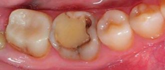
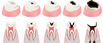
The first stage is characterized by the formation of a light yellow spot on the enamel coating. At this stage, the patient usually does not feel discomfort.
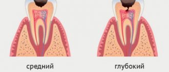
At the middle stage, the destruction of not only the enamel coating, but also dentin, a softer tissue, begins. It is impossible to delay treatment, as caries rapidly affects deep structures. Most often, at the middle stage, external pathological changes are insignificant - there is darkening of the enamel in the affected area, shallow carious cavities, and moderate aching pain. Some patients experience discomfort when the tooth comes into contact with cold or hot food.
Deep caries causes pain when eating sweet, sour or salty foods, as well as when the tooth is exposed to low or high temperatures. If the pain subsides 15–20 minutes after contact with an aggressive factor, this indicates that the disease has not developed into pulpitis. In this case, there is still a chance to save the tooth with full preservation of its functions.
Important!
Advanced forms are not always accompanied by damage to the enamel, although serious destructive processes are already observed inside the tooth. With this course, one of the main symptoms is bad breath, which is caused by rotting food particles accumulated in the cavity.
Enamel caries in children
In different regions, 15-40% of the total child population develops this problem, most often at the age of 9-11 years. Defects are localized both in the cervical region and on the fissures. Since the enamel in children has not yet been fully mineralized, it has less thickness and density than in adults. Childhood caries progresses faster and delaying treatment is dangerous.
Transillumination
The method is intended to identify various types of caries in the initial stage. Based on the heterogeneous light-absorbing ability of dental structures. Transillumination is carried out by shining a light from inside the mouth. Hidden carious cavities are revealed in the rays of light passing through the hard tissues of teeth.
The doctor makes conclusions about the degree and type of the disease based on the visible results. Fissure caries is characterized by a dark, fuzzy shadow, the intensity of which depends on the degree of damage to the fissures: the deeper they are, the darker the shadow. If the caries is approximal, then the carious lesions look like brown hemispheres, delimited from healthy tissue. Transillumination can also detect mineral deposits (tartar).
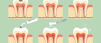
Treatment methods
The following treatment will help restore her strength.
Spot stage
The only reversible form that eliminates the need for drilling and filling. Remineralization therapy is indicated, aimed at restoring the enamel structure. The best results are obtained by Professor Knappvost’s own method – deep fluoridation. The procedure is painless, designed for 10 sessions lasting an average of 30-40 minutes, and takes place in stages:
- Teeth cleaning. Ultrasound if there is tartar. With a brush and paste, if there is no stone.
- Drying the crown surface.
- Direct fluoridation. Using a brush or mouthguard, the teeth are treated with Tiefenfluorid enamel-sealing liquid from Humanchemie.
The sealing liquid contains highly active calcium hydroxide and fluorine. From the effects of the liquid, the pores of damaged enamel are filled with crystals - compounds of fluorine with calcium, copper and magnesium, as well as silicate acid gel. The crystals remain in the pores from six months to two years. During this time, they produce ionized fluoride, which strengthens the tooth surface and increases its resistance to acids.
Surface defects
Tissues softened from damage are removed with a drill. The cavity is filled, as in the treatment of other forms of caries. As a rule, photopolymer filling materials are used for this, which are easily matched by color and hold tenaciously even in small drilled holes.
An alternative to traditional drilling is the infiltration technique. The damaged area is treated with the German polymer preparation Icon. It “seals” the affected pores, thereby making further progression of caries impossible. Icon restores the density and shine of a healthy smile to tooth surfaces.
X-ray examination
Modern dentistry cannot do without this method of diagnosing dental caries. Along with the traditional method, compact devices are now successfully used in practice, making it possible to carry out various types of research. For example, microprocessor-controlled orthopantomographs make it possible to perform literally any diagnostic examination in the field of dentistry. Radiography helps to identify hidden carious cavities on the contact surfaces of teeth, under an artificial crown. Using a radiovisiograph, you can carry out diagnostics without leaving the office. In this case, the patient himself has the opportunity to observe on the screen what exactly is done in the oral cavity during the treatment process.
Prevention
- brush your teeth with a brush and toothpaste after every meal; use pastes containing fluoride - Lacalut Fluor, Splat "Arktikum", Biorepair Total Protection Plus, President Classic, Silca Herbal Complete;
- alternate fluoride pastes with calcium ones - “New Pearl” with calcium”, President Unique, Splat “Biocalcium”, Splat “Maximum”, ROCS;
- use floss to clean the spaces between teeth – this is where food debris usually accumulates;
- do not abuse sweets;
- include in your diet foods rich in calcium, fluorine, vitamin D - fermented milk products, fatty fish, legumes, etc.
Fissure sealing also has a good preventative effect - filling the grooves on the chewing surface of crowns with liquid sealants that protect against bacteria.
You can find more detailed information about fissure sealing on our website.
Examination methods
All methods for diagnosing caries disease can be divided into basic and additional.
The main methods include:
- Questioning (complaints, medical history and life history)
- Inspection
- Probing
- Percussion
- Coloring
- Thermodiagnostics
- Radiography
- Electroodontometry
- Luminescent diagnostics
- Transillumination
Let's consider these methods in more detail.
Survey
First, the patients' complaints are clarified, which are mainly associated with a violation of aesthetics (the presence of a spot or a carious cavity) and with short-term painful sensations from eating cold, hot and sweet foods.
Next, they find out the history of the disease - how long ago the stain appeared, or the cavity formed, how long the tooth has been bothered, whether any treatment was previously carried out and its result.
When finding out the life history, you need to find out about the factors that could contribute to the development of the carious process, find out the general somatic condition of the patient (presence of chronic diseases).
Inspection
This is the main method of examination with which it is possible to detect caries in most cases. You need to examine your teeth sequentially, carefully examining each surface. For these purposes, a dental mirror is used to examine areas that are inaccessible for direct inspection (for example, the distal surfaces of the upper molars). Also, with this tool, rays of light are directed and the cheeks, lips and tongue are pushed back.
The examination should be carried out after cleaning the teeth from plaque and thoroughly drying them using an air gun. Only such conditions make it possible to identify carious lesions in the early stages.
Probing
Using this method, carried out with a probe, the depth of the carious cavity, the density of the affected tissue and pain are examined. Also, using probing, you can determine whether the pulp chamber has been opened - during probing, the patient notes a sharp pain in one point. If sensitivity is detected in the area of the enamel-dentin border of the walls and bottom of the cavity, then this is caries.
Soft walls of a carious cavity indicate an active carious process, dense walls indicate a chronic one.
Percussion
Percussion (tapping on the tooth) for caries is painless.
The following describes additional methods for diagnosing caries:
Vital coloring
It is known that demineralized areas of enamel are stained with special dyes, for example 2% methylene blue. After cleaning the tooth from plaque, isolating it from saliva and thoroughly drying it, apply the substance to the tooth with a cotton swab. After 3 minutes, excess dye is washed off, carious lesions are stained with varying degrees of intensity.
Thermal test
It involves irrigating the tooth with a stream of cold water. As a rule, caries causes a short-term painful reaction that goes away immediately after water comes into contact with the tooth. Sometimes certain difficulties arise when the patient is not able to accurately identify the tooth with increased sensitivity.
Radiography
This method is used to detect caries on contact surfaces. Most often, interproximal radiography (bite-wing) is performed, with which hidden carious lesions can be detected, as well as their size and depth can be assessed. But it is worth keeping in mind that this is an additional examination method; its data must be correlated with visual inspection and probing.
Electroodontometry (EDO)
A test to determine the excitability of the pulp, which in different conditions (normal, inflammation, necrosis) reacts differently to irritation. Normal values for a healthy pulp are 2-6 μA; for caries, this figure does not exceed 8 μA.
Luminescent method
It consists in changing the color of a carious lesion under ultraviolet rays, in contrast to healthy tissue. This method allows you to determine the initial stages of the carious process and identify damaged tissue around fillings (secondary caries).
VITAL COLOR
VITAL COLOR
(lat. vitalis vital, living) - intravital coloring of animal or plant cells and tissues. For this purpose, almost harmless or low-toxic dyes are used. For the first time, vital coloring was performed by the Austrian scientist Unger (F. Unger, 1848), who introduced the juice of red Phytolacca fruits into the white petals of hyacinth. In the histophysiology of animals V. o. with the help of ammonia carmine and sodium indigo sulfate was first used by the Russian scientist N.A. Khrzhonshchevsky (1864, 1866) when studying the function of the kidneys and liver.
V. o. is one of the important experimental methods of cyto- and histophysiology. Sometimes V. o. used in the clinic as a functional test to study the physiological usefulness of the kidneys (see Chromocystoscopy).
With the help of V. o. it is possible: 1) studying the patterns of intake, accumulation and release of substances involved in the metabolism of the body; 2) visual examination of some fiziol, processes (secretion, excretion); 3) marking of certain parts of the body (organs, embryonic rudiments of animals and plants, cells or their organelles); 4) study of physiol, the state of the cell normally and under various irritating and damaging influences; 5) detection of certain chemicals in the cell. substances (nucleic acids, proteins). The most convenient object for carrying out V. o. is cell and tissue culture, however V. o. It is also successfully used for pieces of organs and even small animals and plants, completely immersed in the dye. V. o. It is also carried out by injecting a dye into animal or plant tissue. The distribution of the dye in cells and tissues is usually observed under a light-optical microscope, and in those cases when for V. o. fluorescent dyes (fluorochromes) are used using a fluorescent microscope, which significantly increases the sensitivity of the study.
Rice. 1. Macrophage from rat subcutaneous connective tissue (toluidine blue). Rice. 2. Onion scales with different environmental reactions: a - pH = 4.7 - only the cell membranes are colored; — pH = 7.2 — cell sap of vacuoles is diffusely colored (neutral red). Rice. 3. Leech nerve cells: a - normal, the dye is deposited in the form of granules, the nucleus is not colored; 6—in conditions of paranecrosis, diffuse staining of the cytoplasm, nuclear structures are stained (neutral red).
For V. o. use ch. arr. various aniline dyes used in histol, research methods: acidic (anionic), basic (cationic) and neutral (tsvetn. fig. 1-3). Aqueous solutions of acid dyes do not penetrate normal cells well. Since permeability increases dramatically with damaging stimuli, these dyes (eosin) are used to detect cellular damage. Colloidal solutions of acidic dyes (trypan blue, lithium carmine, etc.) penetrate the cell by phagocytosis and are used by Ch. arr. to study the reticuloendothelial system.
Basic dyes, as a rule, penetrate well into living cells. Their further distribution in the cell depends on its condition. In an undamaged or slightly damaged cell, the main dyes accumulate in lysosomes in the form of clearly defined cytoplasmic granules, therefore acridine orange fluorochrome is widely used for intravital detection of lysosomes. With significant damage to the cell, the accumulation of dyes in the lysosomes stops and diffuse staining of the nucleus and cytoplasm appears. The color change can be reversible - the so-called. paranecrosis (see).
A special place among the main dyes is occupied by Janus green, which has the ability to selectively accumulate in mitochondria; it is widely used for their intravital detection.
Many neutral or weakly ionized basic dyes and fluorochromes (phosphine 3R, benzopyrene) have good solubility in fats and are therefore used for intravital detection of fatty inclusions.
The harmlessness of dyes used for vomiting is relative; they always affect certain cellular processes: Janus green blocks respiration, acridine orange inhibits protein synthesis; long-term exposure to neutral red, acridine orange, toluidine blue, etc. leads to agglutination of ribosomes with the formation of the so-called. crinoma - a basophilic cytoplasmic structure that arises in a cell under the influence of V. o.
See also Histological research methods.
Bibliography:
Gramenitsky E. M. Intravital staining of cells and tissues in normal and pathological conditions, L., 1963, bibliogr.; Zelenin A. V. Interaction of amino derivatives of acridine with a cell, M., 1971, bibliogr.; Levin S.V. and Golfand K.A. Methodology for cytophotometry of vitally stained cells, Cytology, vol. 6, no. 4, p. 525, 1964, bibliogr.
Yu. I. Afanasyev, S. Ya. Zalkind.
Enamel caries
Light and dark spots on teeth are one of the signs of enamel caries. This is a superficial carious lesion that does not affect the internal tissues - dentin and pulp. There is no pain yet, but the smile is already ruined.
Then a carious cavity forms, the tooth begins to react to cold and hot, sour and sweet. A good reason to rush to the dentist is the opportunity to eliminate caries in the spot stage without drilling and filling.
Vital coloring
To perform this diagnostic procedure, the teeth are first thoroughly cleaned, dried and isolated from saliva. Then a swab with a dye solution is applied to them. After 2-3 minutes, the swab is removed, excess dye is removed, and the mouth is thoroughly rinsed with water.
If the tooth enamel is not demineralized, as happens with caries, then there will be no traces of coloring matter left on its surface. If there are carious lesions, then dye residues of varying intensity are visible on the teeth, it all depends on the degree of damage. The more stained the tooth tissue is after removal of the tampon, the more global the degree of demineralization. This method is one of the most accessible and suitable for all categories of patients, allowing to detect the presence of the disease in the early stages.
Causes
Among the versions of the origin of caries, Miller’s theory, presented in 1898, is generally accepted. Miller even then stated that the disease is caused by cariogenic microorganisms that live in the microflora of the oral cavity of every person - Streptococcus mutans and Lactobacillus. They begin to harm only under certain conditions. By consuming the remains of carbohydrate foods, bacteria produce organic acids.
Together with four dozen other microelements, apatites make up 99% of enamel. Without them, it becomes porous, vulnerable, fragile, loses its shine and gradually collapses. This happens so quickly that it does not have time to replenish mineral losses from food entering the human body.
The two most important causes of enamel caries
- consuming excessive amounts of carbohydrate foods – in particular sweets and starchy foods;
- poor oral hygiene: if you don’t brush your teeth every time after eating, leftover carbohydrates will become food for microbes.
An indirect factor leading to the appearance of enamel caries is the buffer capacity and the volume of saliva secreted. Buffer capacity is the ability of saliva to neutralize acids and alkalis, thereby increasing the pH level and creating unfavorable conditions for bacteria. If there is not enough saliva, then the buffer capacity will not be enough to cope with this task.
Luminescent diagnostics
A research method for identifying superficial caries, based on the ability of enamel to change its color under the influence of UV rays. A beam of UV rays is directed at the teeth from a distance of 20-30 cm. Their hard tissues begin to glow. Normally, dentin and enamel emit a bright blue glow. With carious lesions of internal structures or enamel, the intensity of luminescence changes significantly: for example, in areas with chalk spots it is noticeably extinguished. This method of diagnosing dental caries allows you to detect changes inside them at the earliest stages.
Classification and clinical picture
Taking into account the stage of development, there are 2 types of enamel caries:
- At the spot stage – the integrity of the coronal part is not broken, there is no cavity;
- Superficial caries - the lesion has spread into the thickness of the enamel, but the cavity does not reach the dentin.
The earliest sign is dull white spots on the enamel. Initially, they are visible only on the dried surface of the tooth. Such defects are single, located in the cervical zone (at the gingival edge), on chewing surfaces - fissures or between crowns.
Over time, the surface of the spots becomes rough, they change color - from white to yellowish, light brown.
With superficial caries, a cavity no more than 3 mm deep is formed in the enamel. The thickness of the enamel itself ranges from 2.8-3.0 mm. With the appearance of a “hole,” a pathological reaction to chemical, thermal and mechanical stimuli occurs. It becomes unpleasant from tooth contact with sour and sweet, hot and cold. Unpleasant sensations often develop into pain. The same reactions appear when pressing and biting on a tooth.
Electroodontodiagnosis (EDD)
This clinical method is used in dentistry most often for the diagnosis of deep caries. Its essence is as follows: the tooth pulp is exposed to an electric current of minimal voltage until slight pain appears. For a healthy pulp, the values are in the range of 2-6 µA, for a pulp affected by caries - no more than 8 µA. This method also has disadvantages: it does not detect initial caries and does not allow one to determine the depth and extent of the process. The EDI method is not prescribed for children.
Examination and diagnosis
To identify the problem, methods of extraoral and intraoral examination of the oral cavity are used - extraoral and intraoral. Diagnosis is painless, but there may be discomfort when teeth come into contact with the probe.
Matte chalky and brown spots, to the surface of which the probe “does not cling” - a sign of an inactive form of caries in the stain stage. Softened areas of light brown color, in which the probe “gets stuck,” indicate a pathological process.
Vital coloring
The most commonly used is methylene blue (blue). A swab soaked in it is applied to the surface of the teeth, dried and thoroughly cleaned of plaque, for 3 minutes. Damaged areas are painted. After 20-40 minutes the paint is washed off.
Laser diagnostics
Using the DiangoDENT installation, you can also diagnose enamel caries. Demineralized tissues reflect laser waves with a length of 680 nm or more. When this happens, the device notifies you with a signal.
Ultraviolet diagnostics
The affected areas become dark in ultraviolet rays, they contrast with healthy tissues, which are illuminated in blue. The Pluraflex device is used to conduct the study.
Differential diagnosis
When diagnosing caries at the spot stage, it is necessary to exclude non-carious lesions with similar manifestations - hypoplasia and early stage fluorosis, as well as age-related pigmentation.
With hypoplasia, spots appear on the outside of the incisors and canines closer to the cutting edge of the crown. They remain as shiny as healthy enamel. There are many fluorescent spots, they are located chaotically and do not become pigmented from contact with methylene blue. Age-related pigmentation (an organic film on teeth) is easily removed with soft plaque removal instruments.
It is important to differentiate superficial carious lesions from erosion of hard dental tissues and wedge-shaped defects. In both cases, there are clearly defined defects on the crowns. Their walls are dense, the probe does not cling to them.
The main examination method for identifying caries: visual inspection with a mirror
This is the very first and main method used in clinical practice. Conclusions about the condition of a patient's teeth were previously based solely on visual examination data. But this method of diagnosing caries has significant drawbacks: low insufficient information content, as a rule, almost complete impossibility of identifying fissure or proximal caries.
The quality of the visual inspection depends on subjective factors. Mandatory testing conditions include preliminary cleaning and drying of the teeth being examined, and good lighting. This is a suitable method for diagnosing superficial caries, as well as medium or deep caries - at the initial stage of the disease, a visual examination does not reveal defects. Approximal (contact) caries can sometimes be identified by changes in the color of the enamel (it darkens, becomes grayish or chalky), floss gets stuck between the teeth, and the interdental papilla is inflamed.
Enamel caries in children
In different regions, 15-40% of the total child population develops this problem, most often at the age of 9-11 years. Defects are localized both in the cervical region and on the fissures. Since the enamel in children has not yet been fully mineralized, it has less thickness and density than in adults. Childhood caries progresses faster and delaying treatment is dangerous.
Treatment methods
The following treatment will help restore her strength.
Spot stage
The only reversible form that eliminates the need for drilling and filling. Remineralization therapy is indicated, aimed at restoring the enamel structure. The best results are obtained by Professor Knappvost’s own method – deep fluoridation. The procedure is painless, designed for 10 sessions lasting an average of 30-40 minutes, and takes place in stages:
- Teeth cleaning. Ultrasound if there is tartar. With a brush and paste, if there is no stone.
- Drying the crown surface.
- Direct fluoridation. Using a brush or mouthguard, the teeth are treated with Tiefenfluorid enamel-sealing liquid from Humanchemie.
The sealing liquid contains highly active calcium hydroxide and fluorine. From the effects of the liquid, the pores of damaged enamel are filled with crystals - compounds of fluorine with calcium, copper and magnesium, as well as silicate acid gel. The crystals remain in the pores from six months to two years. During this time, they produce ionized fluoride, which strengthens the tooth surface and increases its resistance to acids.
Surface defects
Tissues softened from damage are removed with a drill. The cavity is filled, as in the treatment of other forms of caries. As a rule, photopolymer filling materials are used for this, which are easily matched by color and hold tenaciously even in small drilled holes.
An alternative to traditional drilling is the infiltration technique. The damaged area is treated with the German polymer preparation Icon. It “seals” the affected pores, thereby making further progression of caries impossible. Icon restores the density and shine of a healthy smile to tooth surfaces.
Microscopic carious defects can be treated biologically under the influence of ozone. Ozone kills 99.99% of cariogenic bacteria.
Treatment
Treatment of caries should begin immediately after detection of the lesion. Eliminating pathology at the initial stage usually does not cause difficulties, and there is no need to use anesthesia, since boron does not affect sensitive tissues. In advanced forms, therapy lasts much longer, but it is not always possible to save a severely damaged tooth, and the cost of caries treatment will be significantly higher.
Caries of baby teeth
Treatment of baby teeth differs from that provided for permanent teeth. When carrying out manipulations, the doctor must exercise extreme caution so as not to damage the germs of the permanent tooth.
- If necessary, anesthesia is administered. To do this, the gum tissue is first treated with a freezing gel or spray, and then an injection with a local anesthetic is injected. This is necessary so that the child does not get scared. For injections, syringes with the finest needles are used, so the little patient does not feel pain at all.
- After the anesthetics have taken effect, the doctor carefully removes the tissue affected by caries. Manipulations are carried out using sterile hand instruments.
- Next, an antiseptic treatment is carried out, and then a filling is installed.
Treatment is carried out in sessions that last no more than 30 minutes, since with longer procedures the child gets tired and may begin to be capricious.
Eco-friendly and safe compounds containing fluoride and other minerals that strengthen children's teeth are used as filling materials.
Caries in adults
Treatment of advanced carious lesions in adults is carried out after radiography.
Sequence of manipulations:
- Introduction of anesthesia.
- Drilling out an infected tooth using a drill or laser device.
- Isolation of the pulp (dental nerve) with a sterile lining.
- If there is a need for depulpation, the nerve is first removed, and then the cavity is treated with medications.
- Temporary or permanent filling of root canals and coronal parts.
In case of deep lesions, a medicine that relieves inflammation is placed in the canals and a temporary filling is placed. If after 3-4 days the patient is not bothered by pain, a permanent filling is installed.
With minor lesions, it is possible to treat caries in adults in one session, but this is rare, since many people trigger pathology, which leads to long-term therapy.
In case of inflammatory processes, antibiotics, anti-inflammatory drugs and medications can be prescribed to relieve painful manifestations - antipyretics, painkillers, depending on the symptoms.
In severe forms of caries, the doctor decides to completely remove the tooth along with the root, followed by prosthetics. Such measures are taken in extreme cases when other methods do not bring a positive effect.
Prevention
- brush your teeth with a brush and toothpaste after every meal; use pastes containing fluoride - Lacalut Fluor, Splat "Arktikum", Biorepair Total Protection Plus, President Classic, Silca Herbal Complete;
- alternate fluoride pastes with calcium ones - “New Pearl” with calcium”, President Unique, Splat “Biocalcium”, Splat “Maximum”, ROCS;
- use floss to clean the spaces between teeth – this is where food debris usually accumulates;
- do not abuse sweets;
- include in your diet foods rich in calcium, fluorine, vitamin D - fermented milk products, fatty fish, legumes, etc.
Fissure sealing also has a good preventative effect - filling the grooves on the chewing surface of crowns with liquid sealants that protect against bacteria.
You can find more detailed information about fissure sealing on our website.