October 9, 2017
“Caries is the No. 1 problem of our time,” is how scientists and practicing dentists say about it. Fast food “on the go”, abuse of sweets and carbonated drinks, poor environment and rare, insufficient oral hygiene – all this leads to the rapid spread of caries. And untimely detection of the problem is fraught with complications, including tooth loss.
This is interesting!
Everyone knows that caries is a disease that needs to be treated.
But during the Middle Ages, people considered carious lesions not a problem, but a privilege of the upper class. After all, it was the rich who had the opportunity to limitlessly absorb sweets. Thus, healthy teeth were a sign of poverty. Let's see what types of caries exist today, whether they can be detected on their own, and what methods will help solve the problem.
Types of caries and features of treatment of the disease
Superficial caries
The photo shows superficial caries.
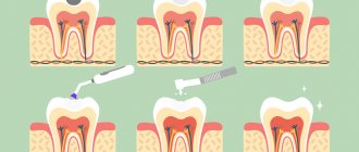
A very mild stage in which inflammation spreads only to the top layer of the tooth - its protective shell, that is, the enamel. This is actually the initial stage, which requires minimal treatment.
Features of treatment : as a rule, it is enough to only grind off superficial caries - with a laser, drill or ultrasound machine. Next, it is important to apply a protective fluoride varnish, which will strengthen the tooth enamel and protect it from further attack by bacteria. But in a number of situations, superficial caries cannot even be treated - it is enough to simply undergo regular examinations with a dentist, who will monitor the spread of caries. It is possible that the tooth will “spend” more than one decade in this condition without complications.
Caries in the spot stage
The photo shows caries in the stain stage.
It is characterized by the appearance of stains on the surface of the teeth: white, black or gray. This is the stage following superficial caries.
Features of treatment : as with initial, superficial caries, it is enough to just grind off the inflamed area. As a rule, even subsequent tooth filling is not required. But in this situation you need to be extremely careful - very often deep inflammation is hidden under caries at the spot stage, which can only be detected on an image or when opening a carious cavity.
Interesting! Did you know that no other technique, except a microscope, today makes it possible to detect caries in the early stages, or to see hidden dental defects? A dental microscope is a high-precision equipment that not only greatly increases the doctor’s visibility, but also allows you to assess the tightness of the filling and achieve its ideal fit during the treatment of caries.
Medium and deep caries
The photo shows medium caries (on the left) and deep caries (on the right).
It is characterized by the transition of carious formation deep into the tooth and damage to its internal tissues - dentin. Sometimes inflammation also affects the nerve of the tooth - in this case we can talk about deep caries, that is, pulpitis. The presented photos very colorfully show the destructive effects of the disease.
Features of treatment : it is the treatment of this stage of caries that is carried out according to the classic scenario - using a drill to remove inflamed lesions and subsequent filling of the tooth. If caries has led to severe destruction of the top of the tooth, then even prosthetics may be required - that is, installing an inlay (instead of installing a large filling) or an artificial crown, which will help restore the shape and appearance of the damaged tooth.
Radical caries
The photo shows root caries.
It occurs near the root, that is, it affects the tooth tissue in the gum area. As a rule, it is a consequence of the accumulation of plaque and tartar on the tooth - and this is nothing more than a breeding ground for bacteria.
Features of treatment : in addition to removing carious formations and subsequent filling, it is also necessary to remove plaque from the teeth - this is done using a laser or ultrasound.
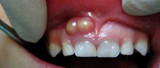
Root caries under the gum
The photo shows root caries under the gum.
A fairly complex type of caries with very specific treatment. Inflammation occurs at the root of the tooth, that is, under the gum. It is caused, like root caries, by the accumulation of plaque and tartar, but in this case the deposits penetrate deep under the mucous membrane and settle in the so-called “gum pockets”.
Features of treatment : removal of carious formations requires peeling of the gums, cleaning of the gum pockets and subsequent filling of the tooth. In fact, this is already a surgical treatment of caries, but it is carried out under anesthesia, so you should not be afraid of pain. As a rule, this stage of caries is characterized by damage to the nerve, which is located inside the root of the tooth, so you will also have to completely remove the dental pulp or try to relieve inflammation of the nerve with medications.
Fissure caries
The photo shows fissure caries.
With this type of caries, bacteria infect the chewing surface of the tooth, and inflammation accumulates in the fissures, that is, grooves or depressions. This is a rather unpleasant disease, since very often, under the surface carious lesion, there is inflammation that goes deep inside the tooth.
Features of treatment : with this problem, very often it is necessary to completely align the fissures, that is, to make the surface of the tooth flatter - naturally, after removing carious formations and filling the cavity. This, of course, from an anatomical point of view is not entirely correct, but it allows you to protect your teeth from a recurrence of the problem of fissure caries.
Interdental caries
The photo shows interdental caries.
It is a complex type of caries, since it is extremely difficult to detect even visually, because enamel damage occurs in hard-to-reach places - between the teeth. The main cause of this disease is food debris that is not removed in time.
Features of treatment : these carious cavities are extremely difficult to remove, since it is often necessary to drill out two teeth at once - the doctor needs access to the inflamed tissues. Therefore, the treatment of interdental caries becomes quite expensive.
Caries of baby teeth
The photo shows caries of baby teeth.
It occurs exclusively in children and is not as rare as one might imagine. Its causes are the abuse of sweets, as well as a lack of vitamins, which leads to weakness of tooth enamel. Treatment of caries of baby teeth must be carried out in order to prevent premature tooth loss (but the removed baby teeth will have to be replaced to prevent changes in the permanent dentition in the future!).
Features of treatment : treatment of baby teeth is carried out according to the classic scenario - removal of carious formations and filling of the damaged tooth. The only thing is that when treating children’s teeth, more gentle drilling methods are used - namely a laser and a piezo apparatus, which allow the removal of inflamed areas without the use of a drill. That is, quietly, more carefully and practically painlessly.
Caries under the crown
The photo shows caries under a removed crown.
It develops when crowns are installed incorrectly, the prosthesis is damaged, or simply the end of its service life. A loose fit of the crown to the tooth and gum, and improper “fitting” lead to the accumulation of food debris under the soft mucosa. And this is an ideal environment for the development of bacteria and microorganisms. Unfortunately, detection of caries under the crown most often occurs when severe pain occurs.
Features of treatment : in order to “get” inside the tooth, the doctor will have to remove the crown, treat the tooth and begin re-prosthetics. But it is unlikely that it will be possible to fix the same crown - you need to create a new prosthesis, taking into account the shape of the healed tooth (the artificial crown, as we have already found out, must fit tightly to it without micro-gaps). In order to prevent caries from developing under the crown, it is important to carefully care for your teeth and gums, as well as undergo regular preventive examinations with your dentist.
Caries under filling
The photo shows caries under a filling.
It is very similar to caries under a crown - it occurs due to a poorly installed filling, when microgaps remain between the filling material and the internal cavity of the tooth. They become clogged with food debris and accumulate bacteria that damage the tooth tissue from the inside. The problem of repeated caries under a filling can be detected during routine dental examinations. Remember that bacteria will very quickly reach the “heart” of the tooth - the nerve, which most often has to be removed.
Features of treatment : treatment of caries under a filling is carried out traditionally - using a drill, access to the internal tissues of the tooth is drilled, the old filling is removed, carious inflammation is cleaned out and a new filling is fixed. But the patient also needs to be prepared for the fact that living tooth tissue is becoming less and less and prosthetics may be required, that is, installing a crown to restore the anatomical shape of the tooth.
Remember that early diagnosis of caries will help to get rid of many serious complications and the need for unpleasant tooth drilling. In addition, prevention is noticeably cheaper than dental treatment. If you notice a violation of the integrity of the enamel, do not try folk and advertised remedies, but consult a doctor. A little attention to your health will ensure a snow-white, healthy smile for life.
Watch a video about how caries develops
Notice
: Undefined variable: post_id in
/home/c/ch75405/public_html/wp-content/themes/UltraSmile/single-item.php
on line
45 Notice
: Undefined variable: full in
/home/c/ch75405/public_html/wp-content /themes/UltraSmile/single-item.php
on line
46
Rate this article:
( 4 ratings, average: 4.00 out of 5)
caries
Periodontitis - forms, classification, photos
It is a consequence of pulp necrosis, which allows microorganisms to enter the hard and soft tissues of the jaw through the dental canal. The inflammatory process includes the peridontal ligament, cementum, root dentin together with the alveolar bone. Signs of periodontitis:
- the occurrence and intensification of pain when pressing on the tooth during the chewing process. There is a constant painful pulsation affecting half of the face;
- increased body temperature, feeling of weakness;
- swelling and tenderness of the gums in the affected area, as well as lymph nodes (mental and submandibular);
- discharge of pus from the root canal;
- facial asymmetry due to edema.
Consequences : lack of medical care leads to independent release of pus through the bones under the periosteum, as a result of which periodontitis becomes chronic.
Chronic periodontitis
A person may not be aware of the presence of the disease for a long time, since it occurs without the slightest symptoms. The reason to suspect the disease is the presence of a history of pain that was in the past, but gradually disappeared and does not remind of itself. A feature of chronic periodontitis is the melting of bone tissue around the affected tooth . Further development of the disease leads to loosening of the damaged tooth, as well as neighboring ones. Consequences: a common result is the loss of several teeth or granuloma .
Granuloma
It is a natural consequence of advanced periodontitis. The disease is marked by the spread of the inflammatory process, which is presented in the form of a small purulent nodule or sac in the area of the tooth root. Consequences: the infectious focus poses a serious danger to the body, as it acts as a catalyst for inflammation. At the initial stage, granulomas are characterized by practically asymptomatic progression. Severe pain gradually arises, which becomes more noticeable when pressing on the tooth or chewing solid food. Redness and swelling of the gums are noted, and tooth enamel becomes darker.
Root cyst
Lack of measures to treat granuloma leads to complications in the form of a root cyst. It forms at the top of the tooth root and is firmly attached to it. It looks like a round cavity located in the bone tissue, inside of which there is purulent contents. The root cyst grows slowly and imperceptibly, and therefore poses a significant danger. It can be visually determined only if it grows strongly, when protrusion of the bone is observed. Consequences: there is a possibility of the cyst growing into the maxillary sinus and nasal cavity. Displacement of teeth to the sides, as well as pathological fracture of the lower jaw.
Periostitis of the jaw bone, tooth
If treatment of root cysts and periodontitis was not carried out or was carried out in bad faith, then the likelihood of periostitis or inflammation of the periosteum sharply increases. The inflammatory process begins to emerge from the periodontal fissure and enters under the periosteum. Surgery is required, which involves making an incision in the mouth under anesthesia to release the pus. Consequences : failure to see a doctor in a timely manner carries the risk of an abscess.
Abscess
An abscess is a collection of purulent tissue in a confined space. The pus begins to put pressure on the root apex area, which leads to the tooth being pushed upward and possible destruction of the bone tissue in the affected area. If the abscess does not open for a long time, then the spread of pus to the periosteum, skin and mucous membrane begins. Consequences : severe and painful swelling occurs.
Phlegmon
It is considered one of the most dangerous forms of abscess. Cellulitis carries with it the risk of serious infection of the body, since infection and purulent accumulations from the swollen area enter the body. In fact, pus begins to spread throughout the surrounding tissues and can reach the neck and even the heart. The following symptoms are characteristic of phlegmon:
- swelling and tenderness of the soft tissue of the gums around the affected area;
- difficulty swallowing and opening the mouth;
- painful sensations behind the eyeball;
- deterioration of appetite and sleep;
- fever and weakness.
Consequences: the disease carries with it a significant risk of death! (we do not publish photos of phlegmon, copy and paste the word into a search engine to see the pictures).
Sinusitis
Inflammatory processes in the oral cavity (pulpitis, deep caries stage, cysts, periodontitis and others) aggravate the risk of single-gene sinusitis (sinusitis). It is caused by the penetration of infection from the affected tooth into the maxillary sinuses (we are mainly talking about infection in the upper molars and premolars, which are practically in contact with the maxillary sinuses). Initially, the infection enters only one maxillary sinus, then it spreads to the other. There is a possibility of getting into the sphenoid, frontal and other sinuses. Sinusitis manifests itself:
- severe headaches;
- swelling of the cheek;
- elevated temperature;
- thick mucous discharge of a purulent nature;
- pathological reaction to light.
If adequate treatment is not carried out in a timely manner, sinusitis can lead to the following consequences :
- transition to a chronic form;
- meningitis;
- sepsis, which is localized near the brain;
- purulent otitis;
- inflammation and swelling of nerve endings.
Tooth loss
Due to improper oral care and delays in seeking medical help, teeth decay and fall out. What are the consequences of losing teeth? Consequences :
- irreversible imbalance in the activity of the oral system. The absence of even one tooth disrupts the position of neighboring teeth; they do not hold so firmly in the gums, which can lead to their loss;
- a decrease in the efficiency of eating, as the degree of chewing and, accordingly, digestion of food deteriorates. A natural consequence is gastrointestinal diseases and metabolic disorders;
- curvature of the dentition, malocclusion, increased risk of diseases of the periodontal tissues;
- loss of aesthetic appeal (change in facial oval, sunken cheeks, visual unattractiveness due to missing teeth).
Bone atrophy
Atrophic processes in the maxillofacial region are a natural consequence of the absence of even one tooth. Its loss means the displacement of an entire row of teeth, a significant decrease in the volume of bone tissue. If atrophy is not stopped by building up bone tissue, then the lips and cheeks begin to “sink” into the oral cavity, and healthy teeth begin to move along the dentition due to loss of support. Consequences:
- the appearance of cosmetic facial defects (wrinkles, sunken lips, cheeks, etc.);
- deterioration of chewing function and the occurrence of gastrointestinal disorders;
- pathological changes in speech;
- displacement of the dentition, loss of healthy teeth.
Avoiding such unpleasant consequences is both easy and difficult. The main thing is to carefully observe oral hygiene and consult a dentist as soon as possible!
Comments
[…] leads to diseases of the teeth and gums such as caries, gingivitis and periodontitis. But despite this [...]
Irrigator Aquajet LD-A8I, reasons why you should choose. (05/05/2018 at 09:00) Reply to comment
[…] tooth enamel and caries are two completely different diseases. However, thin and [...]
Enamel hypoplasia: 6 features of a dangerous disease (08/15/2018 at 12:00) Reply to comment
[…] But these two diseases are completely different. Firstly, caries is associated with the formation of a large number of bacteria, and [...]
Dental fluorosis: 5 facts about the problem that everyone needs to know (08/31/2018 at 09:00) Reply to comment