Surgeon
Bohyan
Tigran Surenovich
Experience 36 years
Surgeon of the highest category, Doctor of Medical Sciences, member of the International Association of Surgeons, Gastroenterologists and Oncologists
Make an appointment
Lipoma (fat) is a benign formation in the form of small nodules, the appearance of which is associated with the accumulation and compaction of excess adipose tissue. The size of the lipoma may increase or remain stable. When exposed to unfavorable factors, pockets of pathogenic microflora can form inside the wen, which makes it a source of other serious diseases of internal organs. The unaesthetic appearance of formations protruding on the surface of the skin is also important. Therefore, in most cases, a decision is made to remove the tumor and its subsequent histological examination to clarify the nature of the wen - benign or malignant.
The area of localization of lipoma is the back, chest, face, limbs, mammary glands, structure of internal organs. The formation can be detected accidentally during diagnostic procedures or during palpation. The edges of the lipoma are dense and clear, the formation is mobile, and its palpation is painless. It is more difficult to detect a lipoma in the tissues of internal organs. However, it is precisely such cases that are recognized as the most dangerous, threatening disruptions in the functioning of the affected tissue area.
Classification
Depending on the internal structure there are:
- Myolipomas are formations from muscle cells.
- Angiolipomas are lipomas with the inclusion of blood vessels.
- Myxolipomas are tumors containing mucus.
- Fibrolipomas are formations containing connective tissue.
It is possible to accurately determine the nature of the formation only after a thorough diagnosis. It should not be postponed for the foreseeable future, especially if the lipoma quickly increases in size and causes discomfort.
What to do and which doctor to contact
If a white tumor appears in the oral cavity, and even if it does not cause you concern or interfere with your life, you should consult a doctor.
Which doctor deals with lipomas in the oral cavity and larynx? Diagnosis and treatment of growths in the mouth is the specialization of a dentist, maxillofacial surgeon, and otolaryngologist.
Any of these doctors can examine and make a preliminary diagnosis. But the treatment will be carried out:
- lipoma on the gum, cheek, tongue, upper palate - dental surgeon;
- lipoma in the larynx, pylorus of the larynx - ENT doctor.
If a suspicious tumor appears in the baby’s mouth, you should visit a pediatrician immediately.
Causes of lipoma
The general pattern of the appearance of wen is the accumulation of fat cells due to a violation of fat metabolism in the body. If measures to treat the problem are not taken in a timely manner, the formation quickly grows, pinching nearby muscles and blood vessels. Among the causes of lipoma that influence the unfavorable course of the pathology, it is worth noting:
- Hormonal disorders, a period of hormonal changes in the body.
- Metabolic failures.
- Disturbances in the diet with a predominance of food of animal origin.
- Diseases of the kidneys and liver, pancreas and thyroid gland.
- Bad habits.
- Diabetes.
- Genetic predisposition.
Also, the formation of excess adipose tissue is affected by physical inactivity, a sedentary lifestyle and refusal of full-fledged physical activity.
Symptoms
The main localization of wen is the place of deposition of adipose tissue. At first it is a soft compaction, which gradually increases in size. The only symptom of a lipoma is the formation of a noticeable tubercle with a movable structure, which can be felt when pressed. There is no pain when palpating the lipoma on the back or arms.
If the wen is localized in the structure or on the surface of internal organs, disturbances in their functioning may indicate pathology. The disease is also accompanied by characteristic symptoms:
- a lipoma in the esophagus causes coughing, nausea and vomiting;
- formations on the bronchi and trachea cause a constant dry cough of a paroxysmal nature;
- lipoma on tendons and cartilage limits their mobility and causes pain;
- a lump in the mammary gland gives pain;
- fatty deposits in the kidneys provoke colic, lower back pain, and increased blood pressure;
- a lipoma in the head causes dizziness and headaches;
- a formation in the neck makes it difficult to swallow food and also causes hoarseness;
- A lipoma in the heart area causes arrhythmia and heart failure.
Are you experiencing lipoma symptoms?
Only a doctor can accurately diagnose the disease. Don't delay your consultation - call
Benign tumors in the mouth must be removed
Benign tumors of the oral cavity are neoplasms in the oral cavity of a benign nature, which are characterized by limited, slow growth. They can be localized on the mucous membrane of the mouth and lips, as well as in the thickness of the jaws and soft tissues.
The most common tumors are those whose origin is caused by glandular, flat, tooth-forming epithelial tissue, but formations from adipose, muscle tissue, connective tissue structures, blood vessels and nerve trunks are also found.
A person notices changes in the mucous membrane of the oral cavity, inflammation and growths almost immediately. Discomfort intensifies when exposed to hot food or friction with dentures. This could be a papilloma in the mouth - a tumor-like formation on the mucous membrane. It is important to mitigate as much as possible the conditions that predispose to malignant degeneration of the inflamed and overgrown epithelium. By visiting a doctor, you can consult on complex treatment.
The proliferation of epithelial tissue in the oral mucosa is caused by the human papillomavirus (HPV). Its particles are present in the bodies of 9 out of 10 adults on the planet. Some people have no idea about this and do not feel any changes at all. Others have to struggle with the unpleasant manifestations of papillomatosis for many years.
What symptoms of papillomatosis in the mouth should you pay attention to?
Regardless of the specific localization of growths on the oral mucosa, several general characteristics of tumors are distinguished. Usually papillomas on the gums and tongue are pink with a whitish tint, their size does not exceed 10 mm. The most characteristic feature is the presence of a narrow or wide base - the so-called “leg”. The upper part is lumpy, with uneven edges.
Feelings of a person who has become the “owner” of a small tumor in the mouth: it interferes with normal chewing of food and talking. Much depends on the specific location of the papilloma. Typically, the localization of formations is associated with the tongue, gums, lips, and lateral surfaces of the oral cavity. There are not only single papillomas, but also multiple growths. Some are soft and mobile to the touch, others have a keratinized surface.
Before removing growths with such characteristic signs, cytological and histological studies of the material obtained from the mouth must be carried out. Laboratory tests can distinguish papilloma from genital warts or epithelioma and determine the degree of oncogenicity of HPV.
Complex therapy of papilloma in the mouth
Specialists need to clearly limit the growth, identify hidden foci of papilloma in the mouth, the treatment of which is not started without consultation with an oncologist, dentist, or ENT doctor. The choice of therapeutic technique affects the final results, but it is difficult to achieve 100% results.
To increase the effectiveness of treatment, it is carried out comprehensively. According to medical research and patient reviews, this approach reduces the number of relapses, that is, the appearance of new tumors in the oral cavity.
Removal of papillomas in the oral cavity
Treatment of small growths of the mucosal epithelium is fairly quick and almost painless. It is easier to get rid of small round formations on a thin stalk. Typically, oral papilloma has an uneven edge in the shape of a cockscomb or cauliflower. Its color is close to the tone of the mucous membranes, sometimes it is darker or lighter. Removal of such tumors is carried out by deep coagulation with radio waves, evaporation with a laser beam, or traditional surgical excision.
Oral fibroma is a benign neoplasm consisting of fibers of mature connective tissue. It is a clearly demarcated rounded nodule on a stalk or broad base, covered with unchanged mucosa. Characterized by slow exophytic growth. Oral fibroma can be located on the inner surface of the cheeks, mucous membrane of the lips, soft palate, gums, and tongue. Diagnosis of oral fibroma is made by inspection, palpation, ultrasound and histological examination. An orthopantomogram, radiography and periodontogram are used to identify inflammatory processes that provoke the formation of fibroids. Treatment of oral fibroma boils down to its excision. Along with papilloma, lipoma, myoma, nevus and myxoma, fibroma is a benign tumor of the oral cavity. It most often occurs in children aged 6 to 15 years. Clinical dentistry considers traumatic and inflammatory factors to be the causes of the formation of oral fibroma, and a hereditary predisposition can also be traced. Quite often, the history of patients with oral fibroma reveals regular biting of the same area of soft tissue of the oral cavity, which precedes its appearance. Factors that provoke the appearance of fibroma also include trauma to the mucous membrane with a sharp tooth edge, a poorly fixed prosthesis or crown; chronic inflammatory processes of the oral cavity (gingivitis, stomatitis, periodontitis, glossitis, etc.) Signs of oral fibroma Oral fibroma has the appearance of a formation rising above the general surface of the mucous membrane with a wide base or stalk. It is painless, has a hemispherical shape and is covered with a mucous membrane of the usual pink color. The surface of oral fibroma is smooth and, unlike papilloma, has no outgrowths. No changes in the mucous membrane in the area of fibroma are usually observed. In rare cases, ulceration is observed over the tumor. In this case, an infection may occur with the development of inflammatory manifestations: redness, swelling, pain in the area of the fibroma. Oral fibroids typically exhibit a slow increase in size. If the fibroma is not traumatized, its size can remain stable for a long time. With constant trauma, malignant degeneration of the tumor is possible. Diagnosis of oral fibroma The characteristic clinical picture of oral fibroma in most cases allows the dentist to make a diagnosis based on examination and palpation of the formation. To determine the depth of growth of the base of the fibroma into the underlying tissue, ultrasound can be performed. In rare cases, usually when there is ulceration or inflammatory changes in the area of the fibroma, a biopsy of the formation is indicated. More often, histological examination of oral fibroma is carried out after its removal. An important point is the diagnosis of the causative factor in the formation of oral fibroma. For this purpose, a thorough dental examination is carried out aimed at identifying inflammatory diseases of the oral cavity, radiography or radiovisiography, an orthopantomogram and a periodontogram are performed. Patients with dentures need to consult an orthopedic dentist to avoid the traumatic effect of the existing denture on the tissues of the oral cavity. Treatment of oral fibroids The most effective treatment for oral fibroids is surgical excision. Pedicled oral fibroids are removed along with the stem using two bordering incisions. The fibroma at the base is excised together with the base using a bordering or arcuate incision. Removal of fibroma on the red border of the lip is made with an incision perpendicular to the passage of the fibers of the orbicularis oris muscle. For large oral fibroids, to prevent deformation of the mucosa, patch closure of the defect remaining after tumor removal is performed. The flap is cut out using a V-shaped incision from adjacent tissues.
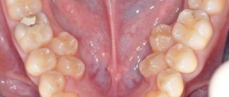
Epulis, popularly called supragingival, refers to benign tumors of the maxillofacial region. Epulis is a tumor-like neoplasm with a diameter of 0.5 to 6-7 centimeters, which is located on the alveolar process of the jaw. The predominant localization of the supragingival is the area of small molars, although in some cases it can occur at the level of any teeth of the upper or lower jaws.
The cause of the occurrence has not been definitively established, but in the vast majority of cases it occurs as a result of prolonged irritation of the mucous membrane of the alveolar process, for example, by the sharp edge of a decayed tooth or from irritation of the mucous membrane by dentures. It occurs quite often in pregnant women; they are often diagnosed with hypertrophic gingivitis. Epulis grows slowly, although pregnant women often experience accelerated growth, apparently caused by hormonal changes in the body. As a rule, patients do not experience any pain, except in cases of injury to the teeth of the opposite jaw. The supragingival partly or completely covers the coronal part of one tooth, and more often than not several teeth at once from the vestibular and sometimes from the lingual surface.
Most often, the tumor has a wide stalk and is covered with ordinary mucosa, without any pathological changes; if the epulis is injured, then areas of erosion and hemorrhage appear. It differs from malignant tumors in that there are no areas of tumor decay. Prolonged growth of the supragingival can lead to destruction of the alveolar process and its spongy substance. In this case, osteoporosis of the bone will be visible on the x-ray.
The color of epulid is slightly different from the color of the oral mucosa - sometimes it has a red-brown or even bluish tint.
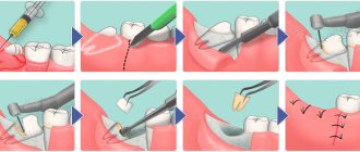
Treatment for epulide is only surgical. Considering that epulis has a growth zone in the periosteum and in the bone, after removing the tumor within the visible healthy mucosa, you need to do a thorough curettage around the tumor and remove the softened bone. Sometimes the tooth itself is also removed if it is located in the area of tumor growth and the alveolar process is destroyed by the tumor process. After removal, in some cases, the supragingival tissue may reappear in the same or other areas of the jaws.
Methods for diagnosing lipomas
The long period of latent progression greatly complicates the diagnosis of soft tissue lipoma in the early stages of development. More often, pathology is detected when the wen reaches a size of 1.5-2 cm and is easily palpable under the skin. To carry out differentiated diagnostics and confirm the benign nature of the formation, you can:
- Blood tests to exclude viral or bacterial infections, as well as the absence of an inflammatory process.
- X-ray of the chest, limbs or abdomen. Allows you to detect a formation, clarify its location, calculate its size and assess the condition of surrounding tissues.
- Ultrasonography. It is used as an additional diagnostic method when the presence of a lipoma is confirmed by other examination methods.
- Computed and magnetic resonance imaging. Effective methods to determine the location and size of the lipoma, as well as confirm its benign nature.
- A biopsy of wen tissue to analyze it for the risk of cells degenerating into a malignant structure.
If the patient has other diseases of the internal organs, the diagnosis of lipoma is carried out taking into account their characteristics, and deciphering the results suggests the mutual influence of pathologies, making it possible to more accurately determine the cause of the appearance of the wen.
Treatment
If small lipomas are detected in a patient, a wait-and-see approach is chosen, which involves ultrasound monitoring several times a year. However, there are often cases when the doctor decides in favor of surgical intervention. Surgery is necessary if:
- The tumor grows rapidly, engulfing neighboring tissues and blood vessels.
- The lipoma rises above the surface of the skin, giving an unaesthetic cosmetic effect.
- The development of a wen gives persistent pain.
- Due to the growth of adipose tissue, the functioning of internal organs and the circulatory system is disrupted.
Removing or destroying a lipoma on the neck or back is possible in the following ways:
- Endoscopic method. It involves removing the tumor along with the capsule through a small incision in the skin.
- Liposuction is the extraction of adipose tissue by softening it and stretching it through a thin needle. Does not leave scars and does not injure the skin.
- Excision of a lipoma is the removal of a wen during a classic surgical intervention.
The optimal method is selected taking into account the general condition of the patient and the size of the lipoma.
What is atheroma?
Atheroma is a cyst of the sebaceous gland that occurs when its duct is blocked. Sebum cannot come out and accumulates inside, it stretches the gland, and it turns into a cystic cavity. This can happen for a variety of reasons, often due to trauma to the skin.
Atheromas most often occur where there is a lot of hair and sebaceous glands: atheromas on the scalp, face, neck, back, genitals.
In its manifestations, atheroma resembles a lipoma: it is also a “bump” that is located under the skin, does not cause pain and easily moves when pressed. But there is one characteristic sign: usually there is a hole in the skin in the area of atheroma through which contents with an unpleasant odor are periodically released. Inside the atheroma there is a substance that resembles cottage cheese or paste in consistency.
Atheroma is a benign formation. It's not cancer. The only thing that is dangerous about it is the possibility of suppuration. An infected and inflamed “bump” swells, turns red, and becomes painful. Sometimes it opens and pus comes out along with fat.
Atheromas can degenerate into malignant tumors, but this happens very rarely.
Diagnosis and treatment of lipoma at the JSC Meditsina clinic in Moscow
The clinic of JSC "Medicine" in the Central Administrative District of Moscow is ready to offer professional assistance to patients with suspected single or multiple cases of lipoma development. The center’s specialists have high-quality equipment and extensive practical experience in using various methods of removing a tumor focus. Each patient is guaranteed attentive attention, referral to a full course of examinations and an individual approach to developing lipoma treatment tactics. Appointments are available on the clinic’s website and by calling the numbers provided.
How are atheromas and lipomas treated?
Atheromas are treated surgically; the type of operation will depend on the size of the formation. Small sebaceous cysts can be removed with a laser. Most often, atheromas are large in size and are removed with a scalpel. The “bump” on the skin is surrounded by two incisions, then the cyst is peeled out and removed along with a small piece of skin. Stitches are placed on the wound.
Atheroma cannot be cured by “sucking out” the contents with a needle and syringe. It is imperative to remove the walls of the stretched sebaceous gland - if they remain, they will begin to produce sebum again, and the cyst will grow again.
If atheroma suppurates, surgical treatment is performed, and the doctor may prescribe a course of antibiotics. Lipomas do not need to be treated. The operation is performed if:
- education is large and growing rapidly;
- the lipoma is in an inconvenient place and is constantly in the way;
- bothered by soreness;
- The patient himself insists on removing the lipoma.
Wen is removed in the classic way, using a scalpel. Relapses are possible, but extremely rare. Typically, atheromas and lipomas are removed on an outpatient basis; hospitalization is not necessary. Anesthesia is also not needed - local anesthesia is sufficient. The operation lasts on average 15–20 minutes.
Make an appointment by phone +7 (495) 120-08-07.