Once upon a time, extraction was the only known way to relieve a person of certain dental problems. And it would be fine if we were talking only about the Dark Middle Ages, but a few decades ago, in Soviet dental clinics, teeth were pulled much more often than now, lacking the technologies, materials and equipment for high-quality treatment and preservation of dental units. But now, fortunately, complete tooth extraction is considered the most extreme measure in dentistry. However, sometimes this is exactly what is necessary.
Purposes of tooth extraction surgery
- Sanitation of the body by eliminating foci of odontogenic infection.
- Elimination of acute odontogenic inflammatory process.
- Elimination of pre-tumor processes caused by chronic trauma to the oral mucosa, tongue and teeth.
- Improving chewing function by creating conditions for dental prosthetics.
- Elimination of psycho-emotional discomfort and improvement of chewing function in the treatment of patients with anomalies of the dentofacial apparatus.
Indications for tooth extraction are divided into two groups: general and local.
General indications include those in which the manifestations of the pathological condition of the body, caused by a diseased tooth as a source of infection, come to the fore. For example, general diseases of organs or systems in the body (endocardium, myocardium, kidneys, etc.).
Local indications can be divided into 3 groups:
- Sanitation indications for tooth extraction: chronic periodontitis, acute periodontitis, the presence of pathological processes around an abnormally located tooth, destroyed milk teeth, etc.
- Sanitation and functional indications for tooth extraction: an incorrectly located tooth that injures the mucous membrane of the oral cavity, supernumerary teeth, when tilted towards the vestibule of the oral cavity or towards the tongue.
- Sanitation and prosthetic indications for tooth extraction: single teeth that prevent the stabilization of a removable denture, tooth roots that cannot be used to support a denture, teeth that have protruded due to the lack of antagonists, tooth extraction for orthopedic indications.
There are no absolute contraindications to tooth extraction. However, for some diseases, the operation should be postponed and appropriate preparations should be made in advance. Temporary contraindications to tooth extraction surgery are:
- cardiovascular diseases during exacerbation;
- acute diseases of the kidneys, pancreas;
- infectious hepatitis (acute and exacerbation period);
- blood diseases occurring with hemorrhagic symptoms;
- acute infectious diseases;
- diseases of the central nervous system;
- mental illness during exacerbation;
- pregnancy (first and third trimesters);
- acute radiation sickness;
- teeth located in the area of a malignant tumor.
To perform tooth extraction surgery, special tools are used: forceps and elevators (levers). The forceps differ in their intended purpose:
- for removing teeth of the upper or lower jaw;
- for removing teeth with or without a crown;
- for tooth extraction in adults and children;
- for removing teeth with limited mouth opening.
All of them have elements such as cheeks, handles and a lock. The use of elevators is based on the implementation of the lever or gate principle.
Elevators can be used as the main tool for removing teeth with damaged crowns (roots), impacted and dystopic, and also as an auxiliary tool. In these cases, the first stage of dislocation is carried out using an elevator, and the dislocation and extraction of the tooth are completed with forceps.
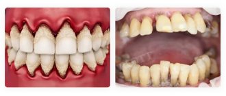
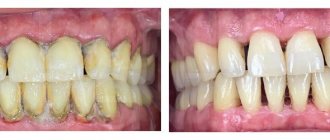
What is periodontal disease?
Periodontal disease is a relatively rare disease characterized by systemic damage to the tissues surrounding the diseased tooth. The causes of the pathology have not yet been established. However, it has been proven that the risk of developing the disease increases in people suffering from diabetes, hypertension, atherosclerosis, osteopenia and insufficient blood supply to the gums.
In the early stages, the clinical picture of periodontal disease includes:
- exposure of dental necks;
- the appearance of wedge-shaped defects on hard dental tissues;
- increased sensitivity of teeth to temperature and taste stimuli;
- itching in the gums.
Advanced forms of the disease are characterized by exposure of tooth roots, fan-shaped divergence of incisors and canines, pathological loosening of teeth and disappearance of interdental papillae. If left untreated, the patient may develop serious malocclusions.
Periodontal disease is often confused with periodontitis. However, there are many differences between these diseases. Periodontitis is an inflammatory disease of the periodontium, accompanied by the formation of gum pockets, bleeding and suppuration from the gums, and loosening of teeth in the early stages of the development of the pathological process. Such manifestations are not typical for periodontal disease.
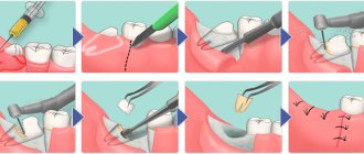
Main stages of the operation
- Syndestomy;
- Forceps delivery;
- Advancing the forceps to the area of the tooth neck under the gingival margin;
- Fixation;
- Luxation (rotation);
- Traction (extraction).
When removing a tooth with forceps, you must follow the following basic rules:
- Apply the cheeks of the forceps only to the vestibular and internal surfaces.
- The longitudinal axis of the cheeks of the forceps must coincide with the longitudinal axis of the tooth being removed.
- Avoid placing the tip of the cheeks of the forceps on the edge of the alveolar process and avoid subperiosteal resection of the edges of the socket.
- When dislocating a tooth, you must remember the possibility of a fracture in the neck or at the apex of the root.
When removing the upper teeth, the patient sits in a dental chair with the back slightly reclined, resting the back of his head against the headrest. The chair is raised to a position in which the tooth to be removed is at the level of the doctor’s shoulder joint. The doctor stands to the right and in front of the patient. When removing lower teeth, the chair is lowered so that the tooth being removed is at the level of the elbow joint of the doctor’s lowered arm. When removing lower incisors, canines, premolars and left molars, the doctor stands to the right and in front of the patient; when removing right molars, the doctor stands behind and slightly to the right.
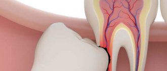
Decayed tooth
The most common indication for surgical extraction of a tooth is its complete destruction. Such situations are not uncommon, and our lifestyle is to blame, as well as ignoring the need for timely treatment. Usually, in such conditions, a specialist prescribes radiography. If it demonstrates that the tooth is indeed completely destroyed, and all that remains is the root with inflamed soft tissue around it, almost every dentist will decide to refer you to a surgeon to perform tooth extraction surgery. However, there remains a chance for salvation. There is a treatment method called depophoresis, which combines elements of electrophoresis and drug therapy. Depophoresis is performed as follows:
- Under the influence of weak pulses of electric current, a medicinal preparation, which is a concentrate of copper ions, is supplied to the roots of the tooth;
- Copper, which has powerful bactericidal properties, penetrates into thin canals and fills them, simultaneously killing all pathogenic flora that parasitize the tooth;
- The medicine settles in the canals for a long time, creating a reliable depot for infection for several years to come.
Since the tooth root becomes sterile after such a procedure, it can serve as a full-fledged support for the installation of prostheses and microprostheses (crowns). A similar treatment method is sometimes used for pulpitis, especially if the patient has hard-to-reach, branched or deformed roots. It is used in the treatment of canal obstruction, when the dentist’s instruments cannot penetrate into the desired place and deliver medications to the internal structures of the tooth.
Factors determining the choice of tooth extraction technique:
- condition of the tooth (degree and nature of destruction of hard dental tissues);
- features of the anatomical structure and position of the tooth, taking into account its group affiliation;
- features of the anatomical structure of the dentoalveolar segment in the area of the tooth being removed;
- The condition of the marginal periodontium is the degree of tooth mobility.
The operation of tooth extraction is the most common in the outpatient practice of a dental surgeon. Like any surgical intervention, tooth extraction surgery may be accompanied by complications that arise during the operation:
- Fracture of the tooth or root being removed. It can be associated both with significant tooth destruction and with structural features of the root or surrounding bone tissue.
- Fracture, dislocation and removal of an adjacent tooth occur as a result of using forceps with wider cheeks than the crown of the tooth being removed, or when elevators are used incorrectly, when the elevator rests on the adjacent tooth.
- Damage to the crown of the antagonist tooth occurs when the forceps slip off the tooth being removed.
- Pushing the root into the soft tissue is possible when removing the third lower molar in case of insufficient fixation with the fingers of the left hand.
- Fracture of the mandible is rare. The development of this complication is facilitated by pathological processes in the body of the lower jaw, which reduce its strength (odontogenic osteomyelitis, benign and malignant tumors.)
- Fracture of a section of the alveolar process. May be as a result of a pathological process in the periodontium.
- A fracture of the tubercle of the upper jaw can occur when the upper 8th tooth is removed.
- Dislocation of the lower jaw can occur when the mouth is opened wide and strong pressure is applied to the lower jaw with forceps or an elevator.
- Damage to soft tissue as a result of instrument slipping.
- Perforation of the bottom of the maxillary sinus can be a result of individual structural features of the upper jaw or due to previous pathological processes in the area of the apex of the tooth root.
- Aspiration of teeth and small instruments. This is facilitated by a decrease in the gag reflex after anesthesia and the position of the patient in a chair with his head thrown back.
Early postoperative complications may also occur. This type of complications includes:
- bleeding, which may be the result of a mechanical injury or a consequence of arterial hypertension or diseases accompanied by dysfunction of the body’s coagulation system;
- alveolar (socket) pain that appears after the cessation of anesthesia in the area of the extracted tooth. This is a natural response to tissue injury;
- alveolitis – a purulent infectious-inflammatory process in the periodontium of an extracted tooth;
- osteomyelitis;
- neuritis of the inferior alveolar nerve.
Procedure for surgical intervention
The operation involving the removal of periodontal teeth is classified as simple and is performed under local anesthesia according to the classical scheme. During surgery, the doctor:
- peels the gums away from the tooth enamel using a smoothing iron or a rasp;
- places forceps on the enamel, advances and closes their cheeks;
- gently dislocates the tooth by rocking (if we are talking about a multi-rooted molar) or rotating around an axis (when removing single-rooted teeth) and removes it from the alveolar socket.
If necessary, the surgeon applies sutures to the postoperative wound. A patient who has undergone surgery is prescribed antibacterial, antihistamine and painkillers.
Care for the socket after surgery
After the operation is completed, the surgeon places a gauze pad on the alveolar socket to stop the bleeding. This turunda can be kept in the mouth for no more than half an hour. After this, it must be removed or replaced with a new one. Dentists also recommend to patients who have undergone surgery:
- refuse food for 3 hours;
- avoid heavy physical activity during the week;
- refrain from hygiene procedures during the day;
- do not chew food on the side of the postoperative wound;
- give up hot drinks and meals for a week;
- do not touch the blood clot formed in the alveolus with your tongue, fingers, toothpicks or other objects, do not disturb its integrity and position.
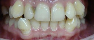
After surgery, it is very important to replace the defect formed in the dentition with a prosthesis. To draw up a prosthetic program, you must contact an orthopedic dentist.
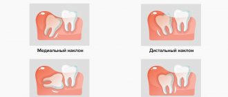
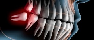
Insidious "eights"
It is known that wisdom teeth are the most capricious teeth in the entire jaw. They begin to cause problems for their owner even at the eruption stage. Without participating in the processes of chewing food, they are something like “extra teeth” in the mouth. They are difficult to clean, so they deteriorate and collapse quite quickly. Some experts are categorical in their judgments about the “eights” - they must be removed if they begin to deteriorate. The arguments are simple:
- A constant inflammatory process in the mouth, spreading over time to the throat;
- High risk of caries recurrence after treatment;
- Possibility of infection of soft tissues and adjacent functional teeth;
- Difficulty in treatment due to poor accessibility.
Other dentists think differently. They counter all of the above arguments with one, but no less powerful argument - wisdom teeth often become excellent supports for prosthetics. However, his modern methods are so diverse that a specialist can cope without the support “eight”. Qualified specialists at Dr. Granov’s dental clinic are waiting for you at their appointment to assess the extent of your problem and make the right choice in favor of tooth extraction or preservation. Call us and sign up for a consultation!