Causes of oral tumors
The origin of benign tumors has not been fully studied, but today there are several possible causes or predisposing factors that influence the formation of formation:
- bad habits - smoking, alcohol abuse;
- oral cavity trauma (including tooth extraction, permanent mechanical trauma to the mucous membrane from sharp edges of teeth or fillings, dental structures);
- viral infection - may cause the formation of some forms of benign tumors;
- disruption of tissue differentiation during intrauterine development of the fetus (as a rule, tumors formed for this reason manifest themselves already in childhood - within 1 year of the child’s life).
Cost of removal of benign tumors of the pharynx and larynx
The prices indicated in the price list may differ from the actual prices. Please check the current cost by calling +7 495 104 8605 (24 hours a day) or at the GMS Hospital clinic at the address: Moscow, st. Kalanchevskaya, 45.
| Name | Price |
| Biopsy of the oral cavity and oropharynx | RUR 9,282 |
| Opening of the epiglottis abscess | RUB 54,999 |
| Opening abscesses, phlegmon of the laryngopharynx | RUB 64,995 |
| Removal of a foreign body from the ear, nose, throat | RUB 6,962 |
| Removal of fibromas, papillomas, pharyngeal polyp (for 1 piece) | RUB 12,495 |
Dear Clients! Each case is individual and the final cost of your treatment can only be found out after an in-person visit to a GMS Hospital doctor. Prices for the most popular services are indicated with a 30% discount, which is valid when paying in cash or by credit card. You can be served under a VHI policy, pay separately for each visit, sign an agreement for an annual medical program, or make a deposit and receive services at a discount. On weekends and holidays, the clinic reserves the right to charge additional payments according to the current price list. Services are provided on the basis of a concluded contract.
Plastic cards MasterCard, VISA, Maestro, MIR are accepted for payment. Contactless payment with Apple Pay, Google Pay and Android Pay cards is also available.
Western standards of treatment (evidence-based medicine)
Continuous staff development
Regular interaction with leading Russian and foreign medical institutions
Modern medical equipment and advanced diagnostic and treatment methods
Unified standard of service
We work around the clock 24/7/365
Make an appointment We will be happy to answer any questions Coordinator Oksana
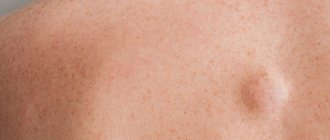
Epithelial tumors of the oral cavity
- Papillomas. Tumors formed by stratified squamous epithelial cells and most often localized on the tongue, lips, palate (hard and soft). They look like a protrusion above the surface of a healthy mucous membrane; the surface can be smooth, but more often the formation is covered with papillary-shaped growths. Most often they are observed in the singular, but multiple forms of the disease also occur. Over time, a tumor of this type becomes covered with keratinized epithelial cells and becomes whitish and also acquires a rough surface.
- Nevi. Most often, an intramucosal nevus is found in the oral cavity, which consists only of ovoid nevus cells located in the thickness of the connective tissue. Typically, the neoplasm has the appearance of a brown formation protruding above the level of the mucosa, on average 4 to 8 mm in size, but nevi of a different pigmentation - light pink in color - are often observed.
- Glands of Serres. Most often, such tumors are localized in the area of the hard palate and alveolar process. They look like hemispherical formations up to 10 mm in size, with a fairly dense consistency and a yellow tint. Such formations often occur in multiple forms in children and, as a rule, go away on their own during the first year of the child’s life.
Features of the rehabilitation period
Endoscopic removal of tumors of the pharynx and larynx is a minimally invasive operation with a short rehabilitation period. After the intervention, the patient is transferred to a day hospital ward, where he will remain under medical supervision for several hours. For two weeks, you need to limit physical activity and exclude thermal procedures (bath, hot bath, sauna).
You also need to adhere to a certain diet:
- exclude coarse, hard, spicy, sour and any other foods that irritate the mucous membrane;
- Avoid hot and cold drinks and food.
Most often, medications are not required after endoscopic surgery. But under some circumstances, an otolaryngologist may prescribe a course of antibiotic therapy, immunomodulatory and antiviral therapy. You can get more detailed information about the removal of benign tumors of the hypopharynx by making an appointment with an otolaryngologist, by phone or online.
Connective tissue tumors of the oral cavity
- Fibroids. A distinctive feature of fibroids is the lack of expression of color - they have the shade of healthy mucous membrane. Most often they are localized on the lower lip, palate, tongue and have the appearance of a round or oval formation with a smooth surface. In some cases, pedunculated fibromas occur.
- Fibromatosis of the gums. Some experts argue that gum fibromatosis is a consequence of the inflammatory process. Such growths have a dense structure and are local or diffuse in nature, that is, they are located around several teeth or cover the entire lower or upper jaw. They are painless, but formation occurs in the gum papillae, which can lead to complete overlap of the tooth crowns. There are difficulties in diagnosing gingival fibromatosis, since the clinical picture is similar to the symptoms of the hyperplastic form of gingivitis.
- Myomas. Formations are formed from muscle tissue cells. There are several forms of oral fibroids:
- rhabdomyomas - such formations are formed by cells of striated muscles, often localized in the thickness of the tongue in the form of single nodes;
- leiomyomas - this type of fibroids is formed from smooth muscle fibers, the most common location is the palate;
- myoblastomas are a consequence of disembryogenesis, therefore they are observed in newborns and have the appearance of a shiny, round formation measuring up to 100 mm.
- Myxomas. They consist of connective tissue and have jelly-like contents. They are more often found on the hard palate and in the area of the alveolar process; they have the appearance of a rounded formation with a papillary, tuberous surface.
- Pyogenic granuloma. It is formed from cells of the mucous membrane, connective tissue elements, and has an elevated shape. It can be caused by increased capillary growth, swelling of the surrounding tissue, and is often formed due to trauma to the mucous membrane. It is characterized by intensive growth and size up to 2 cm, has scarlet, brown, blue-black color.
- Epulis. Located on the gum. There are fibromatous (round shape, location on the vestibular side), angiomatous (finely tuberous surface, location at the neck of the tooth) and giant cell (round shape, tuberous surface, bluish color) epulis.
- Neuromas. They are formed from the sheath of nerve trunks, have a dense structure and are painful. They can reach 100 mm in size and have the appearance of a limited node (there is a capsule inside).
Complications
Complications of benign formations of the ENT organs directly depend on the severity of the process and are caused by disruption of the processes of natural ventilation of the nasal cavities and the free outflow of mucous membranes.
Among the possible complications, the most common are those that occur chronically or frequently recur:
- sinusitis and sinusitis;
- otitis;
- rhinitis;
- tonsillitis.
Complications also include decreased hearing and sense of smell, hypoxia, increased blood pressure and fatigue due to impaired nasal breathing.
Vascular tumors of the oral cavity
- Hemangiomas. Classified into the following types:
- capillary - formations from red to bluish in color, protruding above the mucous membrane (can be of different sizes);
- cavernous - formations with clear boundaries 1-2 cm in diameter, have a bluish color, most often formed on the mucous membrane of the cheeks;
- capillary-cavernous;
- mixed.
- Lymphangiomas. The cause is a violation of embryogenesis, so diagnosis is most often carried out in early childhood. They have the appearance of a limited tumor. Lymphangiomas are classified into the following forms:
- cavernous;
- cystic;
- capillary-cavernous;
- cystic-cavernous.
They are characterized by blanching and reduction in size when pressed; injury to these formations can cause bleeding.
These neoplasms are characterized by a tendency to inflammation: it can often be triggered by trauma to the oral mucosa and exacerbation of chronic inflammatory processes in the upper respiratory tract. The tumor is prone to rapid growth, but its distinctive feature is its spread over the surface without penetrating deep into the mucous membrane. Lymph nodes may also be enlarged. In the absence of inflammation, the formation is not filled with lymphatic fluid, however, experienced dentists diagnose the disease at this stage.
How is the operation performed?
All interventions are carried out via intrapharyngeal access using endoscopic equipment. Local anesthesia is predominantly used. The otolaryngologist carefully inserts an endoscope into the laryngopharynx and carefully examines all parts, assessing the size and location of the tumor. Then, the doctor identifies the tumor and removes it using micro-instruments, while coagulating the vessels.
- cysts, fibromas, neurogenic formations are removed by peeling them together with the capsule;
- for teratomas, the doctor first ligates the base of the tumor, stopping its blood supply, and then excises it, cauterizing the bed;
- local hemangioma, expanding into the lumen of the organ, is excised, followed by diathermocoagulation, laser or cryotherapy. For large hemangiomas growing deep into the tissue, sclerosis or occlusion of the vessels feeding it is used.
Simultaneously with the removal of altered tissues, coagulation of blood vessels is performed to prevent bleeding. Modern endoscopic stands used at the GMS Hospital surgery center allow us to examine in detail the relief of the laryngopharynx mucosa and perform targeted removal of the tumor along with the growth zone. The removed tissues are sent for histological analysis.
You have questions? We will be happy to answer any questions Coordinator Tatyana
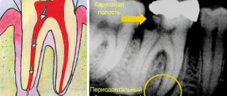
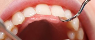
Diagnosis of oral tumors
Benign tumors of the oral cavity are diagnosed through a visual examination by a specialist, and cases of diagnosis are not uncommon during a planned, preventative visit. Also, in the process of treating other diseases of the teeth and oral cavity, a diagnosis can be established, but an accurate determination of the shape and type of tumor is possible only after laboratory analysis - histological examination of tissues. It can be carried out either after complete removal of the tumor, or through a biopsy - partial sampling of material.
To determine the depth of tumor growth, an ultrasound diagnostic method is used - this makes it possible to assess the condition of the soft tissues.
The condition of bone tissue is assessed using radiography.
In case of fibromatosis of the gums, the doctor prescribes an orthopantomogram - a panoramic image to diagnose the destruction of the alveolar process.
Angiography is used to assess the condition of tumors of a vascular nature.
Treatment of the disease
Detection of malignant tumors at an early stage, before the onset of metastases, makes it possible to achieve regressive carcinogen. For this purpose, remote radiation and complex therapy based on brachytherapy methods are used. Not only the local area with the formation is treated, but also the surrounding area.
In later stages, radiation therapy techniques are used before and after surgery. Both the neoplasm and the tissue located around it are removed. However, stage 3 and 4 cancers have a high rate of recurrence and are rarely completely cured.