From this article you will learn:
- a lump has come out on my lip - what is it?
- what does a cyst look like under the tongue,
- treatment of cysts of the lower lip and under the tongue.
A retention cyst is a convex, rounded formation that occurs as a result of blockage of the duct of one of the small salivary glands located in the thickness of the oral mucosa. Most often, a retention cyst of the lower lip occurs and a little less often - under the tongue. Accidental biting of the lip can lead to the appearance of a cyst, which will cause blockage of the duct of one of the small salivary glands and lead to the accumulation of viscous secretion in it.
The cyst on the lip is soft to the touch and is filled with viscous saliva. If you bite into it, saliva comes out, and the cyst itself collapses and disappears for a while, but after the mucosal defect heals, the cyst is usually filled again with viscous salivary fluid. Therefore, when patients complain that they have a lump on their lip or under their tongue, this is a retention cyst. Its appearance will require you to visit a dental surgeon for a 15-minute surgical operation (with 2-3 stitches).
Retention cyst on the lip: photo
The cyst can be located closer to the surface of the mucous membrane (as if rising above it), or it can be located deeper in the tissues. This is why some patients notice that the bump is inside the lip, while others notice that the bump is on the outside.
Content:
- Why does a cyst grow in the oral cavity?
- Types of oral cysts
- Signs of a cyst
- Examination of patients who have a cyst in their mouth
- How to treat
- Treatment of cystic formation at home
- How to reduce the likelihood of developing education
Sometimes, when visiting a dentist, a patient hears a strange diagnosis - mucocele.
More simply, it sounds like an oral cyst. Underneath this disease lies a cavity tumor neoplasm containing mucous contents. It is benign and develops due to obstructed outflow of secretions produced by parenchyma cells. The peculiarity of mucocele is that it does not have a strong epithelial membrane. Usually localized on the mucous membrane of the lower lip or under the tongue. It can also form in the chewing area. Neoplasms of the submandibular zone are very rarely encountered in dental practice.
Conducted studies demonstrate that most often young people under the age of thirty experience mucocele. The disease also occurs in adolescent children.
Why does a cyst grow in the oral cavity?
Most often, doctors are unable to determine the exact cause of the disease. It is believed that its development can be caused by:
- repetitive trauma to the mouth;
- inflammatory pathologies of the oral mucosa;
- congenital obstruction of the excretory ducts of the salivary glands.
Some doctors are inclined to believe that frequently appearing cysts of the sublingual salivary gland indicate a non-standard structure of the latter. There is also a dysembryogenetic version of the origin of tumors. But dentists give the main importance to the traumatic factor. Thus, blisters on the inside of the lower lip often form due to the habit of constantly biting it.
The pathogenesis of the disease can be described as follows:
- The excretory duct of the salivary gland becomes blocked for a certain reason.
- Internal hydrostatic pressure increases. The mucus accumulates and cannot come out.
- Throughout the day, the mucous secretion permeates the surrounding tissues.
- Swelling forms and the blood vessels are compressed. Tissue permeability is impaired.
- A capsule consisting of connective tissue is formed. She is gradually growing.
Diagnosis of pathology of the minor salivary gland
Specialists at the VTV medical center conduct a thorough examination to compile a complete clinical picture. The diagnostic process uses professional instruments and laboratory tests. We also perform a differential diagnosis to rule out hemangiomas, fibromas, or benign tumors.
First, the retention cyst of the lip is studied, and removal is carried out after confirmation of the diagnosis. The most important are the results of ultrasound of the minor salivary glands and other types of scans, which make it possible to determine the size of the cyst and its exact location.
Types of oral cysts
Based on their origin, mucoceles are:
- true;
- extravasal.
The first ones lack their own membrane and are covered with a gland capsule. They occur due to blockage of the duct and accumulation of mucus. The second are post-traumatic. They are formed when the tightness of certain structures is broken and the mucous secretion enters the surrounding tissues.
According to the localization criterion, the neoplasm is classified into:
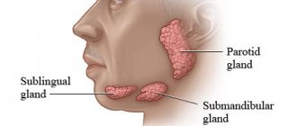
- Sublingual. It is located in the hyoid-maxillary muscles or submandibular region. During rapid growth, it grows very quickly and then causes serious discomfort.
- Submandibular. Located in the lower submandibular region. It feels like a dense ball to the touch. Promotes disruption of the natural mechanism of salivary fluid secretion.
- Parotid. Rarely encountered in dental practice. It can be very painful when you open your mouth wide. It is formed due to impacts, injuries, after which the inflammatory process caused the closure of the salivary ducts.
- Extravasal. Most often found on the inner surface of the lip. Occurs due to mechanical damage. The inside is filled with granulation tissue.
Signs of a cyst
Among the main symptoms of the disease:
- The appearance of an unusual protrusion on the soft tissues of the mouth. It may resemble an abscess. It usually has a bluish color with a burgundy border, but it can also match the tone of healthy gums. The “older” the mucocele, the thinner its walls become. The “bubble” is movable; it is not fused to the surrounding tissues.
- Discomfortable sensations while chewing food. There is a feeling as if there is a foreign object in the mouth that is constantly in the way.
- An unpleasant feeling of constriction of the mucous membrane. In this case, pain does not occur.
The cyst may burst if there is a lot of pressure on it. Spontaneous opening sometimes occurs while eating. The difficulty is that afterwards it forms again - through the passage in the mucous membrane, the cleared cavity is refilled with liquid contents.
Signs, diagnosis and treatment
Manifestations of the disease depend on the location of the cyst. Most often, these are rounded formations that slowly increase in size, elastic and soft to the touch. They do not cause pain at first, rarely exceeding 1 cm in diameter. But in the absence of treatment, cysts begin to seriously interfere not only with eating, but also during conversation. The formation may disappear if, due to an accidental breakthrough, the contents come out. However, over time, the cyst forms again; if its size becomes very large, it acquires a characteristic bluish tint.
For diagnosis, methods such as visual inspection, palpation, and a number of hardware studies are used. Ultrasound, sialography to determine the problem with contrast, MRI and CT are more often performed. Additional examination methods may also be prescribed, for example, cystography of the bladder, probing of the salivary ducts, histology. If the diagnosis is questionable, a biopsy and puncture of samples are performed.
Conservative therapy does not bring any effect; when diagnosing a cyst of this type, surgical intervention is recommended. For this, local anesthesia is used, after which the formation is removed along with the affected gland and membrane to prevent relapses. If surgical intervention is refused, the formation often develops into phlegmon, and a tissue abscess develops.
The conservative method provides only temporary results. To do this, the membrane is punctured and the contents of the cyst are sucked out. But in all cases, the formation appears again, that is, this method of therapy is not recommended, as it is ineffective.
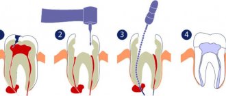
Surgery includes the following steps:
- inspection and surface preparation are carried out;
- local anesthesia methods are used for pain relief;
- the area with the cyst shrinks, which reduces blood flow and ensures stability of the tissue position;
- two oval-shaped incisions are made near the formation, after which the contents are husked;
- the affected lobes of the gland are removed, which helps to avoid complications or relapses in the future;
- the wound is sutured, thin sutures are applied, and self-absorbing sutures are often used.
The contents of the capsule are sent for additional research. This helps to eliminate serious risks, especially if the development of malignant processes is suspected. In case of serious intervention, plastic cystotomy is recommended. Typically, this situation occurs when removing a maxillary-hyoid formation.
Examination of patients who have a cyst in their mouth
Diagnosis of the described disease is simple.
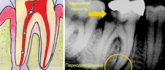
The doctor examines and palpates the abnormal lesion and studies the symmetry of the face. If the mucocele has reached a diameter of more than one and a half centimeters, then its color is blue. When the lesion is opened, viscous yellow contents are released. If the resulting biological material is submitted for analysis, a large amount of salivary proteins and amylase will be found in it. If necessary, the Trommer reaction is performed to confirm the preliminary diagnosis.
During ultrasound diagnostics of the salivary gland, the doctor observes an anechoic formation of a round shape. Its borders are smooth. The fact that the patient has a mucocele is said:
- presence of granulation lining;
- absence of epithelial membrane;
- the presence of mucin and protective blood cells.
If there is doubt about the benignity of the tumor, the patient is referred to an oncologist.
How to treat
It is very dangerous if a person tries to remove a cystic formation on his own and, to do this, puts pressure on it or bites it. Any mechanical influences have a negative effect on the course of the disease. Due to external pressure, the liquid inside the bubble begins to flow beyond its boundaries. Afterwards it accumulates again in the inflamed area. But at the same time, the risk of infection of damaged tissues increases significantly.
Doctors most often treat mucocele surgically. If the “bubble” is localized on the lower lip, two semilunar incisions are made in its projection, after which the internal neoplasm is isolated along with all its contents. Finally, stitches and a sterile pressure bandage are applied.
If the problem concerns the sublingual area, the following can be done:
- cystectomy;
- cystsialadenectomy.
In the first case, only the cyst itself is removed. It is cleaned and cut out. In the second type of surgery, the gland is also removed.
If there is a formation in the parotid zone, a parotidectomy is performed - complete or partial. It involves excision of the cyst and part of the parenchyma. Lesions located in the submandibular area are always removed along with the gland.
If we are talking about treating a child or a weakened elderly person, the dental surgeon may decide to excise only the dome (upper part) of the abnormal structure.
Retention cyst on the lip: treatment
A lump on the inside of the lip is treated in the same way as a lump under the tongue.
There is only one treatment option - surgery, which involves removing the entire cyst (there are no other treatment options). The surgery is very simple and usually does not take more than 15 minutes of the dental surgeon's time. It is performed under local anesthesia with lidocaine solution, and you will not feel any pain at all. It looks like this: a small incision is made on the surface of the mucous membrane next to the cyst, through which the entire cyst is removed, along with its contents. After this, 2-4 sutures are placed on the edges of the mucous membrane, which will need to be removed after about 7 days.
Removal of a retention cyst: video of the operation
During the operation, it is very important not to damage the cyst membrane, because if the walls of the cyst collapse, it is immediately lost in the tissues, and it is almost impossible to remove it entirely after this. If you leave a small fragment of the cyst shell in the tissues, the retention cyst on the lip will appear again.
Treatment of cystic formation at home
Home therapy for oral cysts only makes sense if for some reason you cannot get to a dental surgeon in the next few days. It consists of rinsing with herbal solutions and antiseptics. You can also make compresses with anti-inflammatory herbal medicines.
If the hearth breaks through, it’s too early to rejoice. Most likely, it will soon reappear in its original place. This is how cysts work - their contents expire, but the outer layer remains.
Under no circumstances should you treat a bulge on the gum or mucous membrane as a regular pimple. Any attempts to open it mechanically will not lead to anything good. But the resulting wound can become infected. Then the inflammation will spread to deeper layers in a fairly short time, and it will be much more difficult to cure.
How to reduce the likelihood of developing education
To minimize the risk of mucocele formation, you need to follow the rules:
- lead a healthy lifestyle, quit smoking;
- carefully observe oral hygiene;
- rinse your mouth after every meal;
- eat a balanced diet;
- do not put foreign objects in your mouth;
- get rid of the habit of biting your lip;
- correct malocclusion;
- undergo oral hygiene every year;
- avoid trauma and chemical burns to the lips;
- promptly replace dentures;
- Follow the rules for wearing braces and caring for them.
People who follow preventive measures are much less likely to need treatment for oral cysts. If a lesion has appeared and is growing in size, there is no need to expect it to disappear on its own. This happens very rarely. It will either “deflate” or be filled again with contents and ultimately may grow to such a size that it will not be possible to do without emergency surgical intervention.
Rehabilitation, expected results
After surgery, there is slight swelling and pain at the operation site for two to three days. The doctor may prescribe medications to eliminate such unpleasant symptoms, for example, analgesics, antibiotics to avoid complications. Oral rinses using special antiseptic solutions are also prescribed.
If the healing process is normal, the sutures are removed after a week, after which a pressure bandage is applied. During recovery, it is recommended to avoid solid, spicy foods, too cold and hot dishes. Oral care also requires care to avoid damaging the operated area.
The prognosis for treatment is favorable; if therapy is carried out correctly and the patient follows all recommendations, there are no problems. But in some cases there are risks of complications. These include relapses, damage to the facial nerve or facial muscles when removing a cyst in the ear area. To reduce risks after completion of the recovery period, it is recommended to correct the shape of the tooth and install new orthodontic structures or dentures. In addition, prevention is needed, protection from injuries to the mucous membrane, and compliance with the rules of oral care.