When our teeth suddenly begin to hurt, and pustules appear in our mouth, we begin to swallow medications - self-medicate. But you need to immediately go to the dentist, because these are the first signs of cyst formation.
A salivary gland cyst is a tumor in the maxillofacial area.
It forms in the form of a cavity that is filled with purulent fluid. The development of such a cyst usually occurs in young people, under the age of thirty. Its treatment has its own specifics and is carried out in surgical dentistry or otolaryngology.
Formation of a salivary gland cyst and its causes
The first cause of a cyst is usually an infection and can occur after various diseases that have not been fully treated.
A cyst on the salivary gland is a very serious and dangerous disease that requires urgent treatment. When bacteria and microbes enter the mouth, an inflammatory process immediately begins. The cells become infected and begin to die, creating a cavity. A dense shell appears around it. This is a cyst.
This disease, which is the worst thing, may not be noticed at first. And the disease itself is slowly beginning to develop. An inflamed cyst causes general malaise, headache, and fever. The lymph nodes also become enlarged.
Salivary gland cyst occurs:
- Small
- Sublingual
- Submandibular
- Parotid
Diagnosis of salivary gland cysts
Diagnosis of salivary gland cysts involves determining the nature of the neoplasms; they can be benign or malignant. Determining the nature of the cysts directly depends on their clinical picture. To do this, it is necessary to interview the patient, identify and evaluate complaints, examine and palpate the cyst. During these manipulations, the doctor determines the size, type, location and mobility of the cyst.
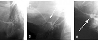
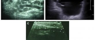
Due to the fact that all cysts have an almost identical clinical picture of the disease, for an accurate diagnosis it is necessary to carry out additional diagnostics with cytological, radiological and biochemical studies.
Cytological diagnosis of the salivary glands involves taking a puncture from the tumor mass. Thanks to this study, it becomes possible to determine the process of tumor development.
X-ray examination allows you to find out how much the salivary ducts are filled with contrast mass. The diagnostic method consists of conventional radiography and contrast radiography of the salivary ducts.
Also, for an accurate diagnosis, the method of differential diagnosis (method of exclusion) is used. This is necessary to distinguish one cyst from another.
Symptoms of the types of cysts listed above:
1. Very often it forms on the inside of the lower lip. It appears less frequently on the inside of the cheeks or other parts of the mouth. Usually it is small in size - up to one centimeter and grows in size very little.
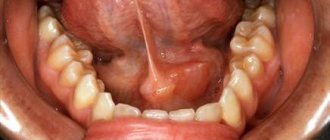
2. Formed at the bottom of the mouth, under the base of the tongue. It usually has a round or oval shape, a small protrusion of a pale blue color. When it enlarges, the cyst causes a displacement of the frenulum of the tongue, which directly interferes with speaking and eating.
3. It stands out as a rounded swelling on one side on the soft tissues in the preauricular area and therefore the person’s face becomes asymmetrical.
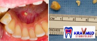
Retention cyst of the lower lip
It is a common pathology of the oral mucosa. It is a benign neoplasm in the shape of a ball. The pathology usually develops due to blockage of the duct of the minor salivary gland. The reason for this may be the most common injury or inflammatory process.
The cyst occurs equally in both women and men. Its appearance does not depend on age. The cyst is characterized by the ability to very quickly increase in size, which affects a person’s daily lifestyle. That is why doctors recommend removing it.
The main reasons for the appearance of cysts
The neoplasm most often forms as a result of mechanical damage and trauma to the lip. These include burns, blows and biting. As a result of constant trauma to the gland, the excretory canal becomes clogged, the secretion begins to linger, and a small tubercle forms. Gradually it fills with liquid and increases in size. The inflammatory process after injury can also lead to the formation of pathology. A retention cyst of the lower lip can occur in a person at any age. The neoplasm is often a consequence of the congenital development of the germinal components of the so-called glandular cells.
In addition to traumatic effects, the cause of a cyst can be atrophy of the excretory ducts. Typically, such a violation is caused by a tumor that directly compresses the duct, or by a scar. The latter narrows the canal, and the accumulated secretion gradually expands the glandular lobe.
How to recognize pathology?
A retention cyst of the lower lip is a capsule of connective tissue with viscous contents inside. Outwardly, it resembles a small ball. The formation is painless, but may cause discomfort while talking or eating. The cyst tends to grow rapidly and can reach up to 2 cm in diameter. The mucous membrane above it usually does not undergo significant changes. Sometimes it takes on a bluish tint, which is explained by the accumulation of contents. The ball is covered with connective tissue, and inside it there is a clear liquid resembling saliva. On palpation the formation is soft. When eating food, the capsule is easily damaged, causing its internal contents to spill out, but after that the cyst fills up again. As a rule, the formation consists of one chamber, but cases of multi-chamber cysts are known.
Diagnosis confirmation
Recognizing a retention cyst is not difficult for a qualified specialist. When pressed with a finger, the formation disappears, but after a while it appears again. A more accurate diagnosis can be made after an ultrasound examination. During the procedure, the doctor evaluates the structure of the cyst, its size and contents. By probing the canals, the width of the duct and the presence of salivary stone are determined. Only after a complete examination can a specialist recommend treatment for a retention cyst of the lower lip.
The need for differential diagnosis In order to accurately make a diagnosis and subsequently prescribe competent treatment, it is important to be able to recognize this pathology among other neoplasms of a benign nature. The following types of salivary gland cysts are distinguished: ranula; submandibular; parotid; minor salivary gland. The ranula is localized above the hyoid-maxillary muscle. There are known cases of penetration into the submandibular region. This formation is large in size. It can displace the frenulum of the tongue, thereby preventing a person from fully eating and having conversations. The submandibular gland cyst is characterized by slow development. During palpation it is easy to detect a round formation. When this cyst grows, it can involve the upper areas of the mouth. In such a situation, bulging of the formation into the sublingual part is usually observed. Pathology of the parotid gland is quite rare. The main reasons for its formation include mechanical damage, inflammatory processes and blockage of ducts. It is the same factors that provoke the formation of another anomaly of the oral cavity called a retention cyst of the lower lip (photo presented in this article). The development of pathology at the initial stage is not accompanied by pronounced symptoms. As a result of mechanical damage, a so-called extravasal cyst can form. This formation is distinguished by the fact that granulation tissue is formed around it.
What treatment is required?
This pathology is treated by a dentist. After confirming the diagnosis, there are two possible solutions to the problem. The specialist may send the patient home, believing that the formation will resolve on its own. The second option involves surgical removal of the retention cyst of the lower lip.
The operation itself lasts no more than 30 minutes and involves the use of local anesthesia. The physician assistant twists his lower lip and holds it tightly. The dentist makes several cuts along the formation, frees the cyst from its internal contents and applies sutures. According to many patients, the operation itself, under the supervision of an experienced surgeon, is almost painless. The main troubles begin during the rehabilitation period, when the retention cyst of the lower lip begins to gradually heal. The operation can be performed using a laser. However, its help is used extremely rarely due to severe bleeding and a high risk of perforation of the gland membrane.
Postoperative period
After surgery, patients are advised to treat the affected area daily with a special antiseptic solution and monitor their condition. Some note that the first day after surgery is the most difficult. Talking, eating - all these simple actions cause painful discomfort, but after about a month everything returns to normal. It should be noted that the duration of the rehabilitation period directly depends on the size of the formation. Many patients report visual distortion of the lip and slight numbness even months after surgery. To avoid negative consequences, it is recommended to remove the pathology at the initial stage of its formation.
Removal of retention cyst of the lower lip
dentistry teethprostheticsemax high-quality dental prostheticscrownstropedic dentistryveneersceramic inlaysprosthetic crownsHollywood smile white teethpearl teethbeautiful smileimplantationwedge-shaped defectsgingival recessiontreatment of children high-quality dentistry baby teeth colored fillings dental implantation Ankylos implantation high-quality implantation without bone tissue augmentation qualified implantation front teeth prosthetics veneers ceramic crowns Imax e.Max zirconium crowns dental prosthetics with crowns depressurization of composite restoration marginal staining re-polishing of fillings professional examination veneer tooth lightening ceramic veneers exclusive prostheticsgold inlayfront tooth single crownDamon bracesimplantTeeth crowdingOrthognathic surgery Victory bracesMEAW technique multi-loop arch IVD Sato meaw orthodonticSplint therapy wedge-shaped defect treatment of a wedge-shaped defect tooth restorationRestoration of teeth re-polishing fillings artistic restorationExtraction of the inlay remove the inlay from the rootAnchor pin preparation for prostheticsadditional canal cyst treatmentProfessional hygiene clean dental treatmentResection of the root apex Apicotomy Retrograde treatmentSingle-stage implantation Tooth implantation ImplantArtistic restoration treatment of caries Teeth cleaning One-stage implantation Ankyloses Implantation of a tooth with a crack cracked root implantation of a front tooth Ranula retention cyst wound removal save tooth tooth preservation alternative to implantation CCPD SST Ankylos Ankylos FDM implant implant wisdom tooth treatment mock-up mock-up prototyping elastics intermaxillary traction braces hygiene products implantation Astra Astra Tech Panov Mikhail Yurievich dental transplantation dental treatment treatment under a microscope treatment under a microscopeCanal treatmentHollywood smiletooth transplantationTooth extrusionTreatment without preparationTreatment without preparationTreatment tool in the tooth canal Panov Mikhail Yurievich tooth saving treatment of canals filling gum recessionSealing fissure resection of the root apexTeeth preservationIntrusionGum recessiontransplantationDental cariesSST
Content:
- Classification of pathology
- Causes of the disease
- Symptoms of the disease
- Diagnostic measures
- How to treat
- What will happen if left untreated?
- What can you do to avoid getting sick?
A jaw cyst is a hollow benign formation localized in the jaw bone.
There is liquid content inside it. Very often, the structure develops unnoticed by a person for a long time and is accidentally discovered during an X-ray diagnosis for another reason. If a cyst in the jaw becomes inflamed, the patient immediately feels it. Suppuration can provoke the occurrence of periostitis, sinusitis, osteomyelitis, and fistula.
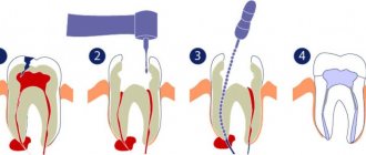
Unfortunately, most often the formation has to be removed surgically. The doctor performs a resection of the apex of the tooth root and at the same time fills the cavity cleared of exudate with a special biological composite composition.
Classification of pathology
Cysts localized in the tissues of the upper or lower row of teeth are very common. With their development, a cavity appears, the walls of which are covered with fibrous tissue, and the inner surface consists of an epithelial layer. The capsule holds clear or cloudy liquid.
Doctors distinguish the following types of pathology:
- Primordial. This is a cyst of the lower jaw. It appears in the area of the third molar. She has very thin fibrous walls. The inside of the capsule is lined with flat epithelium. According to its structure, the tumor may contain one or several small chambers.
- Follicular. Formed before tooth eruption. It can grow in the area of the alveolar margin. Includes cells that intensively produce viscous mucus. Because of this, the internal contents of the anomalous structure are quite viscous. A follicular formation is formed from the enamel organs of the unerupted unit. One or several teeth are found inside it. These may be formed crowns or tooth buds.
- Radicular. It occurs more often than others - in eighty percent of cases. Usually grows above the upper teeth. The diameter reaches from half to two centimeters. Often inflamed. Then the cells begin to hyperplasia, network-like processes are formed that extend into the thickness of the walls. The liquid contents of the radicular structures are rich in neutrophilic leukocytes. Often with this type of pathology, the patient develops sinusitis. This is due to the peculiarities of the localization of the inflammatory site.
- Retromolar. It is formed due to a long-term inflammatory process occurring in the thickness of the soft tissues. It is a consequence of complicated teething.
- Nasoalveolar. It occurs near the nasopalatine canal (the border between the upper row and the wing of the nose).
- Aneurysmal. Very rare in dental practice. Appears only on the lower jaw. This type of neoplasm is still poorly studied. It is known that there is blood or a red-pink liquid inside it. Many scientists agree that the symptoms of an aneurysmal cavity are a consequence of hormonal imbalance.
- Traumatic. Occurs due to recent facial trauma.
- Residual. The result of mistakes made by the doctor during tooth extraction, or the consequence of the patient ignoring the surgeon’s instructions.
The doctor decides how to treat a dental cyst, taking into account the cause of its development. It is very important to understand why the tumor occurred. If the root cause is not identified, the likelihood of relapse will remain high.
What can you do to avoid getting sick?
You can reduce the likelihood of developing the disease by following simple rules:
- Brush your teeth twice a day. Use a high-quality brush and paste during hygiene procedures. Don't forget about the role of dental rinses and dental floss.
- Once a year, remove tartar in the dental office using an ultrasonic scaler. Professional hygiene is the best prevention of most dental ailments.
- Eat a balanced and healthy diet. Eat less sweets. Eat plenty of fresh fruits and vegetables.
- Check your dental health once a year. You need to visit a doctor, even if nothing bothers you.
- Always follow your dentist's instructions.
- Avoid traumatic injuries to the face. When engaging in potentially dangerous sports, always wear special protective equipment.
A jaw cyst is a dangerous tumor. Initially it is benign, but under certain conditions it can become malignant. Fortunately, this happens extremely rarely.
If the tumor grows very quickly, even a jaw fracture is possible. It shouldn't come to this. Treatment must be timely and competent.