Salivary gland stone removal is a procedure indicated for the formation of large stones in the salivary gland. The formation of stones in the salivary gland is a relatively rare occurrence, which, however, can have serious consequences. In the early stages, it is possible to remove the stone from the gland duct on your own. If the resulting stone has become large enough to block it and interfere with the normal outflow of saliva, surgical intervention may be indicated. Sialoscopy The most popular and modern method of surgical removal is sialoscopy. This type of operation is relatively new and became available only with the advent of the latest technologies in the field of medicine. This is due to the fact that the salivary glands are quite complex in structure, and the diameter of their openings does not exceed 0.5 mm.
A modern endoscope is a thin device with miniature systems with a diameter of 0.8 millimeters and a set of even thinner instruments for performing complex medical procedures. Using an endoscope avoids dissecting the salivary gland (previously this was the only way to detect and remove tartar). Using an endoscope, the doctor can see what is happening inside the salivary gland and remove the salivary stone from it with the finest instruments.
Causes of formation of salivary gland stones
When calculi form, the ducts of the salivary gland are blocked, as a result of which saliva does not enter the oral cavity and returns back to the gland. Impaired salivary exchange is characterized by intermittent pain and progressive swelling. In addition, impaired salivary flow can be accompanied by infections.
One of the causes of salivary stone disease is inflammation of the gland. Due to bacterial infections, the parotid gland is damaged, and an inflammatory process begins, blocking the salivary ducts. The disease manifests itself in the form of swelling of the gland, often with purulent discharge that has a specific taste. Most often, the disease occurs in older people who have salivary gland stones. If medical assistance in treating the disease is not provided in a timely manner, there is a possibility of an abscess forming. The inflammatory process of the salivary glands is caused by microorganisms living in the oral cavity - staphylococci. Bacterial infections can result in malnutrition and dehydration.
The progression of viral diseases in humans, such as influenza or mumps, can also cause swelling of the salivary glands. Symptoms of the disease include large swollen cheeks. The appearance of this symptom is provoked by blockage of the parotid salivary glands.
Another reason for the formation of stones in the salivary glands are cysts that form in the oral cavity as a result of injury. Also, the neoplasm may have a congenital pathological nature.
Benign and malignant tumors provoke the occurrence of salivary gland calculi. And in addition to concomitant diseases, the causes of the appearance of salivary gland stones are:
- congenital pathologies of the salivary glands;
- endocrine system disorders;
- smoking;
- hypovitaminosis A;
- impaired metabolism of calcium and phosphorus in the body.
Causes of stones
- Injuries to crowns or teeth. In such cases, the ducts of the salivary glands narrow, therefore, the outflow of saliva is disrupted, which provokes the formation of stones in these areas.
- Inflammation of nearby tissues. In such cases, pathogenic microorganisms enter the duct, which provoke the formation of pus. Due to all this, metabolic disturbances occur, the flow of saliva worsens, and stones appear.
- Salivary gland cyst.
- Bends of ducts and narrowings.
- Vitamin A deficiency.
- Entry of foreign bodies into the salivary gland ducts (bone from food, solid food debris, fish scales, etc.).
- Increased blood clotting.
Symptoms of salivary gland stones
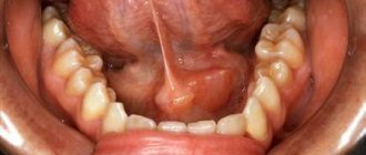
A stone in the salivary gland is a pathology that occurs for a long time without visible symptoms. Often, stones are discovered in a patient by accident during a routine examination in the dental office or simply by touching with your own tongue. A stone in the salivary gland is revealed by its compaction; it causes a delay in the secretion of saliva during meals. The swelling in the area of the gland is painful, the unpleasant sensation goes away after saliva enters the oral cavity. Quite often, the formed stone causes an inflammatory process of the salivary gland, which has characteristic symptoms:
- feeling of dryness in the mouth;
- the presence of a specific taste in the mouth;
- pain in the neck and mouth;
- change in the position of the earlobe and the formation of swelling in its area;
- increase in body temperature.
If a stone in the salivary gland provokes an inflammatory process, then the patient begins to feel fatigue and general fatigue, along with this, the body temperature rises. Dry mouth causes problems with eating and even facial expressions. If you ignore a visit to the doctor for a long time, an abscess may form, characterized by a large accumulation of pus in the area of the salivary gland, which can lead to its breakthrough into the oral cavity.
What types of stones are there?
Mostly, one particular salivary gland is affected; two are affected at the same time very rarely, all of them are never affected. During the operation, either one stone or multiple stones can occur.
The weight of a stone formed in the salivary gland can reach several tens of grams. In addition, stones differ in their shape. They can be oblong, round or irregular in shape. Very often there are foreign bodies in the center of stones.
Salivary stones consist largely of calcium carbonates and phosphates.
Diagnosis of salivary gland stones
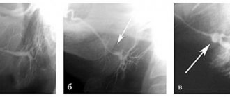
Suspected salivary gland stones are characterized by certain symptoms, but various tests are used to assess the shape and number of stones, as well as their location. The density of the mineralized stone is quite high, so it is quite clearly visible on x-rays. In some situations, an x-ray is not an effective diagnostic method due to the fact that a shadow may fall on the stone or the stone is not sufficiently mineralized. For clearer X-ray results, before starting the procedure, a special substance is injected into the duct, which makes it possible to view the structure and shape of the ducts; the places of ruptures are stones in the salivary gland.
In modern medicine, a diagnostic method in the form of computed tomography is used. With such an examination, stones of the salivary glands are determined, the size of which is less than one millimeter; it is also obvious how many stones are and where they are located. The disadvantage of the method is the inability to determine the condition of the soft tissues.
A clearer diagnostic method is magnetic resonance imaging (MRI). It is used to determine the condition of soft tissues, but it is not able to show the number and location of stones.
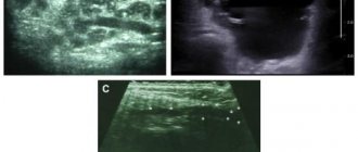
Ultrasound examination is also used as diagnostics, but this method requires a highly qualified doctor.
The most accurate and clear method that gives a complete picture of the disease is sialoscopy. It involves inserting microscopic endoscopes into the salivary ducts, allowing doctors to see a real picture of the processes occurring inside the body.
Diagnosis of pathology
Diagnosis of sialolithiasis is carried out using the following methods:
- After diagnostic measures, an appropriate course of treatment is prescribed
Palpation of the inflamed area;
- X-ray to identify foreign formations in the salivary glands. If necessary, the procedure is carried out with the introduction of a contrast agent into the vein;
- Examination of the salivary canal using a probe. This procedure is prescribed only if the disease is not at the acute stage;
- Sialography. The technique allows you to determine the location of the stone and obtain data on the condition of the duct;
- Cytogram of gland secretion. The procedure allows you to identify the inflammatory process occurring in the gland.
After diagnostic measures, an appropriate course of treatment is prescribed, taking into account the general condition of the patient and the degree of development of the pathology.
Treatment of salivary gland stones
Treatment for salivary stone disease involves removing the stone. Removing stones from the salivary gland occurs in two ways, depending on their location. When a stone is localized at the mouth of the duct, it is dissected and removed into the oral cavity. The effectiveness of the method is high, but various risks are associated with it:
- impaired taste and tactile sensitivity of the tongue due to nerve damage;
- formation of hematitis and bleeding as a result of damage to large vessels;
- worsening of the disease as a result of displacement of the stone deeper into the duct;
- partial removal of stones.
In some situations, stones are located deep in the ducts or in the thickness of the gland. This position of the stones forces them to be removed along with the gland, which leads to serious consequences. The operation requires hospitalization of the patient and the use of anesthesia. As a result of surgery, there is a risk of the following complications:
- tongue nerve damage;
- vascular trauma leading to life-threatening bleeding;
- injury to the facial nerve, leading to impaired facial expressions;
- scar formation.
Modern medicine offers another method of treating salivary stone disease - sialoscopy. With this method, salivary gland stones are detected and removed from the ducts using endoscopes. This treatment takes place with virtually no damage to soft tissues. The manipulation is performed under local anesthesia and does not require the patient to be in the hospital. After 30 minutes after the operation, the patient can be discharged from the hospital. During the operation, the doctor inserts small endoscopes into the ducts of the salivary glands, with the help of which the location and number of stones are determined, then they are removed from the ducts using special instruments. Sialoscopy has a number of advantages over other methods for a number of reasons, namely:
- low level of injuries;
- removing stones from different locations;
- use of local anesthesia;
- no risks associated with nerve injuries;
- complete preservation of the salivary gland.
Complications of pathology
Salivary stone disease is fraught with the following consequences:
- soft tissue abscess;
- phlegmon;
- passage of stone into the duct;
- vascular injury during surgery;
- impaired sensitivity of the tongue due to injury to the lingular nerve, which is possible during surgery.
In order to prevent life-threatening consequences, in case of manifestations of salivary stone disease, it is necessary to contact a specialist in a timely manner.
Prognosis and prevention of salivary gland stones
Depending on the treatment method, prognosis will vary. With modern methods of removing stones that preserve the salivary gland, the prognosis for recovery is favorable. With the radical removal of stones, the microflora of the oral cavity is disrupted, tooth decay is possible and the patient’s quality of life decreases.
To prevent salivary stone disease, it is necessary to eliminate factors that contribute to the formation of stones, as well as normalize metabolic processes in the body, get rid of bad habits and adjust drug treatment.
Forms of the disease
Salivary stone disease can occur in acute and chronic forms.
Acute sialolithiasis is characterized by sudden development. This form is characterized by severe acute pain and fever. In this case, complications often appear in the form of the formation of phlegmon or an abscess.
If the disease becomes chronic, the inflammatory process disappears, but slight swelling remains. When the pathology reaches this stage, mandatory surgical intervention is required.
Prevention
To prevent the development of pathology, it is necessary:
- follow the rules of oral care;
- promptly treat diseases that spread to any organs or systems of the body;
- stop smoking, alcohol;
- consume enough vitamins;
- promptly eliminate congenital ductal anomalies.
Salivary stone disease is characterized by the formation of mineral compounds in the salivary glands. Its danger lies in the possibility of the formation of an abscess and phlegmon, the purulent contents of which can spill into the surrounding tissues. Treatment of the pathology is carried out either conservatively or surgically.
Why is tartar dangerous?
The formation of dental plaque, and then tartar, is possible for various reasons: the involvement of only one side of the jaw, left or right, in chewing; dietary habits, in particular, eating only soft foods; salt imbalance in the body; using low-quality toothbrushes; crooked teeth; smoking. And some people have a congenital predisposition to the appearance of stones. In such cases, even regular care does not guarantee the safety of teeth.
Microbes, which make up 95% of plaque, form acid during their life processes, which, due to tartar, cannot be neutralized by the alkaline environment of the oral cavity. As a result, the enamel is gradually destroyed, and a favorable environment for the formation of caries is formed. Tartar also negatively affects the gums - located between the gum and the tooth, it becomes overgrown with new plaque, which in turn penetrates the gums and causes inflammation. This creates an unpleasant odor in the mouth, gingivitis develops, and with further neglect, periodontitis.
Surgical methods
The simplest surgical method is to remove stones with tweezers if they are located at the mouth of the canal. Lithotripsy is also used - this is the crushing of stones using ultrasound.
If it comes to inflammation and an abscess, then an operation is prescribed under local anesthesia, during which the abscess is opened and cleaned, drainage is installed and the stone is removed. The wound is not sutured.
If a serious pathology of the salivary gland is detected, it is removed - extirpation.