Professional dental care includes the removal of caries, cystic areas and many other procedures. In accordance with the protocol for providing medical care, the dentist is also required to monitor the condition of the salivary glands
. In clinical practice, their damage is quite rare. Most often, poor performance of the excretory ducts occurs due to injury or incorrect oral therapy. Quite often this happens due to insufficient hygiene.
Bougienage or treatment of the salivary gland involves intubation of a dental instrument or probe in order to examine the organ and administer appropriate substances. Saliva contains a large number of enzymes that take part in the processing of food and the removal of metabolites from the body. If appropriate manipulations are not performed, this can lead to an increased likelihood of developing caries, pain when eating and a malfunction in the digestive system.
Diagnostic methods for bougienage of the salivary glands
- Sialometry. Allows you to conduct a quantitative and qualitative study of secreted unstimulated and stimulated saliva. With this technique, a special tube is inserted into the flow part.
- Radiosialography. The study is performed by filling the ducts with special substances. The saliva is then assessed for the presence of inflammation or an autoimmune disease. For contrast diagnostics, iodine-containing substances are often used.
- Pantomosialography. An innovative procedure that involves simultaneous contrasting of four salivary glands. With its help, it is possible to identify hidden pathological processes in the organ.
Additionally, X-ray diagnostics, thermosialography and other methods can be performed. It all depends on the symptoms of the disease and the characteristics of the inflammatory process in the oral cavity.
Indications for use
Bougienage of the ducts of the salivary glands is a procedure aimed at artificially widening narrow areas. Indications for its implementation are:
- strictures formed as a result of sialadenitis of any origin;
- sialolithiasis, or salivary stone disease.
These pathological conditions have the same symptoms, caused by impaired outflow of salivary fluid. You should consult a doctor if the following problems occur:
- feeling of fullness in the mouth during and immediately after eating,
- unpleasant sensations of varying intensity caused by food intake,
- acute pain when eating spicy and sour foods.
Symptoms of impaired saliva flow cannot be ignored. If the liquid released during eating cannot move properly in the ducts for a long time, the salivary glands become denser and stones form in them. If left untreated, the pathological process becomes chronic.
Common diseases of the salivary glands
- Narrowing of the flow channel. An inflammatory process or traumatic injury can lead to scarring of the tissue. As a therapy, a conical probe is inserted into the flow path. The number of sessions can range from 12 to 25 depending on the clinical picture.
- Chronic sialadenitis. In this case, the outlet flow channel does not work fully. This happens most often due to infection, the penetration of a pathogenic bacterial environment. Treatment involves drug therapy using antibiotics of the appropriate spectrum of action.
- Violation of the integrity of the duct. As a therapy, a probe is inserted into the cavity to prevent clogging of the duct and ensure normal patency.
These are the most well-known pathologies. There are other cases in clinical practice. Most often, a narrowing of the flow path with severe swelling is diagnosed. A full course of dilatation allows you to achieve the required therapeutic effect.
Causes of damage to the salivary glands
The salivary glands are damaged very rarely. This occurs during injuries or during surgical treatment of the oral cavity. This often occurs as a result of infection due to insufficient oral hygiene or due to congenital characteristics.
Clinically, the features of the damage look different. It depends on how severe the injury was and the size and condition of the injured area. You can examine the problem area of the oral cavity and assess the condition of the salivary glands at an appointment with our specialist by performing a sialogram.
Features of the procedure
If the flow channels are narrowed, appropriate therapy is prescribed using a probe to bougienage the salivary gland
. The procedure itself is as follows. First, an instrument is selected, a probe of the appropriate diameter, then it is inserted into the flow channel and left there for 12–16 minutes. In this case, the diameter of the probe increases each time. Thanks to this, it is possible to expand the duct as painlessly as possible. Before the procedure, the patient is advised to refrain from eating for 2.5 hours.
In order to consolidate the result, it is recommended to adhere to a special salivary diet; in addition, electrophoresis with potassium iodide and the required physiotherapeutic manipulations may be prescribed. The recovery period takes from several weeks to several months.
The specialists of the AlfaDent clinic will help solve any dental problem. Doctors use high-quality pharmaceuticals and advanced techniques. Timely detection of pathology and its treatment will help maintain the elements of the dental system and salivary glands in a healthy condition. We will help you get rid of any disease painlessly and with minimal time.
Treatment with bougienage for narrowing (stricture) of the excretory ducts
When cicatricial narrowing of the ducts occurs, patients complain of noticeable swelling at the location of the gland. They are also bothered by quite severe pain while eating. These painful and unpleasant sensations temporarily cease within one to two hours. All this is the result of a delay in the flow of saliva.
When eating, saliva is produced quite intensively, but the narrowed duct allows it to pass through very slowly. The narrower the duct, the longer the swelling persists. When the disease is not treated for a long time, after years the gland becomes so dense that it can be unmistakably detected by palpation. Even with visual examination, a soft, painless swelling is detected at the location of the salivary gland. A narrowed duct is detected during probing. A special salivary probe allows you to determine the direction of the duct and the degree of its narrowing.
The main treatment is the bougienage method. For this purpose, probes in the shape of cones with a diameter of 1 to 6 mm are used. Without using any effort, the smallest probe is placed into the duct for 10-15 minutes. On the second day, a larger diameter probe is used. And so every day for two to four weeks, increasing the diameter of the hole. The duration of treatment is determined according to its effectiveness. If bougienage is ineffective, surgical treatment is used. To do this, the mouth of the excretory duct is made behind the narrowing area. A polyethylene tube with a diameter of 2 to 4 mm is placed in the lumen of the duct. Its outer end is fixed to the tooth or to the mucous membrane. The catheter is removed after two weeks.
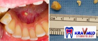
Removing stones from the salivary glands
Removal of stones from the salivary glands is carried out at the dental clinic “Dentist-K” by maxillofacial dentist-surgeon, Elena Vladimirovna Urakova. Elena Vladimirovna, Candidate of Medical Sciences, Associate Professor of the Department of the State Medical Academy answers the most frequently asked questions from patients - from our interview you will learn what the disease is, what methods of treatment exist and how difficult surgical intervention seems to be.
— What is a stone in the salivary gland?
E.V. Salivary stone disease is a pathology that occurs due to the formation of hard stones in the tissues of the salivary gland. The submandibular space is more susceptible to the disease - 9 out of 10 cases occur in this location of the stone.
- How to recognize a stone - are there any typical symptoms?
E.V. The stone clogs the duct of the salivary gland as it grows. This causes severe pain, a feeling of fullness and enlargement of the gland - since the outflow of saliva is hampered. I have seen patients with phlegmons and abscesses - this can result from inattention to unpleasant symptoms.
There may simply not be any obvious symptoms of the disease at the beginning, but even minor pain is a reason to visit the dentist.
— How does a doctor diagnose a stone?
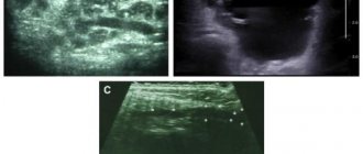
To diagnose a stone in the salivary gland, contrast radiography of the gland ducts, ultrasound and manual examination of the soft tissues of the maxillofacial area are sufficient.
— How difficult is the operation to remove a stone from the salivary gland?
E.V. This is a rather complex surgical procedure. Some stones are difficult to reach, and their location in the parenchyma of the gland is an indication for its complete removal. The procedure is performed under local anesthesia, but only in a hospital.
In the event that the stone is in the first-order flow at the exit, the task is simplified. Stone removal from the salivary gland is performed in the clinic on an outpatient basis, with tissue preservation; the salivary gland continues to function normally after healing. The removal method is chosen by the doctor taking into account the picture of the disease, the location of the stone, and its structure.
Indications for surgical treatment generally include large stone sizes (more than 50 mm) due to the risk of developing infectious complications, as well as the tendency for stone growth. The rehabilitation period includes drainage of the ducts - this helps to avoid complications. Drainage is installed for 1–2 days or in the presence of an inflammatory process until it is completely eliminated.
— Can stones be removed from the ducts of the salivary glands conservatively?
E.V. In some cases, the stone leaves its “habitat” on its own along with saliva - especially at the very beginning of the disease. If this does not happen, a special diet is prescribed - it consists of consuming foods that provoke active secretion of saliva. In addition, self-massage may be recommended - this allows you to remove the stone yourself without resorting to surgery. Of course, you will also have to take medications that help break down stones and increase salivation.
If the stone is located directly at the entrance to the duct, the doctor can remove it using a special tool. In some cases, it is advisable to carry out ultrasound treatment - with the help of ultrasound, the stone is “crushed” into pieces, after which it is removed with a stream of saliva or additional rinsing.
However, the size of the stone can reach several centimeters - in this case, only surgical removal of the stone from the salivary gland duct is indicated.
It is worth noting that non-surgical methods are not used for narrowing of the canals and some other structural features of the tissues that provoke the risk of blockage and complications.
— Which removal method is used more often and why?
E.V. Surgery is the optimal way to get rid of a stone due to its advantages: firstly, it effectively gets rid of the stone; almost 100% of cases of the disease end in complete recovery after surgery. Secondly, the procedure is painless thanks to the use of modern anesthetics. Removing the stone from the salivary gland duct is in most cases the best treatment for the disease.
— How long does it take to recover after surgery?
E.V. The operation to remove a stone from the salivary gland involves a rehabilitation period. Recovery time depends on the individual characteristics of the body - if you follow the doctor’s recommendations, rinse and take medications, after a few days local symptoms in the form of pain and swelling will no longer cause discomfort.
Salivary gland blockage
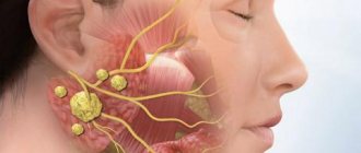
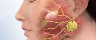
The salivary glands are ducts that secrete a special secretion into the oral cavity - saliva.
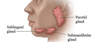
There are paired large salivary glands: submandibular, parotid, sublingual (they are closely related to the digestive system); as well as minor salivary glands: buccal, labial, molar, lingual, glands of the soft and hard palate. They are located in the submucosal layer of the cheeks, lips, palate, and tongue.
Kinds
According to the specificity of the secretion secreted by the glands, they are divided into protein, mucous and mixed. The largest paired salivary glands are the parotid glands, located in the back of the angle of the jaw, in front of the ears. The minor salivary glands are distributed throughout the oral cavity. If there is not enough saliva produced, the oral cavity becomes dry. In this regard, there is a high risk of developing caries, since with a deficiency of saliva, natural protection against such dental disease is no longer created.
Causes
It happens that the excretory duct of the salivary gland is blocked by various mineral deposits, which are also called stone. Blockage of the salivary gland provokes the normal outflow of saliva, as well as an enlargement of the gland. The cause of inflammatory phenomena can be infection with various bacteria and microbes.
When the duct is blocked, the swelling usually intensifies before eating, especially when eating pickled foods that have high acidity. It provokes salivation. When the ducts are blocked, saliva cannot be released freely. This often results in painful sensations.
A complication of blockage of the salivary gland is mucocele - the formation of a cyst, which is caused by the retention of secretions resulting from blockage of the outlet of the so-called Blandin-Nun gland.
Symptoms
Blockage of the salivary glands is manifested by swelling, a change in the color of the mucous membrane of the lips, which acquire a bluish tint. As a rule, this is how inflammation of the minor salivary glands manifests itself. If the major salivary glands are blocked, swelling and pain occur in the neck and cheeks, especially after eating.
Diagnosis and treatment
The Dento Style clinic network provides comprehensive diagnostics and treatment of various pathologies of the salivary glands. Our specialists have an individual approach to each patient. If the salivary ducts are blocked, the patient is examined and the affected area is palpated. Methods such as probing are used. It allows you to identify a salivary stone or a narrowing of the duct.
Hypo- and hypersalvation can be detected using sialometry (a method that allows you to measure the amount of secretion released per unit of time). X-ray and computed tomography are also highly effective.
As a treatment, the doctor prescribes the use of drugs that enhance salivation. In the case of an inflammatory process, antibacterial therapy is prescribed, as well as physiotherapeutic procedures.
The dentist first tries to mechanically squeeze out the stone by pressing on the duct. However, if such manipulation is not possible, surgical intervention may be required.