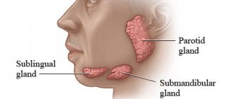
The salivary glands, located in the oral cavity, perform important functions and are directly involved in the digestion process. Saliva produced in the glands prevents infection of the body when eating food, and also participates in the initial stage of breakdown of nutrients. Inflammation of the salivary gland is a common pathological phenomenon that requires special treatment. Most often, the disease occurs in childhood, which is explained by high sensitivity to infections.
Causes of pathology
First of all, it should be noted that the development of the inflammatory process can act as a primary disease, or be a consequence of another pathological process occurring in the body. The salivary glands are paired organs, but often one half is affected. Simultaneous damage to a pair of glands is rare.
Causes:
- Infections.
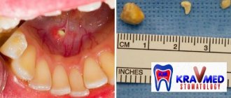
The causative agents of pathology include different types of bacteria and viruses. They enter the glands through the salivary ducts. Rarely, infection occurs through the introduction of infection with the lymph. In children, the disease often occurs against the background of influenza, encephalitis, and pneumonia. Salivary glands - Salivary stones. The formation of salivary stones leads to the blockage of the gland ducts. The salivary gland secreted inside does not come out, as a result of which a stagnation process develops. Because of this, non-infectious inflammation occurs.
- Mechanical damage. In childhood, when a child eats mostly soft foods, it is almost impossible to damage the oral mucosa. However, children tend to place various hard objects in their mouths, which can injure tissue. As a result, inflammation occurs.
- Teething. In some children, the appearance of the first teeth is accompanied by severe inflammation of the tissues, which also extends to the salivary glands. This form of pathology, as a rule, goes away on its own, so treatment is reduced to alleviating the child’s condition by eliminating the symptoms.
Thus, inflammation of the salivary gland in a child occurs due to infection, but there are also other, non-infectious causes of the disease.
How to treat prickly heat in newborns - symptoms, causes, types and prevention
Stage (prevalence) of the disease
After the examination, the stage of the disease is determined, which allows us to develop a treatment plan.
Stage I - tumor up to 2 cm in size, does not extend beyond the gland capsule
Stage II - the tumor size is 2-3 cm, it affects the capsule of the gland, there are signs of mild paresis of individual facial muscles.
Stage III - damage to most of the gland and germination into one of the adjacent structures (skin, lower jaw, external auditory canal, etc.). Presence of symptoms of facial nerve damage.
Stage IV - the tumor grows over a considerable distance into the surrounding tissue. Paralysis of facial muscles on the side of the tumor.
Clinical picture
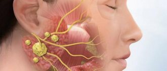
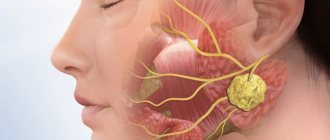
The development of infectious inflammation proceeds slowly, and therefore pronounced symptoms do not appear immediately. In some children, the clinical picture appears only 2 weeks after the infection enters the body. The severity depends on the type of pathogen, as well as the location of the affected glands. In children, the glands located in the area under the ears most often become inflamed.
Main features:
- Deterioration of general condition. Against the background of inflammation, the child’s body temperature increases. The patient becomes moody, sleeps poorly, and refuses food. Possible chills and cold sweat.
- Pain.
When the salivary gland is inflamed, it is painful for a child to swallow food. Also, pain is observed during mouth movements when chewing. When the parotid glands become inflamed, pain occurs when pressure is applied. Baby's pain - Enlarged glands. If the sublingual glands are affected, the patient cannot fully move the tongue, which affects the quality of speech. There is a sensation of a foreign body in the mouth, preventing normal nutrition. The salivary glands under the ears may also increase in size, which becomes noticeable upon visual inspection. A feeling of pressure that occurs simultaneously may indicate purulent inflammation.
- Dry mouth. Due to inflammation, the amount of salivary fluid secreted sharply decreases. As a result, the child experiences dry mouth.
- Hearing impairment. This symptom is often accompanied by inflammation of the parotid glands. The patient perceives sounds worse; noise or hum is heard in the affected ear.
- Local manifestations. In addition to severe swelling, inflammation is accompanied by redness of the tissues. Purulent discharge may appear.
In general, there are various symptoms of inflammation of the salivary gland, and if they occur, you should seek the help of a pediatrician.
Mumps in children
The incubation period for mumps infection lasts from 12 to 26 days. In rare cases, it can last a minimum of 9 days, a maximum of 26 days.
Clinical manifestations depend on the form of the disease. The most common manifestation of mumps is damage to the parotid glands . The disease usually has an acute onset, accompanied by a rise in temperature to 38-39 °C. If cases of the disease are mild, then the temperature is normal or slightly elevated. The level of temperature in the following days depends on how widespread the infectious process is. The temperature curve has a wave-like character (it rises and then decreases). The temperature rises when the infection attacks other salivary glands or organs. Along with the temperature, symptoms of intoxication begin to appear. The child develops headaches and muscle pain, malaise, and loss of appetite. Young children become capricious. Parents observe sleep disturbances.
Among the first symptoms of mumps in children is pain in the area of the parotid salivary gland, which intensifies with chewing and talking. By the end of the first day or at the beginning of the second day (less often), an increase in the parotid salivary glands is observed. At first, the process concerns only one party. The second side is affected after 1-2 days. A swelling is visible in front of the ear, which descends along the ascending branch of the lower jaw and behind the auricle, lifting it up and out.
But the increase cannot always be determined visually; sometimes this requires palpation. Doctors note the softness or doughy structure of the salivary gland upon palpation. Pain occurs on palpation. According to N.F. Filatov, the following painful points are identified: in front of the earlobe, in the area of the apex of the mastoid process and in the place of the mandibular notch.
Within 2-4 days, the parotid glands enlarge. After this, the sizes gradually return to normal. Together with the parotid or after them, the submandibular (submaxillitis) and sublingual (sublingual) salivary glands are subject to the process.
In every 4th patient with mumps, inflammation of the submandibular gland (so-called submaxillitis) is observed. Most often it is accompanied by damage to the parotid salivary glands. It almost never appears at the onset of the disease.
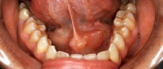
Severe forms of the disease can cause tissue swelling to appear in the gland area and spread to the neck. sublingual inflammation occurs - an isolated lesion of the sublingual salivary gland. In this case, a swelling appears under the patient's tongue. The affected glands are usually enlarged for 5 to 7 days. Then the pain disappears and the swelling subsides. Inflammation ends on the 8-10th day of illness. But there are cases when the glands are inflamed for 2-3 weeks. The temperature periodically rises and falls again.
Damage to the genital organs due to mumps infection
The virus can attack the testicles, ovaries, prostate gland, and mammary glands. Teenagers are susceptible to orchitis (25 cases out of 100). Orchitis leads to persistent dysfunction of the testicles, which means to male infertility in the future. In those who have had orchitis, spermatogenesis is impaired (about 50% of those who have recovered from the disease), and in 1/3 of those who have recovered from the disease, signs of testicular atrophy are recorded. 1-2 weeks after the onset of inflammatory processes in the salivary glands, orchitis may begin to appear. But it happens that mumps infection has a primary localization in the testicles.
Inflammation of the testicles appears due to infection on the epithelium of the seminiferous tubules. Pain receptors are irritated, which leads to pain. The pressure inside the tubules increases, and this threatens to disrupt the microcirculation and functionality of the organ.
One of the initial symptoms of orchitis with mumps is an increase in temperature to 38-39 °C. Often a sick child feels chills. Symptoms of intoxication appear immediately - weakness, intense pain in the groin (attempts to walk cause more severe pain), headache. Localization of pain occurs mostly in the scrotum and testicles. The testicle enlarges and becomes denser. Palpation leads to increasing pain. Reddish vessels filled with blood are visible on the skin of the scrotum, and the skin may also acquire a bluish tint.
The process does not always extend to 2 testicles. The swelling lasts for 5 to 7 days, then begins to subside. After 1-2 months, signs of atrophy are revealed, the testicle decreases in size and becomes softer.
A rare form of mumps is thyroiditis . It usually manifests itself as an enlarged thyroid gland, fever, pain in the neck and tachycardia.
Damage to the lacrimal gland also occurs - dacryoadenitis, which manifests itself as pain in the eyes and swelling of the eyelids.
Damage to the nervous system
Very rarely, central nervous system damage is the only manifestation of mumps. Often the nervous system is affected by infection after the glands. If damage to the central nervous system is the only manifestation, then the salivary glands are almost untouched by the virus and do not hurt. Clinically, the disease manifests itself as meningoencephalitis, serous meningitis, and in rare cases, neuritis or polyradiculoneuritis.
Serous meningitis is most often recorded on the 7-10th day of illness after the symptoms of mumps infection begin to appear less or disappear.
Mumps meningitis has an acute onset and increased body temperature. The patient develops headaches and repeated vomiting. Young children are sleepy and lethargic. In rare cases, on the contrary, agitation, as well as convulsions and delirium, may be observed. From the first days of the disease, meningeal syndrome appears, which manifests itself as positive Kernig, Brudzinsky symptoms (appear as a result of irritation of the meninges, hemorrhage under the membranes). If the forms of the disease are mild, then the meningeal signs are weakly expressed (may be absent). The disease has the following manifestations: single vomiting, headache, slightly elevated temperature. The final diagnosis of mumps meningitis is made based on the results of a spinal puncture.
In some cases, meningitis with mumps can be combined with encephalitis (so-called meningoencephalitis). Cerebral symptoms in such cases occur simultaneously with meningeal symptoms or after two to three days. Clinical manifestations of meningitis include: repeated vomiting, severe headaches, delirium, impaired consciousness, and convulsions. Pathological reflexes and hyperkinesis are also likely. Usually the course of the disease is favorable. Clinical symptoms subside after 3-5 days. The symptoms of meningitis disappear after a week (maximum after 10 days). Cerebrospinal fluid normalizes slowly, changes in it can persist for up to 3-5 weeks.
The healing process may be delayed in some cases (rarely). In such cases, psychosensory disorders persist for a long time, which manifest themselves in decreased memory, increased fatigue, headaches and areflexia (absence of one or more reflexes).
Neuritis and polyradiculoneuritis are rare with mumps infection. When the parotid gland suddenly enlarges, it can lead to compression of the facial nerve, leading to paralysis. In this case, on the side of the affected facial nerve, the function of the facial muscles is impaired: the eyebrow is slightly lowered, the folds of the forehead are smoothed (as well as the nasolabial fold), the palpebral fissure does not close.
In case of mumps infection, lesions of the cochlear nerve with hearing loss have been described.
With mumps, mumps pancreatitis can develop , combined with damage to other organs. The frequency of pancreatitis, as far as can be judged from the specialized medical literature, ranges from 3% to 72%. The diagnosis of pancreatitis is established only by increasing the level of amylase in the blood.
With mumps, pancreatitis, as is correct, occurs 5-9 days after the onset of the disease. Pancreatitis is the only manifestation of the disease in very rare cases.
Mumps pancreatitis in typical cases has an acute onset, manifested by pain. Abdominal pain ranges from mild to very severe. The pain is felt in the epigastric region, left hypochondrium, sometimes it is encircling and radiates to the back, right hypochondrium. In addition to pain, nausea and vomiting also often occur, colds become more frequent, and body temperature rises. When palpating the abdomen, pain and bloating are noted.
A blood test at the height of the disease shows an increased amount of amylase, lipase, and trypsin. Diastase activity is increased in urine. Stool analysis shows a significant increase in the amount of unchanged muscle fibers, fatty acids and extracellular starch. Changes in peripheral blood are not typical.
The course of mumps pancreatitis is favorable. Symptoms begin to subside 10-12 days after the onset of symptoms. First, the pain disappears, then the patient’s well-being gradually improves. But pancreatic function is restored only in the third or fourth week from the onset of the disease.
Diagnosis and treatment
To help the patient, it is recommended to seek the help of a specialist. Self-treatment is carried out if the pathology is mild and not accompanied by aggravating symptoms. The diagnosis is made by a pediatrician or otolaryngologist based on a study of symptoms and visual examination. The treatment method is prescribed in accordance with the established diagnosis, taking into account the age characteristics of the patient.
Why does marbled skin form in a newborn, what to do?
Main methods of treatment:
- Treatment of inflamed tissues.
This is done using an antiseptic substance applied to a cotton swab. They treat the inflamed area, as well as adjacent tissue areas, to prevent the spread of inflammation. Lotions are also made using purified mineral water. This allows you to moisturize the oral cavity and clear the salivary ducts if they are closed. Mouth rinse - Rinse. An effective method of treating oral diseases accompanied by inflammation is antiseptic rinses. The procedure is performed using drugs suitable for a specific age group. It is also possible to use antiseptics in the form of sprays.
- Antibiotics and antivirals. Prescribed for infectious pathology. Administration of drugs is carried out orally. Local or intramuscular administration is also possible. Antibiotics are used only in severe forms of bacterial inflammation, when the benefit of the drug outweighs the potential harm to the child’s body.
- Symptomatic therapy. To eliminate pain, medications that are acceptable in childhood can be used. These include drugs based on Paracetamol or Ibuprofen. It is strictly forbidden to give children medications containing Acetylsalicylic acid and Nimesulide.
- Surgery. It is carried out in cases where conservative therapy does not have the desired effect. Such cases are extremely rare, and usually the severe form occurs in adults, not children, so surgical therapy in a child is a rare exception. Treatment consists of opening the source of inflammation, removing inflammatory exudate and antiseptic treatment.
During therapy, it is recommended not to give the child solid food. It is recommended to include more liquid foods in your diet. If breastfeeding is carried out, it is necessary to temporarily interrupt lactation until the child recovers in order to prevent infection of the mother's body.
Traditional treatment for inflammation of the salivary gland in a child involves the use of antibiotics, antivirals, and antiseptics.
Diagnosis of salivary gland tumors
Inspection and palpation (feeling) can reveal pain, displacement of the tumor and skin, the condition of the lymph nodes, facial nerve and oral cavity.
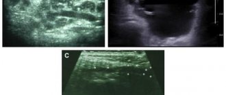
Ultrasound examination (ultrasound) makes it possible to determine the tumor formation, its size, connection with surrounding tissues, and density. Under ultrasound guidance, tumor puncture can be performed.
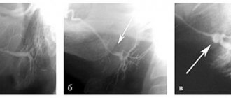
Sialography (radiography with contrast) reveals the condition of the salivary ducts, their compression, displacement, destruction of the gland and ducts.
Computed tomography (CT) is usually used in a common process to assess the relationship of the tumor with surrounding tissues and large vessels and decide on the extent of the operation.
A biopsy (taking a piece of the tumor for examination) makes it possible to establish a final diagnosis, which is necessary for treatment planning.