Why does caries occur in childhood?
It is known that caries is a pathological process that occurs in the hard tissues of teeth under the influence of internal and external factors. Carious lesions arise as a result of the “work” of cariogenic bacteria living in the oral cavity.
In this article
- Why does caries occur in childhood?
- Features of caries of milk teeth
- Symptoms of carious lesions in baby teeth
- Why is caries treated for young children?
- How is caries treated in children?
- Methods for treating childhood caries without drilling
- Invasive treatment of caries
- Do they give anesthesia to children?
- Prevention of childhood caries of primary teeth
By processing carbohydrates that enter the mouth with food, these bacteria produce acid. It is this that contributes to the destruction of enamel, dentin and other tooth tissues. Several factors contribute to the proliferation and active activity of cariogenic bacteria, which ultimately leads to the development of caries in children.
- Improper dental hygiene.
Insufficient cleaning from plaque, infrequent cleaning.
- Eating high in carbohydrates.
Children who eat a lot of sweets and flour, drink sweet compotes at night, and snack on chocolates are more susceptible to developing caries. A favorable breeding ground for microbes is formed in their mouth.
- Reduced immunity.
Weakened immune defense does not allow the body to effectively resist negative influences, so children with weak immunity develop caries more often.
- Features of intrauterine development.
The formation of teeth occurs in the first trimester of pregnancy, and it can be affected by the lifestyle, health, and previous diseases of the expectant mother.
These factors contribute to the fact that tooth enamel begins to lose mineral substances, cariogenic microbes multiply in the mouth, and as a result, dental caries develops.
Bottle caries, the importance of early diagnosis.
Authors : Msefer Souad
Bottle caries (nursing caries) is an extremely dangerous type of caries that can cause the destruction of baby teeth in infants and preschoolers. This is a common, serious disease of primary teeth that begins immediately after their eruption. Until recently, this term referred to caries of baby teeth in very young children caused by prolonged use of a baby bottle during sleep or during the daytime. But in the last few years, the term “bottle caries” has been used much more widely, using it to name diseases that have developed as a result of other factors.
These include: frequent - i.e. more than three times a day - consumption of cariogenic foods (cookies, lollipops, cakes, etc.), children's syrups, non-use of fluoridated toothpaste, and consumption of drinking water with insufficient fluoride content. It has also been found that cariogenic bacteria can be transmitted from mother to child as a result of certain actions, such as when the mother tastes the child's food from the same spoon or tests the temperature of the pacifier by placing it on her lips or putting it in her mouth. Poor maternal oral hygiene is also associated with increased concentrations of microorganisms in the child's oral cavity.
Diagnostics.
This is a serious, sometimes painful disease characterized by early onset and rapid progression. The disease develops in a short time, usually immediately after eruption, and can affect several teeth. As a rule, caries begins with the maxillary incisors at the point of their connection with the gums, then, if timely treatment is not started, the canines become infected, then the molars. As a result, if the patient still does not receive timely assistance, only the lower jaw incisors remain intact. There are four main stages of development of bottle caries.
1. Initial (child’s age - 10-20 months)
At this stage, chalky X-ray contrast foci of demineralization appear on the smooth surfaces of the primary incisors of the upper jaw, and a distinct whitish line can be seen in the cervical region of the vestibular and palatal surfaces of the upper incisors. At this stage, the development of lesions can be stopped. But diagnosing the lesion is possible only after thoroughly drying the affected tooth, so therapists or parents, when examining the baby’s oral cavity, often simply do not notice damage of this kind.
2. Second (child’s age - 16-24 months)
White lesions on the incisors progress rapidly, causing destruction of the enamel, resulting in exposure of dentin, which has a soft consistency and a yellowish tint. Initial lesions are found in the cervical region, as well as on the proximal and occlusal surfaces of the primary molars of the upper jaw. At this stage, the child begins to complain of increased sensitivity to cold, and sometimes parents notice a change in tooth color.
3. Third (child’s age - 20-36 months)
At this stage, large, deep lesions appear on the incisors of the upper jaw, and irritation of the pulp occurs. The child complains of pain when chewing or brushing teeth, as well as periodic attacks of pain at night. The molars of the upper jaw undergo the second stage of the lesion, and in the primary molars of the lower jaw and canines of the upper jaw the initial stage of the disease can be diagnosed.
4. Fourth (child’s age - 30-48 months)
Fractures of the crown of the anterior teeth of the upper jaw are formed as a result of the destruction of the enamel-dentin junction. The incisors of the upper jaw undergo necrosis, the damage to the maxillary molars reaches the third stage. The disease of the second molars and canines of the upper jaw, as well as the first molars of the lower jaw, is at the second stage. During this period of illness, some babies suffer from toothache, suffer from lack of sleep and refuse to eat. The inconvenience experienced by patients is further aggravated by the fact that not everyone can express their complaints.
A positive diagnosis is made based on a parent interview regarding risk factors and a clinical examination of the oral cavity followed by x-rays.
The differential diagnosis is based on observations such as congenital abnormalities in the structure of the tooth: childhood melanodontia, which primarily affects the incisors of the upper jaw, or imperfect amelogenesis of the enamel of all teeth, which is a hereditary disease of dentin and is characterized by dark, brownish color of the teeth and short roots . Enamel hypoplasia, which develops as a result of malnutrition during the perinatal period or vitamin A deficiency, also contributes to high susceptibility to caries and often accompanies bottle caries.
Consequences.
Bottle caries can cause serious long-term or short-term damage at local and systemic levels.
After pulp necrosis, the infection spreads to the periodontal area in one of two clinical forms: acute, characterized by inflammation of the connective tissue, adenopathy and mobility of the affected teeth; and the most common chronic, accompanied by an abscess and damage to the interseptal septum. Depending on the severity of the disease, the infection can penetrate the permanent tooth buds, causing irreversible damage. In children with a low level of immunity, infection often provokes the development of complications.
Contrary to popular belief, the consequences of dental caries in children affect not only the oral cavity. Tooth loss, inevitable in some cases, can cause not only orthodontic and aesthetic problems, but, more importantly, difficulties with pronunciation. In addition, the weight and height of children with bottle tooth decay are usually lower than those of their healthy peers, since difficulties with falling asleep and eating normally caused by infections and pain have a negative impact on the child's growth.
Early treatment is necessary to prevent crown failure and progression of caries. It includes simple techniques for remineralizing enamel, such as topical application of fluoride, the use of fluoride solutions and varnishes.
In conclusion, we note that in the implementation of preventive and therapeutic measures, early diagnosis of bottle caries and identification of risk factors are especially important. This will help avoid complications and consequences of the disease.
Source: Dentistry.by
published 10/11/2011 15:50 updated 22/07/2015 — Dentistry
Features of caries of milk teeth
Saliva at an early age does not have sufficient antiseptic properties, and microbes easily multiply in the oral cavity. At the same time, the degree of enamel mineralization in baby teeth is less than in permanent teeth. In this regard, childhood caries spreads very quickly. If in an adult a carious lesion on permanent teeth can develop from an early stage to a deep one over several years, then in children the same process takes only a few months.
In a child, dental caries often affects several teeth at once. Only children have such types of caries as ring-shaped and planar. The condition of the dental system is also affected by various infectious diseases to which children are susceptible.
Are baby teeth an important stage in development or a temporary phenomenon?
Teeth, the quality of our life largely depends on them - first milk teeth, then permanent teeth. And then - how does it work out? And if at a conscious age one can safely address the statement to teeth with “you value it when you lose it,” then parents, as a rule, treat their children’s temporary “pearls” quite dismissively: “they will fall out...” Due to this undeniable fact and the constant lack of time among parents, insufficient attention is paid to brushing teeth. Let’s add here store-bought juices with a lot of sugar, cookies from loving grandmothers for their pampered children, sweets from attentive guests and the almost complete absence of solid food in the child’s diet - it turns out that a dental surgeon often helps teeth “fall out.”
Statistics show that already at the age of 4, up to 9% of children have extracted teeth. And, unfortunately, by the age of 6-8 years the number of such children reaches 60%.
But complicated caries is not always the reason for the removal of baby teeth.
The reasons leading to the destruction of the crown parts of teeth or their early loss include:
- ineffective prevention or its absence during the prenatal period, during the embryonic development of the fetus and during breastfeeding;
- insufficient prevention during the period of primary, mixed and permanent dentition (timely treatment of primary teeth is of great importance for the prevention of caries complications and the prevention of early tooth loss);
- retention (non-eruption of a tooth), partial or complete primary adentia (absence of tooth buds).
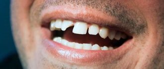
- injuries;
— enamel hypoplasia (incorrect or incomplete development of tooth enamel, the main causes of which are acute infections during pregnancy, hereditary factors, etc.).
When is it necessary to remove baby teeth?
1. Significant defects in the crown of the tooth, in which restoration of the tooth is not possible.
2. Chronic periodontitis (inflammation of the tissue around the tooth), complicated by purulent processes.
3. The period of physiological tooth change (when the permanent tooth is already visible in the oral cavity, but the milk tooth has not fallen out).
Why is it so important to preserve baby teeth until they are physiologically replaced by permanent teeth?
Milk teeth contribute to the correct formation of a correct permanent bite, and therefore important body functions in which the dentofacial system takes part, such as breathing, chewing, swallowing, speech.
Let's look at some facts that prove the importance of dental function .
A child's usual diet contains a large amount of carbohydrates, the breakdown of which requires thorough chewing of food and wetting it with saliva. In the absence of teeth, especially molars, chewing deteriorates, which leads to a delay in the physiological development of the jaws (the performance of the masticatory muscles decreases, the structure of the bone tissue of the jaw areas with extracted teeth is not fully formed) and the body as a whole.
When teeth are lost, the position of the tongue changes, which is placed in the resulting space, promoting the formation of an open bite (the presence of a pathological vertical gap between the teeth of the upper and lower jaws) and interfering with the correct eruption of permanent teeth.
Early loss of natural teeth in children without timely replacement of defects in the dentition leads to various changes in the dental system.
These include the following.
1. Changing the position of teeth adjacent to the lost one towards the resulting space (up, down, forward, backward, around the axis):
a) the absence of upper primary incisors leads to a noticeable flattening of the upper lip, protrusion of the lower lip, and displacement of the lower incisors forward.
b) in children , when they lose antagonists (teeth of the opposite jaw), unlike adults, the teeth very quickly change their position together with the alveolar process until they come into contact with the alveolar process of the opposite side, which is explained by a tendency to growth, which is not yet completed. The dental arches are bent not only in the vertical, but also in the horizontal direction, either towards the tongue or towards the cheek (dentition becomes asymmetrical).
c) with the loss of primary molars or canines , the overlap in the anterior-posterior direction increases, a so-called distal bite is formed, the first permanent molars shift to the center of the jaw, as a result of which the dental arch is shortened.
2. Reduced bite height.
3. Changing the position of the lower jaw forward, backward, to the side.
4. Changes in the relationships of the elements of the temporomandibular joint, which can be asymptomatic in the first period of development of the deformity. If the elements of the joint are not returned to their previous position with the help of orthopedic devices, such conditions often become pathological, such as arthritis, arthrosis, arthrosis-arthritis.
5. Early removal of baby teeth leads to the fact that the permanent tooth germ lying deep in the jaw is covered with a dense layer of bone, which is an obstacle to the eruption of a permanent tooth.
Particular attention should be paid to the first permanent molars , which are most often affected early by caries. They are of great physiological importance for the proper development of the jaw and the growth of the facial skeleton. They bear the main chewing load for up to 12 years, holding the premolars and seventh teeth in the correct position, and preventing secondary bite deformation.
If the first molars move forward due to early removal of primary molars, there may be a shortage of space for permanent canines and they will erupt outside the dentition.
Krolivets Tatyana Grigorievna,
Orthodontist of the first category,
Candidate of Medical Sciences,
Dental clinic "Implantis".
https://implantis.com.ua/
published 09/07/2012 09:49 updated 03/13/2014 - Diseases of the teeth and oral cavity
Symptoms of carious lesions in baby teeth
The manifestations of caries depend on what stage the process is at.
For early caries (the so-called stain stage), a characteristic sign is a light spot or thin line on the surface of the tooth enamel. This is an area of demineralization - the focus of carious lesions. At this stage, it is difficult to notice caries without the help of a dentist, because the child does not show anxiety or complain of pain or discomfort.
When the process enters the superficial stage, a dark pigmented spot forms on the tooth enamel, which has a rough structure. The tooth may react to sweets, and during the examination the doctor discovers areas of softening of the enamel.
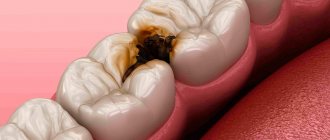
At the next stage of middle caries, dentin is damaged - the bone tissue of the tooth, in which a cavity is formed. The lesion looks like a darkened area, food gets stuck in the hole, and the child complains of pain when eating cold or hot food.
At the stage of deep caries, it is painful for the child to chew, the tooth reacts to any irritants, it can hurt on its own, and partial or complete destruction of the crown occurs. A characteristic type of childhood caries is the bottle form. It affects the dental neck and occurs in infants who often feed from a bottle for a long time. Depending on the form, stage of the disease, and the age of the child, the dentist will decide how to treat teeth affected by caries.
The work of a pediatrician on the prevention of caries in children of the first three years of life
Authors : Delyagin V.M.
Source - RMZh
Introduction
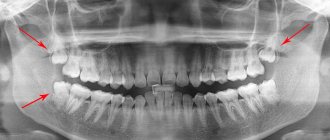
Specialized preventive examinations of children and adolescents by a dentist in our country should be carried out on the recommendation of the Ministry of Health and the Union of Pediatricians of Russia when the child reaches the age of 9 months, 3 years , 4, 7, 8, 9, 12, 14, 16, 17, 18 and 19 years [ 1]. Unfortunately, these deadlines are not always met. In addition, the interval between examinations may be sufficient for the development of serious dental or oral diseases, the treatment of which will require much more effort than if they were discovered earlier. Parents do not consider it necessary to bring their children , especially younger ones, for preventive examinations to the dentist, observing the vicious rule “until it hurts, I won’t go.” The population's oral hygiene habits are not sufficiently developed. As a result, 12% of one-year-old children already have carious teeth. The prevalence of caries in 5-year-olds is 73%. On average, there are 2.84 carious teeth per child. Among all teeth affected by caries, molars predominate (77.5%): molars of the lower jaw - 42.9%, molars of the upper jaw - 34.6%. Less commonly affected by caries are the primary incisors and canines of the lower jaw (3.6% and 3.5%, respectively) and the canines of the upper jaw (4.4%). The share of maxillary incisors in the structure was 12.9%. In molars, carious cavities on the chewing surface predominate (90.2%), damage to other surfaces was much less common (9.8%) [2]. The earliest visible sign of caries is the appearance of white spots on the surface of the tooth. At this stage, caries is reversible. If treatment is not started, demineralization continues and a cavity appears. Not a single healthcare problem is ever solved at the level of so-called “narrow specialists”. Of all the specialists who can really influence this situation, the child most often meets with primary care pediatrician In this regard, the pediatrician’s knowledge of pediatric caries, the ability to conduct an adequate examination, and the ability to convey existing problems to the child’s parents can be decisive in the systemic prevention of caries [3].
Definition of the problem Caries is the most common chronic infectious disease of children and adolescents, caused by the persistence of Streptococcus mutans in the oral cavity on the background of a carbohydrate diet. Infection occurs in early childhood, most often from mothers. Caries is a multifactorial disease. Therefore, prevention should be carried out comprehensively in several directions simultaneously: • reducing the number of cariogenic flora in the oral cavity; • reducing the time of contact of cariogenic flora with the tooth surface; • increasing tooth resistance to caries. Microflora control Str. mutans, a causative factor of caries, is transmitted vertically to a child in early childhood from parents, mainly from mothers. The microbe is a necessary, but not the only condition for caries. The traditional way of prevention is to remove plaque during oral hygiene. However, there are no reliable correlations between oral hygiene and caries. Bacteria are located in peculiar niches, in depressions and microcracks on the surface of the tooth, in the interdental spaces, where caries, in fact, begins. These microbial niches cannot be sanitized with regular toothbrushes or floss. Chlorhexidine. The practice of suppressing microflora with chemotherapeutic agents has long been generally accepted [4]. Local application of chlorhexidine in the form of rinses or, more effectively, gels sharply reduces the number of Str. mutans in saliva [5]. But if chlorhexidine is used in isolation, then after three weeks the acidogenicity of dental plaque is restored [6]. Oral hygiene. Young children need assistance with routine hygiene procedures. Before the age of 1 year, parents should wipe their children's teeth with a soft, damp swab. Starting from 1 year old, you can teach your child to use a toothbrush. You should brush your teeth twice a day, at least once a day before going to bed. Fluoride-containing toothpastes should not be used until the child can independently control swallowing movements. This is usually achieved at 3 years of age. At an older age, in children at high risk of developing caries, special products can be used on the recommendation of a dentist.
Food, household and medicinal factors in the development and prevention of caries Cariogenic bacteria metabolize carbohydrates to form acid, which destroys the mineral structure of teeth. The likelihood of developing caries correlates well with the total amount of carbohydrates consumed and the frequency of their intake [7,8]. Of all sugars, the most cariogenic is sucrose (household white sugar). The consistency of the food also matters. Viscous, thick food with the same content of fermentable sugars is more cariogenic than liquid food that quickly leaves the oral cavity. The minimum amount of sugar required for the development of caries depends on the frequency of its consumption, the hardness of the sugar pieces, and oral hygiene. That is, the leading factors are those that determine the length of time sucrose remains in the mouth. The correlation between the amount and frequency of sucrose consumption and caries is expressed by a wave-shaped curve. At a younger age, with an immature immune system and newly erupted teeth, an increase in sucrose consumption is accompanied by a sharp increase in the incidence of caries. After this rise in older age, there is a decline in the frequency of caries with the same or even greater consumption of sucrose [9]. A number of products protect teeth from caries [10,11]. First of all, it's cheese. The anticariogenic effect of cheese is especially noticeable under conditions of experimental and physiological xerostomia. Physiological xerostomia is common in infants and the elderly. The protective effect of cheese is explained by the presence of calcium lactate, phosphorus, and salivogenic properties. Increased salivation leads to neutralization of acids in the oral cavity. At the same time, the antimicrobial properties of saliva are affected. In addition to cheese, milk, dark chocolate, licorice, and nuts have an anticariogenic effect. The risk of developing early caries increases sharply if a child receives breast milk substitutes, juices, and drinks with fermentable sugars through the pacifier. This same risk group includes children who are freely breastfed, who often receive breastfeeding and fall asleep at the breast [12,13]. Children should never fall asleep with a bottle in their mouth. Another cariogenic product may be oral drugs. Suspensions of ampicillin, amoxicillin, cefaclor, co-trimoxazole and other pediatric forms contain 18–80 percent sugar syrup by volume. Moreover, 28% of the tested drugs contained 2 sweeteners [14, 15]. Along with this, apparently, there are also side effects of the drug itself. Enamel defects and, accordingly, a predisposition to caries may appear later, on permanent teeth [16]. Therefore, parents need to be taught how to clean the child’s oral cavity after each use of a sweet suspension (especially in children who are often ill). The evening dose should be taken before brushing your teeth. General recommendations for parents are: • do not leave a bottle of milk, formula, juices, etc. with a sleeping child; • do not leave a pacifier in the mouth of a sleeping child; • wean your child off the bottle no later than 14 months of age ; • avoid long-term consumption of sweet drinks and sour juices; • do not give candy as a calming aid; • control the amount and frequency of sugar consumption; • give preference to non-cariogenic products; • treat the mouth after taking sweetened medications. The likelihood of enamel degeneration and, accordingly, the development of caries increases in people who swim every day in pools with chlorinated water. Fluorides have long been known as drugs that effectively prevent caries. The main source of fluoride under normal conditions is water. Bottled water, which part of the population now uses, is low in fluoride compounds [17]. Additional doses of fluoride, depending on its content in the drinking water of the region, which effectively prevent caries, are given in Table 1.
Child's own factors Predisposition to caries is determined not only by external factors, but also by the characteristics of the child himself. Saliva. A protective factor is normal salivation. Saliva protects against tooth decay by washing away germs and food debris from the teeth. Saliva has buffering properties, reducing acidity in the mouth; The mineralizing properties of saliva are known; saliva has an antimicrobial effect. The latter effect is achieved through immunoglobulin A (IgA). Already in the first year of life , IgG is detected in saliva. In addition, saliva contains lysozyme, myeloperoxidase, and lactoferin [18]. Under natural conditions, physiological xerostomia occurs at night, when there is no stimulated secretion of saliva. Xerostomia can be caused by antihistamines, anticonvulsants, antidepressants, and antispastic drugs. Tooth resistance to caries development. Tooth enamel is the densest structure of our body. At the same time, it is the first line of defense against tooth decay. Complex drugs are required to prevent caries and increase the overall resistance of the body. As such a drug, along with others, the Kaltsinov vitamin and mineral complex, containing calcium, phosphorus, vitamins A, D3, B6, C, has proven itself well. The main problem that determines the density of enamel is mineralization, which depends on calcium and vitamin D. Hypomineralization is observed in premature infants with intrauterine malnutrition, born from mothers with calcium deficiency and vitamin D deficiency [19,20]. In the minds of doctors, vitamin D deficiency is associated with rickets. However, vitamin D deficiency is clinically diverse and extremely widespread. Even in developed countries, vitamin D deficiency has been reported in 32% of doctors and medical students, 48% of girls aged 9–11 years , and 76% of pregnant women. 81% of children born to mothers with vitamin D deficiency themselves suffer from a deficiency of this vitamin [21]. Vitamin D has a wide spectrum of action [22]. It is a steroid prohormone, a precursor to many active metabolites that have hormonal properties (Table 2). In recent years, much attention has been paid to the non-calcemic role of vitamin D. Along with a discussion of the hormonal component of its action, the effect of vitamin D on T-cell immunity is being studied. Receptors for vitamin D are found on T lymphocytes and macrophages. The highest density of such receptors is found on the surface of immature thymocytes and mature CD-8 T lymphocytes. 1,25-dihydroxyvitamin D3 has been experimentally shown to be able to prevent or suppress autoimmune diseases (autoimmune encephalomyelitis, rheumatoid arthritis, systemic lupus erythematosus, type 1 diabetes and inflammatory bowel disease). The condition for such activity of vitamin D as a hormone was a diet with a normal or even high calcium content. Vitamin D stimulates the production of transforming growth factor α-1 and interleukin-4, which can suppress the inflammatory activity of T cells. This hypothesis is supported by the observation that vitamin D is unable to suppress multiple sclerosis in a model of interleukin-4-deficient mice [23]. Calcium is involved in hemostasis, cell proliferation and differentiation, and intercellular signaling interaction (Table 3). Phosphorus is an essential component of all organs and tissues. Violation of phosphorus metabolism affects the entire homeostasis. The main part of phosphorus is concentrated in the bones (up to 700 g in an adult), less - in soft tissues (up to 200 g). Less than 1% of total phosphorus is found in the extracellular fluid. Phosphorus is the main intracellular anion. Phosphorus is present in high-energy compounds (adenosine triphosphate), determines the structure of the cell wall (phospholipid bilayer), participates in glycolysis, and, due to the regulation of the synthesis of 2,3-diphosphoglycerate, determines the oxygen-binding ability of hemoglobin. Vitamin A, a fat-soluble compound, is involved in the synthesis of rhodopsin, ensuring normal twilight and color vision, helps maintain the integrity of epithelial cells, and regulates body growth. Vitamin A deficiency leads to a decrease in the number of leukocytes, natural killer cells, a decrease in the mass of lymphoid tissue, the concentration of complement, antigen-specific immunoglobulins E and G, tumor resistance, and increased synthesis of interferon-α. Vitamin B6 deficiency is manifested by seborrheic dermatitis, cheilitis, glossitis, peripheral neuropathy, lymphopenia and normocytic anemia (less commonly, microcytic). Infants may develop seizures, especially against the background of hyperthermia. Ascorbic acid is involved in the formation and maintenance of the structure and function of cartilage, bones, teeth, ensures metabolic balance in mesenchymal tissues (connective tissue, osteoid, dentin), affects the formation of hemoglobin, the maturation of red blood cells, and is actively involved in the synthesis of collagen protein - an important structural component of subcutaneous tissue. – fat, cartilage, bone and other types of connective tissue. Enhances the absorption of iron and promotes its inclusion in heme. Increases the activity of liver enzymes. Ascorbic acid is necessary to activate the conversion of calcidiol to calcitriol, an active metabolite of vitamin D, being its synergist. Vitamin C is one of the most effective water-soluble antioxidants. Ascorbic acid is necessary for the rehabilitation of ulcers and the healing of burns. It is involved in the exchange of phenylalanine and tyrosine, ensures the release of folic acid from food and enhances the absorption of iron. Risk groups for the development of caries in infants and young children Identification of children prone to caries is very important for timely and adequate preventive work (Table 4). Children from low-income strata of society, with a low educational level of parents, premature children, low birth weight children, often ill, with systemic pathological processes, receiving many medications with sucrose-based sweeteners, drinking sour juices, with poor oral hygiene, are predisposed to the development of caries. the presence of areas of enamel dystrophy, already formed caries (especially multiple ones), with orthodontic splints that make teeth cleaning difficult. Along with this, aggravating factors are artificial feeding, sleeping with a bottle and food with fermentable carbohydrates.
Literature 1. Rumyantsev A.G., Timakova M.V., Chechelnitskaya S.M. Monitoring the development and health of children (Guide for doctors). – M.: Medpraktika-M, 2004. – p. 219. 2. https://www.dentalsite.ru/new.aspx?id=19333 3. Schafer T., Adair S. Prevention of dental disease. The role of pediatrician. / Pediatric clinics of North America, 2000. – v. 47. – 5. – pp. 1021 – 1042. 4. Tinanoff N. Dental caries: etiology, pathogenesis, clinical manifestations and management. In: Wei S. (Ed.) Pediatric dentistry: total patient care. Philadelphia, Lea & Febiger, 1988. – p. 9. 5. Maltz M., Zickert I., Krasse B. Effect of intensive treatment with chlorhexidine on the number of Streptococcus mutans in saliva. // Scandinavian Journal of Dental Research, 1981. – v. 89. – pp. 445 – 452. 6. Gerardu V., Buijs M., Cate J., Loveren C. Effect of an intensive treatment with 40% chlorhexidine varish on plaque acidogenity. // Clinical oral investigations, 2007. – v. 11. – pp. 77 – 81. 7. Marshall T., Broffitt B., Eichenberger-Gilmore J., et al. The roles of meals, snacks and daily total food and beverage exposure on caries experience in young children. // Journal of Public Health Dent., 2005. – v. 65. – pp. 166 – 173. 8. Marshall T., Eichenberger-Gilmore J., Larson M., et al. Comparison of the intakes of sugar by young children with and without dental caries experience. // Journal American Dent. Association, 2007. – v. 138. – pp. 39–46. 9. Newbrun E. Cariology. 2nd edition. Chicago, Quintessence, 1989. 10. Krobicka A., Bowen W., Pearson S., Young D. The effects of cheese snacks on caries in desalivated rats. // Journal of Dental Researches, 1987. – v. 66. – pp. 1116 – 1119. 11. Moynihan P. Foods and factors that protect against dental caries. // Nitritional Bulletin, 2000. – v. 25. – pp. 281–286. 12. Seow W. Biological mechanisms of early childhood caries. // Community Dental and Oral Epidemiology, 1998. – v. 26. – Suppl. 1. – pp. 28–31. 13. Vachirarojpisan T., Shinada K., Kowaguchi Y., et al. Early childhood caries in children aged 6 – 19 months. // Community Dental and Oral Epidemiology, 2004. – v. 32. – pp. 133–142. 14. Hill E., Flaitz C., Frost G. Sweetener content of common pediatric oral liquid medications. // American Journal of Hospital Pharmacology, 1988. – v. 45. – pp. 135–142. 15. Balbani A., Stelzer L., Montovani J. Pharmacentical expicients and the information of drug labels. // Review Brasilian Otorhinolaryngology, 2006. – v. 72. – p. 400–406. 16. Hong L., Levy S., Warren J., et al. Amoxicillin use in infancy and enamel defects in permanent teeth. // Archive of Pediatric and Adolescent Medicine, 2005. – v. 159. – pp. 943–948. 17. Winkle S., Levy S., Kiritsy M., et al. Water end formula fluoride concentrations: significance for infants fed formula. // Pediatr. Dent., 1995. – v. 17. – pp. 305–309. 18. Tenouo J. Antimicrobial agents in saliva – protection for the whole body. // Journal of Dent. Res., 2002. – v. 81. – pp. 807–809. 19. Eglash A., Kendall S. What vitamins and minerals should be given to breastfeed and bottle-feed infants? // The Journal of Family Practice, 2005. – v. 54. – pp. 20–22. 20. Saraiva M., Bettiol H., Barbieri M., et al. Are intrauterine growth restriction and preterm birth associated with dental caries? // Community dentistry and oral epidemiology, 2007. – v. 35. – pp. 364–376. 21. Adams M. Vitamin D myths, facts and statistics. // Version current at August 14, 2007. https://www.newstarget.com/z003069.html 22. Rumyantsev A.G., Delyagin V.M. Vitamins and microelements for pregnant and lactating women as a prerequisite for healthy offspring. Guidelines. Moscow, MAKS Press, 2007. – 48 p. 23. Abou-Saif A., Lipman T. Vitamins: hormonal and metabolic interrelationship. In: Becker K. (Ed.) Principles and practice of endocrinology and metabolism. Third edition, Lippincott Williams & Wilkins, Philadelphia, 2001. – pp. 1272–1277.
caries, children, pediatrician
published 12/22/2011 16:58 updated 01/24/2017 — Dentistry
Why is caries treated for young children?
There is a myth that baby teeth with caries do not need to be treated, as they will fall out anyway. In fact, dentists unanimously say that treatment of childhood caries at any age, both on milk and permanent teeth, is mandatory.
There are several reasons why caries of baby teeth should be treated before the age of 5-6:
- The baby tooth preserves space for the permanent tooth and stimulates its growth.
- It takes part in the chewing function and, therefore, in the digestion process.
- Baby teeth affected by caries are a source of chronic infection in the child’s body. In addition, the root of the baby tooth is located next to the germ of the permanent tooth. If caries in children is not treated, the inflammatory process can affect permanent teeth even before they erupt.
- Healthy baby teeth are necessary for a child to develop high-quality speech skills and correct pronunciation of sounds.
- If parents do not treat their child's caries, this can lead to untimely loss of baby teeth. Because of this, other teeth in the row shift, permanent teeth begin to cut in the wrong place, which leads to malocclusion.
- Early loss of a baby tooth can delay the eruption of a permanent one.
This is why it is so important to preserve baby teeth until they fall out naturally. And this can only be done if baby teeth are treated properly in a timely manner.
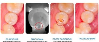
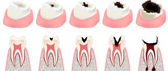
Types of caries
You can learn to distinguish caries on baby teeth by stages from a photo, or by knowing a description of external signs:
- Initial caries of baby teeth is noticeable on the enamel as a matte white spot. It is treated by strengthening the enamel with fluoride compounds, which can be quickly eliminated if detected in a timely manner.
- Surface. The spots are still light, but pain is already possible when eating.
- Average. Carious cavities become deeper - the tooth enamel is already destroyed, pain appears when cold or hot.
- Deep caries. Requires an urgent appointment with a dentist and treatment, since the infection has already reached the dentin and can spread to the pulp. If you ignore caries of baby teeth at this stage, the risk of root infection and the appearance of a baby tooth cyst increases significantly.
Dolotova Marina
Little patients of Aza&Buka rarely encounter deep caries. After all, we, doctors, do everything necessary for this - we make appointments on time, teach how to brush teeth and eat right. And if caries occurs in baby teeth, we treat it as early as possible!
How is caries treated in children?
What to do if a child has caries? Parents often ask dentists this question. And the answer is to treat. It is not so important when a child develops caries, whether he is one year old, 5 years old or 12 years old; dental disease is treated at any age. The only difference is how exactly dental caries are treated.
Different treatment methods are divided into two main groups - conservative and surgical. Conservative ones allow you to quickly and painlessly stop the carious process without drilling the tooth. Such techniques are most suitable for children, but they are effective only in the early stages of caries.
Surgical treatment involves drilling the carious cavity with a drill, removing the affected tissue and installing a filling. In case of deep or moderate caries, it will not be possible to cure a tooth without preparation.
How to identify milk caries
It is quite simple to identify caries in young and older children. White or brown spots appear on the teeth, a painful reaction to hot and cold is observed, and the child may develop bad breath. When these first symptoms of caries appear, it is already worth sounding the alarm, since the carious process is developing very rapidly. It is characterized by almost instantaneous damage to several teeth, and if measures are not taken, the entire dentition may be affected. Of course, it is often difficult for a child to articulate that his baby tooth hurts. He may refuse to eat or chew on only one side. This should also alert parents and encourage them to take their child to pediatric dentistry.
Methods for treating childhood caries without drilling
A child has been diagnosed with a carious spot, that is, initial caries - what to do in such a situation. The doctor may offer several options.
- Fluoridation.
A special composition with a high concentration of fluoride is applied to the surface of the tooth enamel. The mineral substance helps strengthen the enamel, restore its structure, and helps stop the further process of tissue destruction.
- Remineralizing therapy.
The teeth are coated with compounds containing phosphorus, calcium, magnesium and other minerals. The treatment promotes the remineralization of enamel, restoring its strength and structural characteristics.
- Ozone therapy.
Another modern non-invasive method in which children's teeth are treated with ozone. It destroys different types of cariogenic bacteria, preventing their proliferation in the oral cavity.
- Silvering.
Previously, this method was very popular, today it is used less frequently due to the fact that it affects the aesthetics of the teeth. After silvering, white baby teeth acquire a distinct dark color. The essence of the method is that tooth enamel is coated with compounds containing silver. They have an antiseptic and bactericidal effect, forming a protective film on the surface that prevents the spread of infection. Silvering does not cure caries, but it helps slow down the process of tooth decay.
Invasive treatment of caries
If there is a hole in a tooth, it is treated as if it were permanent, using an invasive method. First, using a drill, the dentist prepares the carious cavity and removes softened, pigmented tissue. Rinses, dries and disinfects the tooth.
After sanitation, a filling is placed inside the cleaned cavity. Today, light-curing filling composites are the most popular. They are applied in layers, and at each stage they are illuminated with a special lamp for curing. Light-curing fillings have good strength, long service life and do not shrink, which reduces the risk of developing recurrent caries under the filling.
After installing the filling, the doctor adjusts it to the bite, grinds and polishes it, giving it the correct anatomical contours.
Do they give anesthesia to children?
Many parents are concerned about how to painlessly treat teeth in children under 5 years old.
Non-invasive methods are absolutely painless. If a cavity needs to be prepared, anesthesia may be required. Children most often undergo two-stage anesthesia. First, the gum area is treated with an anesthetic gel, which numbs the injection site, and then an anesthetic injection is given.
In some cases, it is advisable to use sedation - when the child is introduced into a relaxed state of half-asleep using a special gas. Choosing the optimal method of pain relief and treatment is the responsibility of the dentist.
Anesthesia in pediatric dentistry
What type of anesthesia will be used during treatment depends on the degree of advanced disease. Most often, doctors use a spray that makes the gum tissue insensitive.
If the problem is serious, injectable painkillers are used. But so that the little patient does not feel pain during the injection, the doctor first lubricates the area where the needle is inserted with an anesthetic. This approach to pain relief makes even complex dental procedures not at all scary and as comfortable as possible for the little fidget.
General anesthesia in pediatric dentistry is used only for the most severe illnesses or if it is not possible to persuade the patient to open his mouth and sit quietly in a chair. Parents should not be alarmed if the doctor recommends general anesthesia. It involves the use of a modern, safe drug with a minimum number of side effects.
Prevention of childhood caries of primary teeth
Caries of primary teeth can be prevented if you listen to the recommendations of dentists and follow preventive measures for this dental disease.
- Take care of your oral cavity from the moment the first tooth appears.
Previously, it was believed that young children needed to brush their teeth from about one year old, when they grew eight front incisors. Modern dentistry has revised this approach, and today it is recommended to brush children’s teeth as soon as the first baby tooth has erupted. This usually occurs between 6-9 months of age. It is clear that a baby cannot take care of the oral cavity on his own, so this task falls on the shoulders of the parents. The first teeth are cleaned with special fingertips or a baby brush without toothpaste. The brush should have a small cleaning head and ultra-soft bristles. As they approach the age of one year, they begin to brush their teeth with fluoride-free toothpaste. As the child gets older, the size of the head and the stiffness of the bristles may change. For example, for a child aged 5 years, you can purchase a toothbrush with medium-hard bristles.
- Follow the rules for brushing your teeth.
A child may develop caries even with regular brushing if this process is left to chance. Children under about 7 years old cannot brush their teeth properly, so parents should help them and monitor the process. Proper oral care involves brushing your teeth twice - in the morning after breakfast and in the evening before bed. The hygiene procedure should not take less than 2-3 minutes. It is important to ensure that the child thoroughly cleans each tooth on both sides and cleans the chewing surfaces. A large number of bacteria accumulate on the tongue and the inside of the cheeks, so at the end of cleaning you need to teach your child to remove plaque from these surfaces.
- Routine visit to the dentist.
Few children like to treat tooth decay. Modern methods make it possible to do this quickly, painlessly and without drilling, but only if the disease is detected at an early stage. This is why it is so important to take your child to the dentist twice a year, even if the child has no complaints. The doctor will check the condition of your teeth and give recommendations for care. Regular visits accustom your child to visiting the dentist and help get rid of fear. In the future, such children sit in the dental chair without any problems and have their teeth treated.
Follow these preventive measures and you may not have to treat your child’s caries.
Prevention of caries
Many pediatric dentists offer painless and fun treatment. But it is easier to prevent than to treat. Simple preventive measures will help preserve the integrity of baby teeth for as long as possible:
- Daily oral hygiene. As soon as the first tooth appears, you need to purchase children's toothpaste and a brush. Grooming should be done twice a day - morning and evening, and gradually become a healthy habit for the child.
- Proper nutrition. It is necessary to include foods high in calcium (kefir, cottage cheese, cheese, etc.), as well as vegetables and fruits, in the child’s diet. Harmful sweets and fast carbohydrates, on the contrary, should be excluded or reduced as much as possible in the daily menu.
- Preventative examinations with a doctor. A pediatric dentist should be visited 2-4 times a year. This will allow you to monitor dental health and reduce the risk of caries in your child.
It is recommended that a child’s first meeting with a dentist take place after the incisors appear – at 1-1.5 years. This will be a preventive examination, during which parents will be told the principles of caring for children’s teeth and advised on the optimal scheme for monitoring the growth of temporary units.
Watch an informational video on this topic