Often, a slowly growing exostosis that does not cause pain remains invisible to both the patient and the specialist. It is discovered by chance, when the lump is already palpable under the skin or during an X-ray examination.
The increase in osteochondral exostosis occurs in childhood and adolescence. Like the bone itself, the growth grows slowly and stops growing along with the growth of the musculoskeletal system. Particular attention should be paid to the continued increase in the size of the tumor when a person’s growth stops - sudden rapid growth of the tumor can be caused by the transition of the disease to the malignant stage. This is the most serious complication of exostosis, possible in 1% of cases.
Qualified removal of exostosis in the NEARMEDIC network will help get rid of osteochondroma and prevent its dire consequences. High-tech diagnostic and treatment equipment in our clinics allows us to reduce examination time, guarantees accurate diagnosis and successful treatment.
To sign up for exostosis treatment, call us at +7 (495) 6 171 171; or fill out a simple form on the website.
Causes and symptoms
A tooth extraction operation causes osteophytes to appear if the dentist does not smooth out the edges of the socket. Injuries and old fractures, improperly connected damaged parts of the jaw also lead to the formation of exostoses.
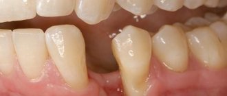
Symptoms depend on size and location. Small irregularities do not bother patients and do not cause pain. Their most common location is the area of the median suture. Osteophytes do not interfere with opening the mouth. The mucous membrane over them is healthy, pale and mobile. There are no noticeable deviations.
If the growth of exostoses continues, the mucous membrane becomes thinner. The risk of damage increases if a prosthesis is installed. Injury can occur from damaged dental crowns.
When palpated, osteophytes are felt as dense growths that are not connected to the soft tissues around them. If they are located near the articular process, they cause pain. Growths near the joints prevent the mouth from opening wide. The mental part of the lower jaw is displaced, the occlusion is broken. Regional lymph nodes are of normal size and cannot be palpated. General condition is good.
Tori of skeletonized maxillae
Of the 600 examined skeletonized maxillae, in 90 (15%) we found tori of various shapes, sizes and locations. The height, length, and width of the palatine ridge were measured. Characteristics of the severity of the bony elevation of the hard palate are given according to a 3-point system. The torus, which belongs to the first point, is characterized by its small size and height (up to 15 mm). It can be flat, tuberous or pointed ridge and is noted at various parts of the median suture. Such dimensions of the palatal eminence were present in 36 skeletonized upper jaws. Typically, such a bone thickening in the patient’s mouth is detected only by palpation. As a rule, a weakly defined ridge does not cause any particular difficulties during prosthetics. If a slight imbalance of the prosthesis appears, it is easy to correct the base. With the second score, there is a significant severity of palatal elevation. In this case, the height of the bone protrusion varies between 10-20 mm. Such bony elevations were found on 22 skeletonized jaws. The shape of the torus can be ovoid and is often located in the middle of the hard palate. There are bony protrusions of the main localization on the horizontal plate of the palatine bone with a small transition of the posterior part of the palatine processes along the intersection of the transverse and sagittal sutures of the upper jaw. At the third point of the torus, a large swelling of the bone was noted. The protrusion covers a large area of the hard palate, reaching a height of more than 20 mm. Large torus sizes were found in 32 skeletonized maxillae.
The results of measuring the length and width of the skeletonized upper jaws and the horizontal plate of the palatine bone made it possible to distinguish them into 3 groups. Group I included jaws with a short hard palate along the midline: 150 upper jaws, of which 18 were toothless. The main feature of this group of jaws is their short length along the sagittal suture (33-44 mm); there are almost no large bony protrusions. As a rule, the arch of the palate of the first group of jaws is mainly wide-bottomed with a flat surface - a good prerequisite for success in prosthetics. Group II includes 240 skeletonized jaws of medium size (45-48 mm in length) with different shapes of the roof of the palate, of which 52 are toothless. More often, this group includes jaws with a deep dome-shaped arch of the palate, which is an unfavorable factor for fixing the prosthesis. Group III consisted of large maxillae (from 45 to 56 mm or more): 210 jaws, of which 30 were skeletonized toothless maxillae. Basically, this group of jaws has unfavorable conditions for the anatomical retention of a removable denture. In the clinic, bone protrusions are found not only in the middle of the hard palate (torus), but also in the area of the tubercle, the vestibular surface of the sockets of extracted teeth of the upper jaw.
Removal of exostoses on the lower and upper jaw
There is no conservative treatment for osteophytes. The operation is performed for the following indications:
- rapid growth and pain,
- difficulty eating or talking.
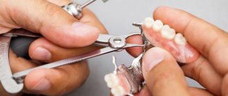
To remove exostosis on the upper jaw, the following manipulations are performed.
- A midline incision is made along one line, with release incisions in front and behind.
- Peeling of the mucoperiosteal flap.
- Removing the build-up in one block or sawing and removing in parts.
Sometimes cutting with a bur or mill is possible.
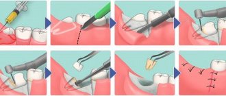
Next, the dentist checks the condition of the wound, smoothes its edges, returns the flap to its place and applies interrupted sutures. To prevent local accumulation of blood, apply an iodoform bandage. Silk threads are used to secure it.
To remove exostoses on the lower jaw, the dentist makes a trapezoidal incision. Moves back the mucoperiosteal flap and cuts down the protrusion, smoothing the surface. Next, he restores the mucous membrane and applies sutures. If the spines are small and there is little bone tissue, then a tunnel is formed. Hydroxyapatite is placed in it. The material is an important part of bones and tooth enamel. It helps to build up the skeletal system for further treatment. The doctor covers the stitches with a bandage.
Why do bone growths form on the gums?
Benign formation - exostosis - on the lower jaw
Exostosis in anatomy - a bone located on the outside, a common dental pathology. The neoplasm does not degenerate into a malignant tumor, but requires constant medical supervision. Painless growths in the form of small round or sharp lumps can be located at the base of the tooth, on the palate, or under the tongue. The ICD-10 code is K10.
With exostosis, there is a sensation of a foreign object in the mouth, the mucous membrane changes color. Sometimes the mobility of the lower jaw is impaired, and facial asymmetry is observed.
Reasons for the appearance of the torus of the lower jaw:
- congenital defects of the jaw structure, malocclusion;
- genetic pathologies that are accompanied by impaired bone growth;
- strict diets;
- long-term inflammatory processes, abscesses in the oral cavity;
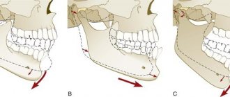
- injuries and fractures of the jaw followed by improper reposition of broken jaw fragments;
- hormonal imbalance, diseases of the endocrine system;
- periodontal displacement due to improper tooth extraction;
- edentia – partial or complete absence of molars;
- herpes, other viral infections;
- dysplastic growths along the edge of the jaw are a consequence of osteoma of the skull bones.
Lines of small bumps on the upper and lower jaw
Bone spines in the mouth are formed from cartilage tissue, less often affecting the base of the jaw. Externally they look like small white tubercles, similar to foci of inflammation. Most often, several symmetrical growths appear or a line of small neoplasms is formed.
Solitary single osteochondral exostosis is a sedentary neoplasm. Occurs due to injury and inflammation or during puberty. Multiple exostotic chondrodysplasia - numerous spines in different areas. This genetic disorder is most often diagnosed in infants, children, and adolescents.
From a psychosomatic point of view, bone growths in the mouth indicate a fear of losing support or not providing adequate assistance to others. The problem arises among people who underestimate themselves and cannot build their own lives on their own.
How to get rid of bone growth in your mouth
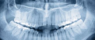
Before removing the growth, an x-ray of the jaw is taken.
Bone spines in the mouth on the right or left are often discovered by chance during a dental examination. The main method of differential diagnosis is x-ray and study of the medical history.
Exostosis of the jaw resolves on its own in 50% of cases. This happens when you change your diet, eliminate vitamin deficiency and disrupt salt metabolism in the body.
If the growth on the jaw grows, begins to hurt, and interferes with chewing and talking, it is surgically removed in the clinic. Excision of the spine is also carried out before prosthetics. Contraindications – disorders of the endocrine system, adrenal glands, diabetes, poor blood clotting. There are no medications or folk remedies for removing osteophytes.
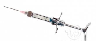
The operation is simple and is performed under local anesthesia. After treating the oral cavity with an antiseptic, a small incision is made on the gum, the base of the growth is cut off with a laser or bitten with dental forceps.
Using a special attachment on a drill, smooth out all sharp corners on the bone. Stitches and wound healing ointment are applied.
Treatment at home
After removal of exostosis, it is necessary to maintain oral hygiene.
Further treatment takes place at home. Before the wound heals completely, you need to provide the oral cavity with proper care. Every day, rinse your mouth with Miramistin, Chlorhexidine or soda solution. Procedures cannot be performed on the first day after surgery. After a day, passive rinsing is indicated - take the solution into your mouth, hold for 5-7 minutes without performing active actions. Hydrogen peroxide and potassium permanganate should not be used for mouth rinsing.
Cotton pads soaked in Levomekol or Solcoseryl ointment should be applied to the incision. Sometimes doctors prescribe antibiotics to prevent bacterial infections from developing. If the immune system is functioning normally, the recovery period is 7 days.
You can also rinse your mouth with folk remedies based on herbs with antiseptic and anti-inflammatory effects:
- a collection of equal parts of sage and mint;
- a mixture of lemon balm and oak bark;
- oregano and chamomile;
- nettle and birch;
- Golden mustache;
- eucalyptus;
- soda and salt - dissolve 1 tsp each in 250 ml of warm water.
To prepare any herbal infusion, pour 2 tsp. raw materials 200 ml of water at room temperature. Simmer in a water bath for 20 minutes. Strain and use warm.
To prevent suture dehiscence, you can only eat soft pureed food, soups and broths for 2 weeks after surgery. Avoid hard and viscous foods, hot and cold foods, smoking and alcohol.
Diagnosis and treatment of exostosis at NEARMEDIC
At the beginning of the development of pathology, the disease is usually asymptomatic. But as the growth increases, uncomfortable pain may occur and dense formations may be visualized. Due to significant deformation of the bones, physical changes also appear.
Diagnosis of exostosis
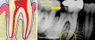
In some places, neoplasms can be felt under the skin, and the doctor can determine their presence already at the first appointment. To confirm the diagnosis, an x-ray examination is prescribed. As a rule, in the picture the size of the growth is several millimeters smaller than in reality, since the cartilage tissue is not visible.
In some cases, a specialist will prescribe a biopsy, especially when the formation rapidly increases in size. To exclude the possibility of malignant degeneration of cells, tissue biosamples are collected and sent to NEARMEDIC’s own laboratory for further cytological examination.
Treatment of the disease
The most effective treatment for exostosis is surgery. It consists of deep resection (excision) of the growth, capturing part of the healthy bone. Surgery is performed under local anesthesia or general anesthesia, depending on the location and size of the exostosis.
After removal of cartilaginous exostosis, a lasting recovery occurs.
The use of modern medical equipment, the application of cosmetic internal sutures, and minimal tissue trauma during surgery at NEARMEDIC allows the patient to return to their normal lifestyle in the shortest possible time.
How dangerous are growths on the gums?
A large growth can cause erosion of soft tissues in the mouth.
Due to the absence of pain, elevated temperature, and signs of inflammation, patients consider exostosis to be a harmless disease. But growths can provoke the development of complications.
- As the osteophyte increases, the tongue does not have enough room to maneuver, problems arise with the pronunciation of certain sounds, and the person begins to whistle.
- Due to rubbing of the inner surface of the cheek against the thorn, erosions are formed that do not heal for a long time, phlegmon, abscesses.
- Some osteophytes grow constantly, reaching the size of a chicken egg.
- The functions of the jaw joint are impaired.
- In advanced cases, there is a constant aching pain in the jaw.
A sharp bony protrusion on the upper or lower jaw interferes with prosthetics and leads to permanent destruction of fillings. If the neoplasm passes upward through the cartilaginous plates, chronic rhinitis and sinusitis develop.
There are no specific methods for preventing exostosis. People who have had jaw injuries need to visit the orthodontist 2-3 times a year. It is necessary to regularly examine the mucous membranes of the oral cavity, check the integrity and elasticity of the gums. You should not forget about the rules of dental care, use high-quality toothbrushes and toothpastes.