Author: Frank Netter
Intraoral anesthesia ensures painless dental procedures. A variety of techniques have been developed, all of which require a thorough understanding of the anatomy of the head and neck to maximize the success of the application and minimize possible complications. Injections should not be given in infected areas or in areas of inflammation, and the use of topical anesthesia will help reduce the pain caused by the needle.
Classification
• Local anesthesia (infiltration)
• Conduction anesthesia
Embryology
Rice.
1. Frontal section of the head of a human embryo (according to L.I. Falin): a - seven-week-old embryo (1 - nasal septum, 2 - tongue, 3 - palatine process of the upper jaw, 4 - ventral cartilage of the lower jaw); b — embryo of eight weeks (1 — nasal septum, 2 — palatine processes of the upper jaw have taken a horizontal position and are located above the tongue, 3 — the tongue has moved down, 4 — ventral cartilage). The formation of the palate begins at the 6-7th week of intrauterine development with the formation of lamellar projections - palatine processes - on the inner surface of the maxillary processes (see Face).
The latter are initially directed downward, later taking a horizontal position (Fig. 1, a, b). At the end of the 8th week. During intrauterine development, the edges of the palatine processes fuse with each other and with the nasal septum. Fusion begins from the anterior parts of the palatine processes and gradually spreads posteriorly. In the posterior part of the oral cavity, the palatoglossal and velopharyngeal arches are formed from the palatine processes.
Anatomy
The palate is divided into the anterior section - the hard palate (palatum durum) and the posterior section - the soft palate (palatum molle).
Solid sky
It is represented by the bony palate (palatum osseum), covered with a mucous membrane with a submucous base, the severity of the cut in different parts of the solid N. is different. The bony palate is formed by the palatine processes of the upper jaws (processus palatinus maxillae) and the horizontal plates of the palatine bones (laminae horizontales ossis palatini). The right and left halves of the bone N. are connected by a median palatine suture (sutura palatina mediana), along which a palatine ridge (torus palatinus) protruding towards the oral cavity often runs. At the anterior end of this suture there is an incisive fossa (fossa incisiva), into which the incisive canal (canalis incisivus) opens. In the posterolateral areas of the bone N., at the junction of the upper jaw and palatine bone, a large palatine opening (foramen palatinum majus) is formed. In the horizontal plate of the palatine bone next to the greater one there are small palatine foramina (foramina palatina minora). All openings lead into the greater palatine canal and further into the pterygopalatine fossa (see). The palatine grooves (sulci palatini), separated by the palatine spines (spinae palatinae), run forward from the greater palatine foramen.
Along the midline of the mucous membrane of the hard N. there is a suture of the palate (raphe palati), on which behind the incisors, corresponding to the incisive fossa, is the incisive papilla (papilla incisiva). On the sides of the anterior section of the suture there are transverse palatine folds (plicae palatinae transversae), more pronounced in children.
The submucosa is present in the lateral sections of the N., on the border with the soft N.; in the area of the suture and at the transition of the N. mucous membrane to the gum, it is absent. In the anterior sections of the N., the submucosa contains a small amount of adipose tissue, penetrated by thick tufts of dense fibrous connective tissue, between which vessels and nerves pass. In the posterior sections of the solid N., this layer is occupied by the mucous palatine glands. The shape of the bone N. is related to the shape of the skull and face.
Rice. 2. Schematic representation of the muscles of the soft palate: 1 - muscle that strains the velum palatine, 2 - muscle that lifts the velum palatine, 3 - hook of the pterygoid process, 4 - palatoglossus muscle, 5 - uvula muscle, 6 - velopharyngeal muscle.
Soft sky
It is represented by the palatine aponeurosis, into which the muscles of the soft palate and pharynx are woven. With calm breathing and muscle relaxation, the soft palate hangs vertically, forming the so-called. palatine curtain (velum palatinum). In the middle of its rear edge there is a protrusion - a tongue (uvula). Soft N. includes the following muscles (Fig. 2): the muscle that strains the velum palatini (m. tensor veli palatini), the muscle that lifts the velum palatini (m. levator veli palatini), and the muscle of the uvula (m. uvulae). The terminal parts of the palatoglossus muscle (m. palatoglossus) and the velopharyngeal muscle (m. palatopharyngeus) are woven into the soft N. The muscle that strains the velum palatine is a pair, it begins with wide muscle bundles from the spine of the sphenoid bone (spina ossis sphenoidalis), from the membranous part of the Eustachian (auditory, T.) tube (tuba auditiva), from the scaphoid fossa (fossa scaphoidea) and the medial plate of the pterygoid process (lamina med. processus pterygoidei). The muscle bundles, converging, descend vertically downwards, the resulting tendon is thrown over the pterygoid hook (hamulus pterygoideus). Then, taking a horizontal direction, these tendon bundles, together with the tendon bundles of the opposite side, form the palatine aponeurosis, which is attached to the posterior edge of the hard N.
The muscle that lifts the velum palatine, also paired, starts from the lower surface of the pyramid of the temporal bone, anterior and medial from the external opening of the carotid canal (canalis caroticus) and the cartilaginous part of the Eustachian tube; approaching the midline, it intertwines with bundles of the muscle of the same name on the opposite side.
The uvula muscle is a paired muscle that starts from the N. aponeurosis and ends at the tip of the uvula; shortens and raises the tongue. The palatine muscle is a continuation of part of the bundles of the transverse muscle of the tongue (m. transversus linguae), at the root of the tongue it rises along the posterior part of the side wall of the mouth and is woven into the soft palate; the muscle forms the thickness of the palatine arch (areus palatoglossus), when contracted, it lowers the palatine curtain and reduces the diameter of the pharynx.
The velopharyngeal muscle is a steam muscle, located in the lateral wall of the pharynx, starting from the posterior wall of the pharynx and the thyroid cartilage of the larynx and, moving upward, is woven into the lateral sections of the velum palatine. The muscle forms the velopharyngeal arch (areus palatopharyngeus) and, when contracted, lowers and pulls back the palatine curtain and narrows the pharynx. Between the arches are the palatine tonsils (see).
Soft N. is covered with a mucous membrane, which has a submucosa containing mucous and mucoserous glands.
Rice. 3. Schematic representation of the blood supply and innervation of the palate: 1 - incisive canal, 2 - nasopalatine nerve, 3 - greater palatine nerve, 4 - greater palatine foramen, 5 - lesser palatine foramina, 6 and 7 - branches of the lesser palatine nerve, 8 - palatine tonsils , 9 - small palatine arteries, 10 - greater palatine artery, 11 - anastomosis with the incisive artery.
Blood supply
the palate (Fig. 3) is carried out by the maxillary artery (a. maxillaris) and the facial artery (a. facialis). The descending palatine artery (a. palatina descendens) departs from the maxillary artery, and from it to the hard nerve through the greater palatine foramen - the greater palatine artery (a. palatina major). This artery lies in the groove at the site of the transition of the hard N. to the base of the alveolar process, gives off branches to the mucous membrane of the hard N., and its terminal branches anastomose with the incisive artery (a. incisiva), emerging from the incisive canal. The incisive artery is terminal. It is formed from the posterior nasal lateral and septal arteries of the nose (aa. nasales post, laterales et septi), extending from the maxillary artery.
In addition, the small palatine arteries (aa. palatinae minores)—branches of the descending palatine artery—exit the hard palatine from the small palatine foramina, located posterior to the greater palatine foramen. The soft N. is supplied with blood through the ascending palatine artery (a. palatina ascendens), which extends from the facial artery.
Venous outflow occurs through the palatine vein (vena palatina), which originates in the thickness of the soft tissue, passes through the bed of the palatine tonsil and most often flows into the facial vein. Other veins drain into the pharyngeal venous plexus.
Innervation
carried out by the second branch of the trigeminal nerve due to the greater palatine nerve (n. palatinus major), emerging from the greater palatine foramen, and the small palatine nerves (nn. palatini minores), emerging through the small palatine foramina, as well as the nasopalatine nerve (n. nasopalatinus), exiting through the incisive foramen. The motor innervation of the soft nerve is carried out by the branches of the IX and X pairs of cranial nerves. The muscle that strains the velum palatine is innervated by the mandibular nerve (n. mandibularis).
Lymphatic drainage
occurs to the deep cervical lymph nodes (nodi lymphatici cervicales profundi), retropharyngeal nodes (nodi lymphatici retropharyngei), as well as to the submandibular lymph nodes (nodi lymphatici submandibulares).
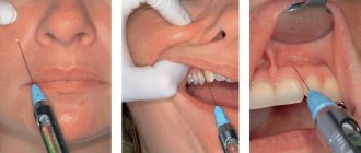
Technique of palatal (palatal) conduction anesthesia (injection site and needle direction)
The patient, with his head raised, fixed on the headrest when he is sitting in a chair, or on a bolster when he is lying on the operating table, opens his mouth wide. In one way or another, the location of the greater palatine foramen is determined and an injection is made approximately 10 mm in front of it. The need to inject at a distance of 10 mm anterior to the greater palatine foramen is due to the fact that it is impossible to inject the needle vertically in the palate (the lower jaw gets in the way), but you need to direct the needle obliquely from the front and from bottom to back and up. Only by injecting at the specified distance can the target be easily achieved. It should be noted that using such an injection site is very convenient, since it is there, 10 mm in front of the greater palatine foramen, that there is a fairly clearly visible retraction, which can be used to determine the injection site for a given anesthesia.
Thus, if there are teeth, we make an injection medially from the middle of the alveolus of the upper second molar, when there is already an upper third molar, and medially from the alveolus of the upper first molar in the absence of a wisdom tooth (the distance from the middle of the alveolus of one molar to the middle of the alveolus of the other molar is approximately 10 mm). If there are no upper molars, we inject 15 mm in front of the posterior edge of the hard palate, since the large palatine foramen, as we already know, is 5 mm in front of the posterior edge of the hard palate, and we need to inject a needle 10 mm in front of this hole. Directing the needle obliquely from the front and from bottom to back and up, reaching contact with the bone, 1/3 ml of anesthetic solution is released, after which anesthesia occurs.
We recommend releasing the numbing solution only when the needle clearly hits the bone. Sicher does not consider it necessary to stick the needle all the way to the bone on the sole grounds that the tissue covering the palate in the place intended for injection is very loose and easily permeable to the solution, so that the latter, if released at some distance from the bone, will still soon reach before it and will get on the nerve. This is of course true. But precisely because the tissue here is very loose, it is possible for the anesthetic solution to spread not only deeper, but also to the sides, especially back - towards the soft palate, and this can lead to temporary unpleasant sensations for the patient. As is known, behind the anterior palatine nerve are the middle and posterior palatine nerves, which give the soft palate not only sensory, but also motor branches.
Histology
The mucous membrane of solid H. is covered with stratified squamous keratinizing epithelium. In the epithelial layer, the basal, spinous, granular and stratum corneum are clearly distinguished. The stratum corneum is formed by several rows of completely keratinized cells (without nuclei). Glycogen is not normally detected in the epithelium of solid N., but it can accumulate here when the process of keratinization is weakened (for example, when wearing laminar dentures for a long time). The basal and spinous layers are characterized by high activity of redox enzymes. The connective tissue basis of the mucous membrane of solid N. consists of fairly dense connective tissue; Some of the bundles of its collagen fibers are directly woven into the periosteum of the palatine bones, especially in those areas where there is no submucosa, due to which the mucous membrane is tightly fixed to the bone. In the area of the palatal suture and when the N. passes into the gum, the submucosa is absent; throughout the rest of the solid N., a clearly defined submucosa is revealed in the mucous membrane. In the anterior section of the N., on the sides of the palatine suture, the submucosa is represented by an accumulation of adipose tissue, and in the posterior section by an accumulation of small mucous glands.
The mucous membrane of the anterior surface of soft N. is covered with multilayered squamous non-keratinizing epithelium. The cells of the spinous layer of the epithelium contain a large amount of glycogen; They are also characterized by high activity of enzyme systems. The lamina propria of the mucous membrane consists of relatively thin interwoven bundles of collagen fibers; At the border with the submucosa there is a massive layer of elastic fibers. The submucosa is represented by loose connective tissue, which contains the end sections of small mucous glands. The posterior surface of the soft N. is covered with multirow ciliated epithelium, characteristic of the respiratory tract. Both surfaces of the uvula in adults are covered with stratified squamous non-keratinizing epithelium, rich in glycogen. In newborns, on the posterior surface of the uvula there is a multi-row ciliated epithelium, which is replaced by multi-layered epithelium during the first month of life.
Pathology
Developmental defects.
The most common of these is congenital cleft of the upper lip (outdated name “cleft palate”), often in combination with a congenital cleft of the upper lip. Congenital underdevelopment of the soft N. or uvula is also observed. According to M.D. Dubov (1960), per 1000 newborns, at least one is born with a cleft lip or cleft. The causes of congenital facial clefts, including the palate, are not well understood; Various assumptions have been made about the influence of unfavorable factors on the development of the fetus during the period of facial formation.
In the USSR, according to the accepted classification of malformations of the N., proposed by M. D. Dubov, clefts of the N. are divided into two main groups: through clefts passing through the alveolar process, hard and soft N., and non-through clefts of the N., with The loose alveolar process is developed normally.
Rice. 4. Oral cavity of a child with a cleft palate: a - through bilateral cleft passing through the lip, alveolar process of the upper jaw and palate; b - through left-sided: cleft.
Through clefts are unilateral (to the right or left of the midline) and bilateral (Fig. 4, a, b), when the connection of the premaxillary bone with the nasal septum and maxillary bones is absent on both sides. With a unilateral cleft, the nasal septum and premaxillary bone are connected to the palatine plates on only one side. With bilateral through clefts of the N. and upper lip, the premaxillary bone moves forward, which complicates surgical treatment.
Non-through clefts of the N. are divided into complete (the apex of the cleft begins at the alveolar process and passes through the hard and soft N.) and partial clefts (the cleft of the soft and part of the hard N.). Partial ones include hidden, or submucosal, clefts, in which the cleft of the muscles of the soft N. or the cleft of the uvula, and sometimes part of the hard N. is covered with a mucous membrane.
With N.'s clefts, especially with through ones, the respiratory and nutritional functions of newborns are sharply impaired; when sucking, part of the milk is poured out through the nasal passages, it is aspirated into the respiratory tract, and nasal breathing is disrupted (with this malformation, there is a high mortality rate in newborns). With age, children with cleft N. experience speech disorders—dysarthria (see) and nasality (see), in which children become withdrawn and lag behind in their studies. The development of the upper jaw is often disrupted - narrowing of the upper dental arch, changing the shape of the face, retraction of the upper lip, etc. As a rule, due to the lack of a normal muscular apparatus, an expansion of the middle part of the nasopharynx is formed.
Treatment of the cleft is surgical. If surgery for a lip defect is indicated in early childhood (see Lips), then it is recommended to begin surgery for cleft lip at the age of 4-7 years. Ensuring proper nutrition and breathing is achieved by using devices for separating the oral cavity and nose - obturators (see Obturators). Children with N. clefts are under medical supervision from a number of specialists: pediatrician, dentist, otolaryngologist, speech therapist. The prognosis for N. clefts, especially through ones, in newborns is not always favorable; high mortality is observed.
A malformation is also a narrow high N.—hypsistaphylia; It is believed that this defect occurs as a result of mouth breathing with hypertrophy of the pharyngeal tonsil (see Adenoids). Treatment is carried out using orthodontic methods (see Orthodontic methods of treatment).
In the absence of positive results, surgical treatment is possible, usually ending successfully.
Sometimes there is congenital isolated underdevelopment of the soft N., mainly the uvula, as well as the palatine arches, which negatively affects the act of swallowing, and subsequently the pronunciation of certain sounds. Surgical treatment is lengthening of the soft N. (staphyloplasty). The results are favorable.
In adults, an impacted tooth may be found in the area of the transition of the alveolar process into the palatine process of the upper jaw. Surgical treatment: removal of an unerupted tooth using a chisel.
Damage
. In everyday conditions, N. can be injured by sharp objects (fork, bone, pencil, etc.). Treatment consists of suturing the wound of soft N.
Burns are often observed - hot food or chemicals. substances, but they do not reach a large extent.
Treatment: antiseptic and protein rinses.
Gunshot wounds of N., as a rule, occur in combination with wounds of the nasal cavity, maxillary sinus, and upper jaw. Surgical treatment of the wound of the N. is carried out with sutures on the detached flaps of the mucous membrane of the hard N. and on the soft N. To protect the surgical field and maintain the dressing, an individual protective plate is made of quick-hardening plastic.
In the vast majority of cases, outcomes for N. injuries are favorable. Staged treatment - see Face.
Diseases
.
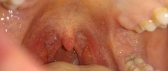
The mucous membrane of N. is usually affected by stomatitis (see). In newborns and weakened children of the first year of life, so-called N. may be observed. aphthae of newborns (see Aphthae), as well as thrush (see Candidiasis). Oral candidiasis often develops in older people, especially those who wear dentures. The mucous membrane of soft N. is involved in the pathological process in scarlet fever, measles, especially in diphtheria. Inflammatory infiltrate and swelling of the soft tissue often accompany tonsillitis and phlegmon of the pterygomaxillary and peripharyngeal space. Rice.
5. The oral cavity of a patient with an abscess of the hard palate: a - abscess (indicated by an arrow); b - plaster model of the palate (the dotted line shows the cut line of the abscess). The source of the purulent process in the area of hard N. is usually an infection emanating from the upper lateral incisors or first upper premolars; less commonly, the inflammatory process is associated with periodontitis of the palatal roots of molars. Pus usually accumulates under the periosteum, forming a hard N. abscess (Fig. 5, a and b). The mucous membrane in this area becomes hyperemic. Swelling and hyperemia sometimes spread to soft N. Pain is noted, eating is difficult, and body temperature rises. Fluctuation is determined 2-3 days after the onset of the disease. With a periosteal abscess, due to the detachment of soft tissue from the bone within the abscess, necrosis of the bone tissue may form.
More often, the purulent process in the area of the hard N. is purulent periostitis (see) or osteomyelitis (see) of the palatine process of the upper jaw; when making a diagnosis, it is necessary to differentiate it from an abscess due to periodontal disease (see), with a dental cyst (see), emanating from the apex of the root of the second incisor. Surgical treatment: an incision is made to the bone along the N. parallel to the alveolar edge. It is advisable to excise a small triangular section of the mucous membrane along with the periosteum for a more reliable drainage of pus and to prevent bone necrosis.
In cases of severe diphtheria or with damage to the vagus nerve, paralysis of the soft muscles of the N. may develop.
Tuberculosis of the mucous membrane of N., as well as its other localization in the oral cavity, is observed with active pulmonary tuberculosis. Small infiltrates or small tubercles of gray-yellow color appear on the mucous membrane. They can disintegrate, resulting in the formation of superficial (less often deep) ulcerations of irregular shape, with undermined edges; their bottom is covered with small flaccid pink-yellowish granulations or a grayish purulent coating, surrounded by miliary tubercles. Ulcerations are significantly painful. At the same time, damage to the submandibular or submental lymph nodes is observed. Anti-tuberculosis treatment (see Tuberculosis).
Hard chancre, or primary syphiloma, localized on soft N., has the appearance of a limited superficial ulcer. In the secondary period of syphilis, the mucous membrane is affected, tubercles appear, located focally in the form of a semiring. The mucous membrane thickens and turns red. Tuberous syphilide of the mucous membrane can resolve, leaving tender scars, or form ulcerations of irregular shape, the bottom of which is covered with gray disintegrated tissue.
Rice. 6. Oral cavity of a patient with tertiary syphilis: a perforation is visible on the hard palate (indicated by an arrow), formed as a result of the disintegration of the gumma.
The development of gumma is rare. With gumma, a diffuse, dense, slightly painful swelling with blurred boundaries is determined in the periosteum; the mucous membrane is swollen, hyperemic, and sometimes there is intense night pain. Subsequently, the swelling increases in diameter to 3-4 cm or more, gradually softens and opens into the oral cavity. In some cases, perforation of the solid N. may occur (Fig. 6). With the development of gumma in the thickness of bone tissue (gummy osteomyelitis), extensive bone destruction is often observed. There is severe pain and impaired sensitivity in the area innervated by the nasopalatine nerve. Often a communication is formed between the oral cavity and the nasal cavity or maxillary sinus. When healing occurs, radiant scars remain on the N.
The results of serol tests are important for diagnosis. The main one is general antisyphilitic treatment (see Syphilis). Surgical intervention to close the bone defect is indicated only after general treatment of syphilis.
Actinomycosis can sometimes develop under the mucous membrane on the alveolar process of the upper jaw. In this case, the infection usually spreads from the inflammatory-changed area of the mucous membrane, the edges in some cases forming a canopy over the not completely erupted upper wisdom tooth (so-called pericoronitis). A persistent inflammatory infiltrate is formed. The course, diagnosis and treatment are the same as for other localizations of actinomycosis in the maxillofacial region (see Actinomycosis). In the vast majority of cases, N.'s diseases (with the exception of untreated syphilis) end successfully.
Tumors
. In the area of hard and soft N., benign and malignant neoplasms are observed, emanating from soft tissues, and in some cases growing from the bone tissue of the alveolar and palatine processes of the upper jaw, maxillary sinus, nasal cavity, and nasopharynx. Sometimes tumors developing from the soft tissues of N. cause secondary changes in bone tissue (usures) or grow into the bone.
Fibroma of hard and soft N. usually protrudes above the surface; sometimes it, like a polyp, is located on a short and thick stalk. When wearing a plate denture, this neoplasm may have a flattened shape.
In the area of hard and soft N., especially on the uvula, cavernous hemangioma (see) and lymphangioma (see) are found, neurofibroma (see) is rare, neuroma (see) is even more rare.
Papilloma is observed relatively often; usually it is localized on the uvula, palatine arches, and less often on the hard palate. Often papilloma is multiple.
In the area where the mucous (small serous) glands are located, benign tumors develop - adenoma (see), adenolymphoma (see), mixed tumors and malignant ones (mucoeidermoid, cylindroma, sometimes glandular cancer). As they grow, neoplasms can cause thinning of bone tissue, and malignant ones can destroy bone, growing into the maxillary sinus and nasal cavity.
Sarcoma (see) usually grows from the upper jaw, nasopharynx (primary ones are extremely rare). Primary cancer (see), emanating from the epithelium of the mucous membrane of the hard N., is also observed extremely rarely. Its occurrence in some patients is preceded by leukoplakia (see). In most cases, cancer affects N. secondarily (from the maxillary sinus, nasopharynx). These tumors have common wedges and signs (swelling, density, injection of vessels of the mucous membrane above the tumor), so they are difficult to differentiate. Cancer is characterized by rapid ulceration and density of the edges of the ulcer.
Adamantinoma (see), osteoblastoclastoma (see) sometimes develop from the alveolar process of the upper jaw, and chondroma (see), osteochondroma, osteoma (see) from the palatine bone. they manifest themselves as bulges of varying sizes.
Diagnosis of many tumors is often possible only with rapid press biopsy during surgery. X-ray examination of malignant neoplasms reveals foci of clearing of bone tissue with unclear boundaries. Benign tumors are enucleated, and sutures are placed on the N. in the longitudinal direction. For malignant tumors, the tumor is excised within healthy tissue with resection of adjacent areas of bone; For cancer, combination treatment is more often used. The prognosis for malignant tumors is unfavorable.
Acquired defects arise as a result of gunshot wounds, trauma, after surgery for congenital clefts and malignant tumors of the upper jaw. Relatively rarely, defects are observed after gummous syphilis. Acquired defects are characterized by the presence of significant cicatricial changes along the edges, varied shape and localization. After uranostaphyloplasty, a significant number of unsuccessful outcomes are observed - residual defects of the N. and shortening of the soft N. Treatment is surgical. Its results are satisfactory.
Complications of palatal anesthesia
- Paresis of the soft palate. If the anesthetic solution gets into this area, temporary paresis of the soft palate may occur. The patient in this case complains of a feeling of some kind of plaque on the soft palate, the presence of a foreign body in this place, as with a sore throat. Paresis of the soft palate in some patients causes coughing and vomiting, which sometimes ends in vomiting when the stomach is full. If you bring the needle close to the bone, the anesthetic solution enters the less loose tissue and cannot spread back so easily. It should also be noted that paresis of the soft palate can occur not only in cases where the needle is not brought to the bone, but also under the following circumstances: firstly, when the injection site or the direction of the needle is taken incorrectly (more medially and backward); secondly, when too much solution is released during this anesthesia. It must be remembered that the limit here is 1/2 ml. Usually they limit themselves to an even smaller amount of anesthetic solution.
Paresis of the soft palate due to the entry of an anesthetic solution into the motor nerves of his muscles does not pose anything dangerous, since it passes quite quickly without any trace.To eliminate these unpleasant sensations and prevent vomiting, the patient is given a little water to drink. Sometimes these unpleasant sensations go away after several deep breaths of fresh air.
- Vascular injury. This complication occurs here quite often, but due to the absence of any more or less large vascular branches in this area, it is not of particular importance. Vascular injury occurs here more often than with conductive anesthetic injections in other places, because near the greater palatine foramen there is a dense network of vessels, one of which can easily be penetrated by a needle. A vessel injury is recognized by slight bleeding from the injection site after removal of the needle. This bleeding stops quite quickly on its own or is stopped by pressing the injection site with a gauze pad.
- The appearance of ischemic (blanched) areas of facial skin. Sometimes the anesthetic solution during palatal conduction anesthesia through the pterygopalatine canal enters the pterygopalatine fossa and acts on one of the vessels lying in it, which, as is known, are connected with the vessels of the facial skin; As a result, temporary ischemia appears over a fairly large area of the face.
An anesthetic solution can affect the vessels of the pterygopalatine fossa through the pterygopalatine canal not only when it penetrates into the bloodstream due to injury to a blood or lymphatic palatine vessel. The anesthetic solution can leak between these vessels up to the pterygopalatine fossa without stopping them. The possibility of penetration of the anesthetic solution through the pterygopalatine canal was confirmed by our experiments on corpses using the injection of colored gelatin at the greater palatine foramen. After opening and dissecting the pterygopalatine canal, we found threads of colored gelatin in the canal itself between the vessels and nerves.Adrenaline contained in the anesthetic solution, penetrating one way or another into the pterygopalatine fossa, can cause contraction of a large vessel that gives branches to the skin of the face. Hence the ischemia of the facial skin area. By the contours and location of the area of ischemia on the face, one can judge which vessel was exposed to adrenaline.
Ischemia of areas of the facial skin occurs either immediately after injection and even at the beginning of it, when it is associated with vascular spasm due to contact of the needle with the vessel, or after 1-2 minutes after injection, when it occurs as a result of the vasoconstrictor effect of the adrenaline contained in the anesthetic solution .
Ischemia in certain areas of the skin of the face does not last long, especially when it is associated with a spasm of the vessel from the touch of a needle, and most often completely disappears after approximately ½ - ¾ hours. In rare cases, blanching of skin areas remains for 2-3 hours.
The sensitivity of pale areas of skin decreases very slightly. Most often, ischemia with this anesthesia covers the corner of the mouth, half of the upper lip and nose, the front of the cheek, the inner corner of the eye and the infraorbital region on the corresponding side. More extensive areas of facial skin ischemia are also observed, including the supraorbital region. In the first case, we are apparently dealing with the entry of an anesthetic solution into a vessel when it is wounded, and in the second, with the leakage of the solution between the walls of the vessels without wounding them. It must be assumed that the anesthetic solution, seeping between the vessels without injuring any of them, can narrow several vessels, affecting their walls, and as a result cause ischemia of a larger area of facial skin. However, even with infiltration anesthesia on the palate, even in the area of premolars and canines, ischemia is possible in large areas of the facial skin.
It should only be added that, since blanching of an area of facial skin is usually caused by injury to a vessel and the entry of an anesthetic solution into it, sometimes when post-injection ischemia appears in a particular area of facial skin, other complications are observed (the harmful effect of the anesthetic solution on the body and the lack of anesthesia). These complications will be discussed in detail below.
Let us note, by the way, that with this injection there is no need not only to enter the pterygopalatine canal with a needle, but even to go very close to the opening itself: we do not need to interrupt the conduction of the nerve in the part lying in the canal, but it is enough to wash the nerve with the solution immediately upon exit it from the greater palatine foramen. To relieve pain from surgical interventions in the maxillofacial area, as already mentioned, we use the perineural method of conduction anesthesia; The solution reaches the nerve by diffusion. Knowing this, when injecting at the greater palatine foramen, we try to release the solution in front of the opening. Anesthesia of the anterior palatine nerve will also occur, but the danger of paresis of the soft palate and injury to the vessel is reduced (the latter is explained by the fact that the closer to the front, the smaller the vessels and are less likely to be injured).