Home » All about modern dentistry » Pain relief in dentistry » Pain relief in the upper jaw For various operations on the upper jaw, dentists prefer to use infiltration anesthesia. This method is most convenient because the thin layer of the compact lamina of the alveolar process of the upper jaw is porous. If you use modern painkillers that have a high diffusion ability, then with this method of their administration the desired anesthetic effect is achieved.
Impregnation of tissues and alveolar processes with an anesthetic is carried out by injection, injecting the solution under the mucous membrane at an angle of 40-45o in the projection of the upper areas of the dental roots along the transitional fold of the vestibule of the oral cavity in the area that is supposed to be operated on. If this area is too large, the needle is moved along the transitional fold and the anesthetic solution is slowly injected. Because rapid administration of the substance can cause pain in the patient.
To anesthetize the palatal tissues, an injection of an anesthetic drug is made into the angle formed by the palatine and alveolar jaw processes, at a distance of 10-15 mm from the edge of the gum.
Regional (conduction) anesthesia is used quite rarely; it includes anesthesia on the tubercle of the upper jaw, in the area of the greater palatine, incisive and infraorbital foramen.
Intraoral anesthesia
The patient's mouth is in a half-open state, the doctor inserts a mirror into the oral cavity, moving the cheek to the side. This ensures an excellent overview of the arch of the vestibule of the mouth, as well as tension in the mucous transitional fold near the molars. The needle is inserted into the mucosa above the projection of the apexes of the teeth at the level of the 2nd and 3rd molars. If there are no molars, then the needle should be behind the zygomaticalveolar ridge and move up, back and inward. The insertion angle should be 45°. It is necessary to control that the needle always moves along the bone with the beveled surface of its tip. During this movement, the anesthetic should be gradually injected. This method will help prevent injuries to the vessels of the pterygoid plexus. Having deepened the needle by 2-2.5 cm, the anesthetic solution is deposited. Thus, the molars located on the side of the vestibule of the mouth, the mucous membrane, periosteum and the outer posterior bone wall of the maxillary sinus are perfectly anesthetized.
Non-injection methods
For short, small-scale interventions, it is sufficient to superficially numb the soft tissues; this can be achieved using non-injection methods of local anesthesia in dentistry. These include:
- Application method . Applying a special gel or spray containing a certain anesthetic in a small concentration to the desired area of the oral mucosa. This method is often used in pediatric dentistry in the treatment of baby teeth, when treating gingival margins, opening small superficial abscesses, and also to prepare the patient for injection-type local anesthesia (to numb the injection site).
- Physico-chemical method . Injection of the required amount of anesthetic into soft tissues using electrophoresis. It is usually used for trigeminal neuralgia.
- Physical method (rarely used in recent years). Anesthesia of a specific area by applying a freezing reagent (chloroethyl), exposure to a laser beam or electromagnetic waves of a certain frequency.
All other techniques for performing local anesthesia in dentistry belong to the injection group, that is, they require an injection in a certain area of the oral cavity or even outside it.
Extraoral anesthesia
Having made a puncture with a needle in the area of the lower anterior corner of the cheek bone, the needle is then directed upward at an angle of 45° and again towards the tubercle of the upper jaw, bringing it to the bone. And then the anesthetic solution is deposited. The anesthetic effect occurs in approximately the same time as with the intraoral method of anesthesia.
However, during the process of anesthesia, a hematoma appears on the tubercle of the upper jaw, which is a consequence of injury to the veins of the pterygopalatine plexus by the needle.
Computed tomography studies of the path of solution propagation during tuberal anesthesia confirmed that such complications are most likely.
This tomogram, which was taken 5 days after intraoral anesthesia, shows that there are contours of a hematoma on the tubercle of the upper jaw in the pterygopalatine fossa. Approximately 40-60% is allocated to the fact that this cellular formation will fester and turn into phlegmon.
When using the tuberal method of anesthesia, injury to the veins of the pterygopalatine plexus is almost impossible to avoid, and there is also a fairly high risk of complications, in particular if the intraoral method is used. All this threatens the health and even the life of the patient. That is why it is recommended to use this type of anesthesia extremely rarely.
Conductor technology
Conduction local anesthesia in dentistry is used when infiltration anesthesia is ineffective (for example, if volumetric manipulations on the lower jaw are to be performed), when several teeth need to be treated simultaneously, or a long-term intervention is expected. In this case, the anesthetic solution is supplied to the branches of the nerve innervating the area of the upcoming intervention. The drug can be administered endoneurally (directly into the nerve, used rarely and for special indications) or perineurally (next to the nerve, so that the solution gradually permeates the nerve fibers, the most commonly used method of administration). There are also extraoral and intraoral methods of performing the injection - the doctor decides which method to choose in a particular case.
Depending on the site of administration of the anesthetic drug and the branches of the nerves that are to be blocked, the following types of conduction local anesthesia in dentistry are distinguished:
- Infraorbital – anesthesia of the branches of the infraorbital nerve.
- Palatine – blocking impulses from the greater palatine nerve.
- Tuberal - blocking impulses from the superior posterior alveolar nerves.
- Mental, or mental – anesthesia of the mental nerve.
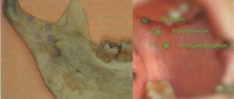
- Mandibular - blocking impulses of the lower alveolar and lingual nerves in the lower jaw. One of the varieties is considered to be torsal anesthesia (blocking the branches of the lingual, inferior alveolar and buccal nerves).
- Incisive – blocking impulses from the nasopalatine nerve passing in the incisive canal.
Typically, the conduction technique requires preliminary preparation of the patient for local anesthesia - applying the anesthetic by application method to numb the injection site, as well as explaining to the patient how best to position his head for this or that type of administration of the anesthetic drug.
Anesthesia of the infraorbital nerve
The purpose of this anesthesia is to block the branches of the inferior orbital nerve, which forms the lesser “crow's foot” in the area of its exit from the bony canal, as well as the middle and superior anterior alveolar branches.
The result of such anesthesia, performed at the lower orbital foramen, is pain relief:
- — Reztsov,
- - Fang,
- — Premolars,
- — The gums adjacent to the premolars in the area of the vestibule of the mouth,
- — Bone tissue of the alveolar process and nasal septum,
- — Shells of the mucous and bone structures of the walls of the maxillary sinus,
- - Skin of the infraorbital region of the lower eyelid and wing of the nose,
- — Mucous membrane and skin of the upper lip.
The projection of the infraorbital foramen, which is oriented towards during an anesthetic injection, is located 5 mm below the edge of the orbit, which corresponds to the axis drawn through the center of the eye pupil when the eye looks forward.
Indications for conduction anesthesia
- Implantation.
- Carrying out surgical intervention (removal of cysts, granulomas, ulcers and benign neoplasms).
- Therapy of dental diseases.
- Preparation before surgery.
- Removal of teeth or roots.
- Inflammation in the oral cavity.
- Carrying out preventive procedures to prevent periodontal diseases and caries.
- Inability to use general anesthesia.
Intraoral access
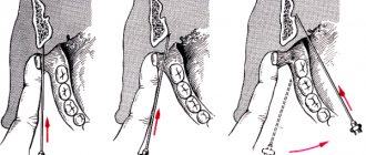
Using the thumb and forefinger of the left hand, push the upper lip up and out, and with the middle finger hold the projection of the infraorbital foramen. With this type of access, it is located at the intersection of two axes. One of them, horizontal, runs 5-7 mm below the lower orbital margin, and the second, vertical, runs on the corresponding side along the axis of the second upper premolar. The needle must be inserted, retreating 5 mm from the upper edge of the attachment of the transitional fold between the lateral and middle incisors. Then it is pushed upward, forward and outward in the direction of the lower orbital foramen until it stops at the bone. And only there the anesthetic solution is released.
Prices for tooth extraction at Apex-D dentistry
| Name | rub. |
| Opening an abscess | 500 |
| Abscess drainage | 350 |
| Removal of a single root tooth | 2000 |
| Removal of a double-rooted tooth | 2500 |
| Removal of a three-root tooth | 3000 |
| Removal of the third molar “wisdom tooth” of the upper jaw | 3000 |
| Removal of the third molar “wisdom tooth” of the lower jaw | 3500 |
| Root separation | 350 |
| Hemisection | 1500 |
| Osteotomy | 1500 |
| Socket curettage | 300 |
| Tamponade with iodoform turunda | 300 |
| Alvaugille | 300 |
| Neocones | 300 |
| Stitching | 750 |
| Periostotomy | 500 |
| Hemostasis | 200 |
| Cystectomy | 3000 |
| Cystectomy with resection of the apex of one root, frontal group | 4500 |
| Cystectomy with resection of the apex of one root, chewing group | 5500 |
| Cystectomy, resection of each subsequent apex | 1500 |
| Cystotomy | 1500 |
| Cyst removal | 1000 |
| Closed curettage | 1000 |
| Open curettage of one tooth area | 1500 |
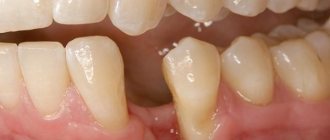
Modern dental practice makes it possible in most cases to prevent tooth extraction. The doctor’s rule is to prescribe tooth extraction surgery only as a last resort, when all available treatment methods have been tried and it is no longer possible to save the tooth, or in the event that the problematic tooth is the cause of the development of more serious complications.
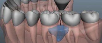
Anesthesia at the greater palatine foramen
This method blocks the innervation of the greater palatine nerve, thus achieving an anesthetic effect on the mucous membrane on the desired side of the hard palate, as well as on the alveolar process from the palate from the third molar to the middle of the short part of the canine. The anesthetized area can reach the lateral incisor, as well as the vestibular surface in the area of the third molar. In some patients the area extends to the second premolar.
The greater palatine foramen is located in the horizontal plate of the palatine bone and its pyramidal process at the base of the alveolar process, 5 mm anterior to the border of the soft and hard palate. Above the hole in the mucous membrane there is a small depression. The projection of the hole onto the mucous membrane of the hard palate is located at the intersection of two mutually perpendicular lines. The one that runs vertically goes through the middle of the line that connects the crest of the alveolar process and the center of the upper jaw, and the horizontal one runs through the middle of the coronal part of the third molar.
Anesthesia technique. The mouth should be opened wide, the syringe needle is directed from the opposite corner of the mouth and inserted 1 cm forward and inward in the direction from the projection of the opening of the palate into the mucous membrane. The needle is advanced until it comes into contact with the bone, and 0.5 ml of solution is injected. After a couple of minutes, the analgesic effect occurs. In the case when the solution is injected directly near the greater foramen of the palate, as well as into the opening of the pterygopalatine canal, the effect captures the posterior nerves of the palate emerging from its lesser foramen. As a result, the soft palate is numbed. Injecting the solution into this area may cause nausea and vomiting. Another side effect that may occur due to excessive injection of a solution under pressure is necrosis of the soft tissues of the hard palate. This can occur in patients with vascular atherosclerosis.
Infiltration technique
Infiltration local anesthesia is most often used in dentistry. In this case, the normal transmission of nerve impulses at the site of injection of the anesthetic is blocked. Experts distinguish two types of infiltration anesthesia:
- Direct – impulse transmission is blocked directly at the site of drug administration.
- Indirect - the anesthetic gradually penetrates into the tissue surrounding the injection site, due to which the “freezing” area increases.
Infiltration anesthesia is successfully used to anesthetize manipulations on the upper jaw; on the lower jaw, the effect of anesthesia in some cases may not be sufficiently expressed. This difference in effectiveness is due to the anatomical features of the alveolar processes of both jaws. If on the top the compact plate of the alveolar process is thin and there are many small holes in it (which facilitates rapid and sufficient diffusion of the drug into the bone tissue), then on the bottom the compact plate is thicker and noticeably denser, and there are much fewer holes in it. Therefore, during volumetric manipulations on the lower jaw, the doctor usually chooses another method to numb the area requiring treatment.
The main advantages of infiltration anesthesia in dentistry can be considered:
- Rapid achievement of analgesic effect.
- Safety for the patient, since low concentrations of anesthetic solutions are sufficient to achieve the required effect. In this case, if necessary, you can increase the amount of anesthetic without any harm to the patient’s body.
- The drug is eliminated from the body quickly.
- In addition to the desired nerve, nearby fibers of neighboring nerves are also anesthetized, so the treatment is painless and completely comfortable.
A variation of the infiltration method is intraligamentary (intraligamentous) anesthesia. The anesthetic solution is injected using a very short needle directly into the periodontium under measured pressure (the area of distribution of the anesthetic depends on the magnitude of the pressure). With this technique, the soft tissues of the oral cavity will not become numb, so it is often used in the treatment of baby teeth in young children - so that the child, who does not feel the numb areas of the soft tissues, does not accidentally injure them.
Anesthesia at the incisive foramen
Such anesthesia is carried out by neutralizing the nasopalatine nerve in order to anesthetize the anterior region of the mucous membrane of the hard palate in the area of the anterior teeth.
The incisive foramen is located between the front incisors 7-8 mm from the edge of the gum at the intersection of the lines that connect the distal edges of the median palatal suture and the necks of the canines.
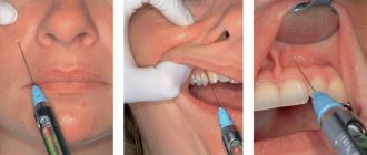
Anesthesia technique: the patient is in a chair, his head is thrown back, his mouth is open wide. The needle is inserted into the mucous membrane near the incisive opening to a depth of 3-4 mm. The anesthetic solution is released slowly. The process of inserting a needle into the papilla itself is very painful, so thin needles are used for such injections, and additional pain relief is performed. The anesthetic effect is achieved within a few minutes.
General principles of pain-relieving procedures
In order for local anesthesia in dentistry to bring the expected effect and not become a source of complications, it is important for the doctor to follow certain principles of its implementation:
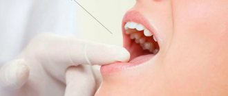
- You should first assess the patient’s condition and find out if there are any allergic reactions to painkillers.
- The correct choice of anesthetic drug and place for its administration.
- The use of exclusively sterile preparations that are compatible with oral tissues.
- The temperature of the solution for administration should be close to normal human body temperature.
- The rate of drug administration should be minimal, and the patient should not experience any unpleasant sensations (burning, itching, pain) during the process.
- Only sharp needles should be used to avoid tissue injury.
- The area of the upcoming injection must be pre-treated with an antiseptic.
- The injection should not be unexpected: the patient must be prepared for local anesthesia.
Use in dental practice
An anesthetic drug is injected into an area located a short distance from the site where surgery will be performed.
Conduction anesthesia can be performed in one of two ways:
- Central. The doctor determines the main trunks of the nerve endings for subsequent administration of the medicine into one of them.
- Peripheral. The anesthetic is injected into one of the branches of the main nerve of the stem system.
High-quality pain relief involves delivery of the solution to its destination. We are talking about a canal - a bone opening through which the nerve ending passes.
Side effects and complications
The cause of complications that arise during infraorbital anesthesia is non-compliance with the technique of administering the anesthetic, which determines the requirements for the qualifications of the doctor performing the procedure. Incorrect administration can lead to damage to the eyeball, bleeding or hematoma formation. In addition, possible problems include:
- Disruption of the natural blood circulation cycle in the area under the eye socket;
- Swelling of the tissues of the lower eyelid;
- The appearance of the effect of diplopia - splitting of visual perception;
- Post-traumatic neuritis, as well as blockade of the muscles of the eyeball.
It is also worth considering that when the lower wall of the canal located under the orbit is perforated, there is a possibility that the solution will enter the sinus of the maxillary region.
Strict adherence to the protocol at all stages of pain relief allows you to avoid negative consequences. You can prevent the formation of a hematoma in the area where the needle is inserted by pressing the area where the vascular bundle exits for 2-3 minutes.