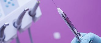
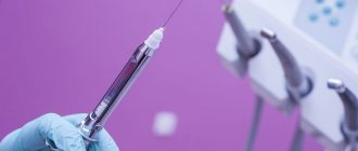
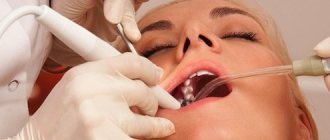
Infiltration anesthesia is one of the types of local anesthesia, in which the anesthetic is administered using a syringe into the soft gingival tissue. In dentistry, it is one of the most common, as it can ensure painless dental procedures for sixty minutes or more. Among dental patients, it is called “freezing” because after its administration, depending on the area of anesthesia, numbness is felt in the tongue, upper/lower lip, cheek, oral cavity or part of the face.
Specialists of the dental department of CELT use different types of infiltration anesthesia in the treatment of pulpitis, deep caries, and tooth extraction. The process uses various new generation drugs that have proven themselves well. Their diversity allows you to choose the one for which the patient has no contraindications. The active substances of their composition are quickly eliminated from the body. You can find out the price of infiltration anesthesia in the price list, in the “Services and Prices” section. To avoid misunderstandings, check the numbers with our information line operators or during a consultation with a doctor.
Consultation with a dentist-therapist - 1,000 rubles.
Infiltration anesthesia, conduction anesthesia - 500 rubles.
At CELT you can get advice from a dental specialist.
- The cost of a consultation with a dentist-therapist is 1,000
Make an appointment
Infiltration anesthesia in dentistry
The operating principle of infiltration anesthesia is to stop the nerve impulse that is transmitted from the pulp to the brain. It is manifested by numbness of the treated area: sensitivity is restored as the components of the drug are broken down in cellular tissues. The effect is achieved almost instantly and covers only a certain area, and the drugs used are safe for the human body, since the concentration of active substances in them is minimal.
In order to eliminate the risk of an allergic reaction to the anesthetic in the patient, the dentist conducts preliminary testing. Having individually selected the drug, he determines its dose and injects it into the tissues by injection, following a certain technique. Distributed throughout the cells, the anesthetic stops nervous activity and eliminates pain during dental procedures. It can be entered:
- Under the skin;
- Under the oral mucosa;
- Into the tissue surrounding the bone (periosteum).
Depending on the method of administration, infiltration anesthesia is:
- Direct – targeted introduction into the area that will be subjected to dental procedures;
- Indirect - injection of the entire area adjacent to the pathological focus with penetration of the active components deep into the tissue.
How it goes
Regardless of the area of application of infiltration anesthesia (on the skin or mucous membrane), manipulations are carried out according to the same technique:
- Treating the puncture area with an antiseptic drug.
- Convenient placement of the specialist in relation to the patient (on the right side of the client).
- Exposing the transitional fold of the patient's mouth by pulling back the cheek or lip. The specialist performs the action with his hands or using a special tool.
- Place the tip of the needle on the transitional fold, maintaining an inclination angle of 45° (relative to the alveolar ridge). The bevel of the needle should be directed towards the jawbone.
- Carefully insert the needle all the way into the bone tissue. The immersion depth can be 5-15 mm, depending on the jaw area.
- Inject the drug by applying gentle pressure with your finger on the syringe plunger.
Infiltration anesthesia: application
Indications
- Treatment of pathological conditions of teeth and gums (caries, pulpitis, etc.);
- Removal of teeth of varying complexity;
- Implantation operations;
- Prosthetics;
- Opening the abscess and draining it;
- Removal of tumors on the oral mucosa;
- Surgical dental intervention in the oral cavity.
Contraindications
- Identification of the patient’s individual intolerance to the components of painkillers;
- A pronounced inflammatory process in the oral cavity (anesthesia may not give the desired result);
- The patient has a mental illness;
- Neoplasm of malignant nature.
Which anesthetic is right for you - summary
- For bronchial asthma or high allergies, an anesthetic without preservatives is needed (usually sodium disulfite is used in anesthetics, which is needed to stabilize epinephrine or adrenaline). Therefore, the anesthetic “Ultracain D”, which does not contain any preservatives, is best suited for such patients.
- If you have thyroid disease or diabetes, in this case you also do not want to use anesthetics containing vasoconstrictor components - adrenaline, epinephrine. The drug of choice, for example, Ultracaine D, Scandonest or Mepivastezin. But, choosing between these three anesthetics, I would give preference to the first.
- If you have high blood pressure and heart disease - with moderate hypertension and compensated heart disease, the optimal choice is anesthetics containing a concentration of epinephrine (adrenaline) - 1:200000. This can be anesthetics “Ultracain DS” or “Ubistezin 1:200000”. In case of severe hypertension, decompensated heart disease, it is necessary to use anesthetics that are completely free of adrenaline and epinephrine. Then, for example, “Ultracain D” will do.
- If you are a healthy person - if you do not have the above diseases, then you can safely administer anesthetics containing epinephrine/adrenaline in a concentration of 1:100,000. Moreover, a person weighing about 70 kg can be given up to 7 carpules of anesthetic inclusive. An example of such anesthetics is Ultracain DS Forte, Ubistezin Forte and analogues.
→ The most powerful analgesics for toothache
Types of infiltration anesthesia
Classification into species depends on the area of influence.
| Species name | Injection area | When is it shown? |
| Intraosseous | Bone in the area between tooth roots | Removal of one, two, three teeth. |
| Intraseptal | Interdental bony septum |
|
| Intrachannel | Pulp | As an adjunct to intraligmentary anesthesia. |
| Intraligmentary | Between tooth root and bone |
|
| Subperiosteal | The area between the gum and the base of the tooth root |
|
Anesthesia according to Vishnevsky, which combines infiltration and conduction techniques, deserves special mention. It is called “creeping infiltrate” because it stops not only receptors, but also the transmission of nerve impulses along the endings of nerves located in the treated area. An excellent effect is achieved through layer-by-layer administration of the drug: after each stage, the dentist cuts the next layer of tissue with a scalpel. It has the ability to quickly stop bleeding if it occurs and minimizes the risk of damage to blood vessels and nerves.
Non-standard anesthesia techniques. Anesthesia over the periosteum. Anesthesia according to Go-Gates
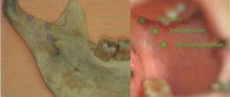
During the survey, the most commonly used methods of pain relief in the upper jaw were noted by practitioners to be infiltration anesthesia, incl. supraperiosteal (many people call it incorrectly “subperiosteal”), conduction anesthesia, incl. palatal and incisive, intraligamentary and intrapulpal. Intraligamentary anesthesia, which we classify as periodontal methods of anesthesia, and intrapulpal anesthesia, have much in common in the upper and lower jaws and will therefore be discussed together at the end of our publication. Conduction anesthesia in the upper jaw is called tuberal anesthesia. We recommend using external anesthesia according to Egorov; Infraorbital or infraorbital anesthesia is performed mainly for blockade in the treatment of trigeminal neuralgia and for manipulation of the maxillary sinus. When working with articaine-type drugs, there is no need for conduction anesthesia on the upper jaw, because These drugs have a very high diffusion ability and a maximum concentration among other anesthetics - 4%. Conduction anesthesia can be completely replaced by infiltration (periosteal) anesthesia.
Anesthesia over the periosteum
Anesthesia over the bone is an infiltration type of local anesthesia and is ensured by good diffusion of a 4% ultracaine solution through soft and bone tissue to the blocked nerves.
Since the lateral surface of the alveolar process of the upper jaw is thin and formed by porous bone tissue, solutions penetrate through it quite easily. Therefore, effective blockade of the alveolar nerves of any tooth in the upper jaw can be achieved by creating an ultracaine depot at the apex of the tooth.
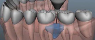
The technique of infiltration anesthesia of a tooth in the upper jaw practically does not depend on its location. The needle is inserted into the transitional fold between the tooth being anesthetized and the tooth located more medially, and advanced to a place located slightly above the apex of the tooth being anesthetized, where 0.5 ml of solution is slowly injected, preventing swelling of the mucous membrane. For ease of use of the drug, ultracaine carpules have the necessary graduation to clearly determine the administered dose of anesthetic. In each case, the depth of the needle is determined by the length of the tooth root. This length is 12-14 mm for any tooth, with the exception of the canine, whose root is 2-3 mm longer. Another guideline for determining the depth of the needle is the length of the tooth, which includes the length of the crown and the length of the root of the tooth. By using this guideline, you can avoid errors in determining the depth of immersion, which is caused by exposure of the neck of the tooth as a result of periodontal processes. The average length of a tooth in the upper jaw is 21-23 mm, while the length of the central incisors is 1-3 mm longer, and the length of the canines is 4-5 mm longer. Due to these dimensions, a thin needle with a diameter of 0.3-0.4 mm and a length of 16-25 mm should be used to administer anesthesia over the periosteum. When inserted, the tip of the needle should be oriented with its cut towards the bone to direct the injected solution into the bone tissue. You should not try to insert the needle under the periosteum, because its rich innervation will lead to a sharp increase in the pain of not only the needle insertion, but also the injection of the solution under the periosteum, as well as post-injection complications. Due to the high diffusion ability of ultracaine, introducing the solution under the periosteum will not create significant advantages compared to its deposition under the mucous membrane. Anesthesia of the first upper molar, which is located at the base of the zygomaticalveolar ridge, has the following features. Since in this place the lateral surface of the bone plate is thick, preventing the diffusion of the solution, the injection is carried out at a distance from this place on both sides of the zygomaticalveolar crest. Injection of a local anesthetic solution at the apex of the second premolar can effectively block the nerves of the medial buccal root of the first molar. To anesthetize the distal buccal root, a slightly larger amount of anesthetic (up to 1 ml of ultracaine) should be injected directly behind the zygomaticalveolar ridge at its apex. During surgical interventions, there is a need for additional anesthesia of the mucous membrane on the palatal side of the alveolar process, even when working with ultracaine. For this, in addition to the injection at the root apex above the periosteum on the vestibular side, the solution is also injected from the palatal side. As a rule, the location of the solution in this case is the angle formed by the palatine and alveolar processes of the upper jaw, opposite the tooth being anesthetized. The exception is the incisors, in which such a place coincides with the incisive foramen, as well as the second and third molars, where the large palatine foramen coincides with such a place, where the solution should be injected.
It is necessary to point out one more feature that occurs when anesthesia is administered over the periosteum of the maxillary canines. Since the length of the root of the canines is large, the projection of their apex onto the anterior surface of the upper jaw is located next to the infraorbital foramen. Therefore, the injected solution can penetrate into the infraorbital canal, which will lead to blockade of the anterior superior alveolar nerves, which innervate the incisors and tissues of the anterior surface of the upper jaw.
In conclusion, it should be noted that the use of modern highly concentrated solutions of local anesthetics (4% articaine solution) obliges the doctor to use small quantities (0.5-0.6 ml of ultracaine solution). Therefore, when performing anesthesia over the periosteum, the doctor is required to more accurately orientate and determine the injection site than was previously recommended, when the technique of a similar anesthesia method involved the injection of several milliliters of solution with the formation of an infiltrate over the apexes of several teeth. In this regard, we can say that the modern technique of infiltration anesthesia is closer in requirements to the technique of conduction anesthesia: this technique is based on the targeted administration of a smaller amount of solution. Thus, anesthesia over the periosteum provides technically simple, effective and safe pain relief. It would not be difficult for us to once again note the importance of performing an aspiration test.
The main indicators of anesthesia when working with ultracaine are presented in Table 1.
Table 1.
The main indicators of anesthesia when working with ultracaine on the upper jaw using supraperiosteal anesthesia.
| Groups of teeth | Recommended cannula (diameter and length) mm | Anesthetic quantity ml | Latent period sec | Duration of anesthesia min | Success of anesthesia % |
| Incisors and canines | 0.3-0.4 long 25 | 0,4-0,5 | 60-70 | 20-25 | 97-99 |
| Premolars | 0.3-0.4 long 16-25 | 0,4-0,5 | 60-80 | 20-30 | 99 |
| Molars | 0.3-0.4 long 16-25 | 0,5-0,7 | 100-120 | 20-35 | 98-99 |
List of procedures: surgery on soft tissues, preparation of hard tissues for a crown, cystectomy, opening of subperiosteal abscesses, treatment of periodontitis and depulpation, removal of one or more teeth, resection of the apex of the tooth root, osteotomy, restoration, implantation surgery, as well as all dental interventions for sanitation of the oral cavity.
Anesthesia on the lower jaw
The peculiarity of the anatomical structure of the lower jaw is that the outer and inner surfaces of the body of the lower jaw consist of dense layers of compact substance, which has practically no holes. Under these conditions, the diffusion of local anesthetic solutions deep into the bone tissue to the alveolar nerves is difficult. Therefore, for effective pain relief, most dentists use conduction anesthesia. To effectively and safely perform conduction anesthesia, it is necessary to have a good knowledge of the anatomical features of the relative positions of sensory nerves and blood vessels. To avoid getting into the vessel, developing bleeding or hematoma, it is necessary to strictly perform the aspiration test, which was described above.
Infiltration anesthesia can be successfully performed in patients up to 55-60 years old in the range from 3 to 3 teeth in the form of anesthesia over the periosteum, the scheme of which we described above. For surgical interventions on soft tissues, it is possible to use submucosal anesthesia, and for working with canals, intrapulpal anesthesia. Of all the widely known methods of blocking the mandibular nerve, the most effective is the one proposed in 1973 by the Australian dentist and practitioner Gou_Gates (GAEGow_Gates, 1973). According to various researchers, effective pain relief when using this method is achieved in 90-98% of cases, which is noticeably higher than when using other methods. Positive aspiration tests range from 1.6% to 1.9% of cases, which is almost 10 times less than with other methods of anesthesia.
Local post-injection complications (hematomas, difficulty opening the mouth) occur so rarely that the authors do not even estimate them as a percentage of cases. In addition, with one injection of 1.8-2.2 ml of local anesthetic solution using the Gou_Gates method, it is possible to achieve anesthesia not only of the lower alveolar nerve, but also of the lingual, mylohyoid, auriculotemporal nerves, and also in 65-75% of cases the buccal nerve.
The greatest difficulty, in our opinion, when studying this anesthesia arises when orienting the direction of immersion of the needle into the tissue. To overcome this <11> complexity, we propose to use the following manual technique, which seems to us more practical (S.A. Rabinovich, O.N. Moskovets, 1999). Holding the syringe in the right hand, the index finger of the left hand is placed in the external auditory canal or on the skin of the facial part of the head immediately in front of the lower border of the tragus of the ear at the intertragal notch. Using the sensations of the index finger of the left hand to control the movement of the head of the condylar process onto the articular tubercle as the patient opens his mouth wide, the neck of the condylar process is determined and the needle is directed to a point in front of the end of the index finger, which will also correspond to the direction to the tragus.
The described manual technique does not require careful visual control, construction of spatial images and is successfully performed with satisfactory coordination of movements, as well as bringing the index fingers of two hands together with eyes closed. Thanks to the choice of the injection point and target point, as well as the movement of anatomical structures when the mouth is opened wide, the entire path of needle advancement is devoid of both muscles and large neurovascular bundles. The only major blood vessel, the internal maxillary artery, remains below the path of the needle, located in the notch of the mandible (Bennett CA, 1984), and with the mouth wide open, pressed against the bone. Thanks to these anatomical features, bringing the needle to the neck of the condylar process is not accompanied by significant tissue trauma and does not cause post-injection complications. Thus, the analysis of the features of the method of local anesthesia of the mandibular nerve according to Goe-Gates explains its high efficiency and safety, and also allows us to clarify a number of provisions in the technique of its implementation.
Technique for performing a mandibular nerve block according to Go-Gates in our modification:
1. The patient is placed in a horizontal or semi-horizontal position. This position is not only convenient for anesthesia using the Gou-Gates method, but also more physiological for the prevention of emergency conditions in the patient due to possible reflex changes in the tone of blood vessels. 2. The dentist is positioned on the right side of the patient. The more precise location of the dentist will be determined by which side of the patient is expected to undergo anesthesia. If it is on the right side of the patient's jaw, then it is more convenient for the dentist to stand in a position that corresponds to the 8 o'clock position on the dial, which is easy to imagine when standing at the patient's feet. In this case, the patient’s head should be asked to turn towards the dentist in order to clearly see the pterygoid-maxillary depression on the side of the injection in the depths of the open mouth. 3. If anesthesia will be performed on the left side of the patient’s jaw, then it is more convenient for the dentist to stand in a position that corresponds to the 10 o’clock position on the same dial. In this case, the patient’s head should be asked to turn slightly away from the dentist for similar reasons. 4. With the patient’s mouth open, treat the mucous membrane at the site of the intended injection in the pterygomaxillary recess, first drying it and then anesthetizing it with a topical anesthetic. The anesthetic should be applied pointwise, removing its remains after 2-3 minutes. 5. An additional technique to reduce tissue trauma and prevent vascular reactions during injection may be the following. Before piercing the mucous membrane, ask the patient to take a deep breath and hold it. Holding your breath will reduce the number of possible unwanted movements of the patient while advancing the needle to the target point. Preliminary additional ventilation of the lungs during a deep breath will increase blood oxygen saturation and lead to a slight increase in heart rate due to the cardiorespiratory reflex, which will increase blood flow to the vessels. 6. Taking the syringe in the right hand, place it in the corner of the mouth opposite to the injection side, retracting the mucous tissue of the cheek on the injection side with the thumb of the left hand placed in the mouth. The patient is asked to open his mouth wide, controlling the degree of its opening according to the exit of the condylar process onto the articular tubercle. The movement of the condylar process, as already described, is traced by sensations under the index finger of the left hand, placed in front of the tragus or in the external auditory canal. The needle is directed into the pterygo-maxillary space, medial to the tendon of the temporal muscle, to the place where application anesthesia was previously administered. 7. After a deep breath and holding the breath, the patient makes a puncture of the mucous membrane and the needle is advanced slowly until it stops in the bone - the lateral part of the condylar process, behind which is the tip of the index finger of the left hand. The depth of needle advancement is on average 25 mm. Reaching the bone corresponds to the placement of the needle tip at the target point. If this happens, then the needle is slowly withdrawn to the mucosal surface and the orientation of the needle and its advancement to the target point are repeated. 8. If the target point is successfully reached, retract the needle 1 mm back and be sure to perform an aspiration test. If the aspiration test is negative, 1.7 ml (1 carpule) of anesthetic solution is slowly injected, focusing on the patient's condition. 9. After introducing the anesthetic solution, the needle is slowly removed from the tissue. The patient is asked not to close his mouth for another 2-3 minutes so that the local anesthetic solution permeates the surrounding tissues in the anatomical ratio in which they come when the mouth is opened. As already noted, in most cases with this anesthesia the buccal nerve is also blocked. However, in our opinion, it is always advisable to additionally anesthetize the buccal nerve before the intervention, without causing anxiety to the patient in case of insufficient block of this nerve. Thus, the given description of the method of local anesthesia of the mandibular nerve according to Go_ Gates, as well as the developed manual technique and additional clear extra- and intraoral landmarks, a small number of local and systemic complications and a high percentage of positive results - 98% (when working with ultracaine) will be, as we hope to contribute to the wider dissemination of this effective and safe method in our country.
It should be noted that the incisors and canines on the lower jaw are anesthetized using supraperiosteal anesthesia, and the main indicators of anesthesia when working with ultracaine are presented in Table 2.
Table 2.
The main indicators of anesthesia when working with ultracaine on the lower jaw when using the mandibular nerve block according to Gou_Gates with our improvements (premolars and molars) and infiltration anesthesia in patients under 50 years of age (incisors and canines).
| Groups of teeth | Recommended cannula (diameter and length) mm | Anesthetic quantity ml | Latent period sec | Duration of anesthesia min | Success of anesthesia % |
| Incisors and canines | 0.3-0.4 long 16-25 | 0,6 | 1,5 | 25-30 | 97-98 |
| Premolars | 0.3-0.4 long 42 | 1.7 (carpule) | 7,1 | 45-60 | 98 |
| Molars | 0.3-0.4 long 42 | 1.7 (carpule) | 6,7 | 45-60 | 98 |
List of procedures: surgery on soft tissues, preparation of hard tissues for a crown, cystectomy, treatment of periodontitis and depulpation, removal of one or more teeth, restoration, opening of subperiosteal abscesses, implantation surgery, resection of the apex of the tooth root, osteotomy, as well as all dental interventions for sanitation of the oral cavity and other dental surgical interventions
Pros and cons of infiltration anesthesia
| Pros: | Minuses: |
|
|
Drugs
Articaine-based products are considered effective and safe:
- Ubistezin. Contains epinephrine, a vasoconstrictor that can provide a long-lasting and stable anesthetic effect.
- Ultracaine. Available with or without epinephrine. Epinephrine is contraindicated in patients with diabetes, hypertension, asthma, and anyone who is prohibited from taking medications with epinephrine and epinephrine.
- Septanest. Contains epinephrine and preservatives that are not found in ubistezin and ultracaine.
- Orablock. The drug is similar to ubistezin and septanest.
Novocaine and lidocaine are not used: they are more toxic compared to anesthetics based on articaine. Novocaine, in addition, is powerless against purulent inflammatory processes.
Reviews about our dental therapists
I am very grateful to Evgeniy Borisovich Antiukhin for removing my three eights.
Especially considering that the lower tooth was not the simplest (it was located in an embrace with a nerve). The removal took place in 2 stages, one tooth under local anesthesia, two under general anesthesia. I had no idea that wisdom teeth could be... Read full review Sofia
28.12.2020
Words cannot express my gratitude to Elena Nikolaevna Kiseleva. This is the best doctor in the world. I got an appointment after many years of being ignored by the dentist’s office and with a bitter experience of treatment in another paid clinic, the mistakes of which had to be corrected in the first visits. Thank you for this... Read full review
Roman Stanislavovich Sh
25.07.2020