Hello! Today we will talk about a procedure used in dental practice, x-rays. And the most common question we hear from our clients is how safe is this procedure?
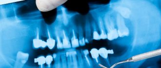
Our clinic performs two types of X-ray examinations using a radiovisiograph and a computed tomograph. Using them, we can take a picture of one or several teeth, as well as a panoramic picture of the entire oral cavity.
What is the difference between a radiovisiograph and a computed tomograph? On a radiovisiograph we see all teeth in a two-dimensional projection. It's essentially a photograph of a tooth. On a computed tomograph we can see all organs in a three-dimensional projection.
What can a doctor see on an x-ray?
He can see:
- total number of teeth;
- anatomical features of the structure of the maxillofacial region;
- level, bone height;
- possibility of installing implants.
X-rays are also done for the purpose of establishing a diagnosis, when the doctor cannot make one based solely on the patient’s complaints.
X-rays are also done to monitor the doctor’s work. It shows how:
- canals are sealed;
- tooth removed;
- an implant was installed.
Conservative (medicinal) treatment of dental granuloma
This technique is used only for those cases in which the development of granuloma has not yet progressed far. The essence of conservative therapy for tooth root granuloma will be the intake and use of various drugs - antibiotics, antiseptics, drugs that have an anti-inflammatory effect. All medications are prescribed by a dentist and the treatment process takes place under the strict supervision of a specialist. This treatment option for granuloma is effective with early diagnosis of the disease and, if you strictly follow the doctor’s instructions, the granuloma will resolve within 1-2 weeks.
In some cases, conservative treatment of granuloma is complemented by root canal therapy. Working with tooth canals for root granuloma is indicated:
- For constant toothaches, the intensity of which does not subside from taking painkillers;
- Swelling of the gums, spreading to the cheek and jaw;
- Treatment of tooth canals is clearly indicated when large granulomas are detected - from five millimeters.
What is an x-ray?
This is one of the types of radiation. If you think that X-ray radiation is present only in the X-ray room, then we can say right away that you are very mistaken. Let's remember physics lessons. We receive small doses of radiation every day:
- outdoors, from a natural source of radiation - the sun;
- at home from household sources located in the apartment: from a TV, various gadgets, a refrigerator, etc.
Radiation is measured in sieverts. What is the rate of radiation that a person can consume? Acceptable is 1000 sieverts per year.
Now let's remember mathematics and calculate how many X-rays can be taken for a person. A targeted image of one tooth is equal to 2-3 microsieverts. You can take 300-500 x-rays per year. A panoramic photograph (all teeth) is equal to 16-18 microsieverts. You can take 60-70 x-rays per year.
Now let's look at computed tomography. 1 photo taken on this device is equal to 60-80 microsieverts. You can take 16-20 x-rays per year.
We are now considering the maximum dose limits for X-ray equipment. Our clinic has a radiovisiograph and a computer tomograph of the latest generation made in Germany. The load on the devices is reduced to a minimum.
Previously, when taking an x-ray, the exposure time (you hear it as a beep) was approximately 2-3 seconds. Now this time has been reduced to 0.05 seconds.
Now let's talk about household radiation. If you watch TV for 3 hours at a distance of less than 2.5 meters from the screen, you get approximately 0.5 millisievert, i.e. Watched TV for 5 days, get 1 x-ray. If you sit in front of a computer monitor for more than 3 hours, you get 1 microsievert, 1 x-ray. We flew by plane to Turkey, where the flight is 3-3.5 hours, you get 10 millisieverts or 5 targeted shots. And add radiation from TVs, refrigerators, microwave ovens and you will understand that you will not receive terrible radiation in the X-ray room.
Symptoms of periodontitis
- continuous throbbing pain, both with and without contact with the tooth;
- a sharp unpleasant odor from the mouth that cannot be gotten rid of;
- swelling of the gums;
- feeling of heaviness and mobility of teeth;
- a sharp change in the color of the enamel - darkening or yellowing;
- accumulation of pus in periodontal pockets.
purulent fistula on the gum with chronic periodontitis
Can X-ray examinations be performed on pregnant women?
In the first half of the term, the study is done only according to strict indications. In the second half of the term, you can do as much research as you like.
Another interesting fact. People often ask X-ray technicians to wear more protective lead aprons. Let's say right away that this is useless. You receive the same dose of radiation from covered and exposed parts of your body.
And remember that a doctor will never order an x-ray just out of curiosity. X-ray is one of the types of diagnostics of the area of proposed treatment and the anatomical features of the oral cavity.
Come to our clinic and we will make every effort to make your stay comfortable and enjoyable!
Kinds
There are many types of periodontitis, and dentists distinguish several classifications - depending on the source of inflammation, the stage and form of the disease.
By localization:
- Marginal – a type of periodontal lesion at the gingival margin. Most often it occurs due to traumatic factors - even a minor chip can provoke the disease.
- Apical - inflammation in the apical part of the root. It is often misdiagnosed as a form of pulpitis (nerve inflammation).
By stage:
- Acute form - can be of serous and purulent type. The second type is dangerous due to serious complications, further development of purulent inflammation in nearby tissues, development of abscesses or phlegmon of the oral cavity.
- The chronic form is divided into fibrous, granulating and granulomatous periodontitis. Often it is practically asymptomatic, but exacerbations with severe pain are periodically possible.
Complications of periodontitis
With improper treatment or untimely diagnosis, the patient develops extensive purulent inflammation - multiple ulcers, abscesses and fistulas appear. All this leads to the development of such dangerous diseases as:
- flux;
- periostitis;
- dental cyst;
- osteomyelitis;
- extensive blood poisoning (sepsis).
The possible consequences of each of the complications are very serious. They often lead to hospitalization, surgery and a long rehabilitation period.
complication of dental periodontitis - purulent fistula on the skin
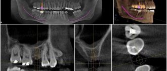
How does the procedure and decoding work?
To take a 3D photograph of teeth, a standing or sitting patient needs to bite a special plate and fix his position in the device using a fixing stand. During the entire scanning time, you must remain absolutely still.
The tomograph sensor makes a series of revolutions around the patient’s head for 8-20 seconds, producing about 200 images in different projections. Processing digital data takes 5-15 minutes, after which the information is written to a disk or flash drive. No preparation is required, you just need to remove all metal jewelry from your neck, ears, and hair before the procedure.
Summary
To summarize what has been said, we repeat that the occurrence and development of caries is influenced by many factors, including lifestyle, eating habits, and personal hygiene. It is difficult to diagnose caries on your own, so it is better to visit a doctor regularly for a preventive examination. The only treatment for caries these days is surgical. You should not rely on traditional methods. The sooner you see a doctor, the fewer problems will arise during treatment.
The article material was approved by the doctor: Konishchev Vitaly Konstantinovich Oral and maxillofacial surgeon, dentist-implantologist
14 years
Diagnostics
In some cases, using a mirror and analyzing one’s own feelings, the patient can make a diagnosis of “caries” himself. However, there are cases that only a specialist can correctly assess. We are talking about the initial stage, when there are practically no symptoms, but the doctor, using modern diagnostic tools, will detect the first signs of the disease. At later stages, it is important to distinguish caries from other dental diseases, which is also the responsibility of a certified physician. We will consider the symptoms that the patient himself should pay attention to.
The first symptom is a change in the color of tooth enamel. Spots of dark brown or, on the contrary, too white color appear on the surface of the tooth. Take a special look at the areas around the fillings. Also, do not ignore the fact that the tooth has begun to react to hot and cold. Another bad sign is damage to the dental floss when using it: caries may develop on the invisible side of the tooth. If you notice suspicious symptoms, you should immediately consult a doctor. Unfortunately, statistics show that only ten out of a hundred patients can independently detect caries.
Prevention
To avoid any form of periodontitis, you must follow simple recommendations from dentists, which apply to most clinical cases:
- You need to brush your teeth at least twice a day - the brush should not be too hard, so as not to injure the gums or introduce infection into the soft tissues.
- After each meal, use mouthwash - if you don’t have a special liquid on hand, just rinse your mouth thoroughly with warm boiled or filtered water.
- Visit the dentist at least 2 times a year - a scheduled appointment allows you to identify most diseases in the early stages, begin treatment in a timely manner and avoid serious consequences.
- Have your teeth professionally cleaned and tartar removed annually – even minor plaque creates conditions for the proliferation of pathogenic bacteria.
General advice for everyone includes giving up bad habits. Smoking and alcohol contribute to the deterioration of dental health, as well as their attractiveness and natural whiteness.
Possible harm
Cone beam dental computed tomography is the safest and fastest diagnostic method. Thanks to the use of a conical X-ray beam, the radiation dose received during the study is 10 times less than when using spiral CT. And the pulsating mode of the X-ray beam further reduces the radiation dose. The three-dimensional computed tomograph SOREDEX Scanora 3D is one of the safest devices in terms of X-ray radiation dose - only 0.035 m3v.
However, despite the safety of the study, CT also has contraindications. If we just talk about dental tomography, it is not performed during pregnancy (in the 1st trimester). 3D dental x-rays with contrast are prohibited for pregnant and lactating women, patients with endocrine disorders (diabetes mellitus, thyroid pathologies), renal failure and intolerance to iodine-containing drugs.
Treatment of caries
Treatment of dental caries is surgical, depending on the degree of damage.
At the initial stage, the stain stage, it is easiest to treat a tooth. The doctor removes the stain and applies a special mineral paste to the tooth, including sodium fluoride and calcium gluconate.
Treatment of superficial caries depends on its location. The tooth may need filling. The same remineralization is carried out as in the previous stage.
In case of average caries, the affected area of the tooth is removed, medicinal treatment is carried out and a filling is placed.
Deep caries requires the same treatment, only it affects a larger area. Finally, the patient may need a crown on the treated tooth.
Treatment of complicated caries depends on the type of complication. When treating pulpitis, for example, it is necessary to remove the nerve. Antibiotics may be prescribed. In some cases, the only thing that can be done is to remove the tooth.
Of course, scientists all over the world are working on how to stop the destructive effects of caries using medicinal, that is, non-surgical, means. Now interesting data have appeared on the treatment of caries with special hormones. But these methods are still at the experimental stage and have not entered into general practice.
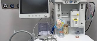
What equipment is used
To carry out 3D diagnostics, a three-dimensional computed tomograph SOREDEX Scanora 3D with advanced functionality is used. This is the latest generation equipment, which allows you to obtain three-dimensional images of the anatomical structures of the maxillofacial region in a few seconds, with the least radiation exposure for the patient.
The program analyzes the obtained multiplanar sections and builds them into a 3D model, thanks to which the specialist is able to accurately assess the condition of the dental system, detect all pathological processes occurring in this area and competently plan a treatment regimen.
A virtual 3-dimensional model of the scanned area can be recorded on any digital media (CD, flash drive), which allows the attending physician, if necessary, to view diagnostic data or involve related specialists in the analysis of the received information.