Questioning and examining the patient gives the doctor only part of the information about the real state of the dental system. To clarify the suspected diagnosis, in many cases it is necessary to undergo an X-ray examination. But the very mention of it bothers some people. Is this procedure really dangerous to health and what do its results tell the doctor? Below are answers from experts to such questions.
Why does a dentist refer a patient for dental x-rays?
An x-ray gives the doctor the opportunity to most accurately assess the condition of the problem unit or all organs of the oral cavity. And also study the clinical picture in detail. In particular:
- detect foci of inflammation that have not yet manifested themselves;
- identify neoplasms in the bone and soft tissues of the dental system;
- establish the signs and stage of development of periodontitis or periodontal disease;
- establish linear dimensions, volume, structure of bone tissue before implantation;
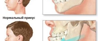
- more accurately assess the correctness of occlusion (tight closure of the dentition);
- detect anomalies of the dental system organs;
- determine the position of erupting wisdom teeth.
As a rule, radiography is prescribed before the start of orthodontic treatment, when planning implantation and prosthetics, before and after filling the tooth canals. This study is also used to evaluate the results of conservative and surgical treatment.
Free consultation
30-40 minutes
inspection and diagnostics
treatment plan and cost
Make an appointment
Recommendations for a nursing woman
Resumption of feeding directly depends on the medication used for pain relief. If it takes several hours for the drug to be eliminated, then the milk must be gradually expressed. In more complex cases, antibiotics may be prescribed. At the same time, medications are selected that do not enter milk or are safe for the child’s body.
In addition to temporarily stopping feeding, you should:
- do not eat for 2 hours;
- Do not eat hot food for 1-2 days;
- for chewing, use the side of the jaw opposite to the free socket;
- refuse to rinse the mouth (to prevent the healing clot from falling out and bleeding);
- do not visit the bathhouse;
- do not touch the socket of the extracted tooth;
- Brush your teeth especially carefully, trying not to touch the socket.
During a personal consultation, our dentists will tell you in detail how the procedure occurs. Don't be afraid of tooth extraction - it's a safe procedure if it's done by professionals.
What types of X-ray diagnostics are used in dentistry?
The main types of images obtained during x-ray photography of the dental system:
- Sighting. Provides information about the condition of one or more teeth and adjacent tissues. This helps the specialist assess the condition of dentin, root canals, periodontium, and blood vessels.
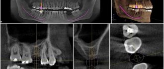
- Panoramic (OPTG, orthopantomogram). It is a detailed flat image of the jaws, teeth, joints, and sinuses. This image helps to detect carious areas, defects in fillings, impacted teeth, and cysts. And also draw up a correct plan for implantation, prosthetics, and treatment.
- Occlusal. This is the so-called bite radiography, which is often used when examining children, as well as when it is impossible to obtain clear results using targeted radiography.
In addition, photographs can be film or digital, when the resulting image is transmitted using a special sensor to a computer monitor. In the second case, the study is called radiovisiography.
Price for dental x-ray
As for the cost of x-ray examination of patients, it all depends on the clinic, the equipment used for diagnosis, as well as the type of image required. On average, prices for a dental photograph in Moscow start at 250 rubles, and the cost of a panoramic photograph of the jaw ranges around 1,300 rubles. Where to take dental x-rays is up to you. Many clinics have adequate equipment and their patients do not need to go elsewhere, while others offer x-rays as a separate service.
How is dental x-ray examination performed?
The photo is taken in a special room with lead-protected walls and floors. The doctor asks the patient to remove metal jewelry and accessories, as they may distort the results of the study. The vital organs of the body are protected by an apron with lead inserts. Features of the study depend on the method used.
When receiving a targeted image, the doctor directs an x-ray beam to the area of the jaw being examined from the side of the face or to an area of the oral cavity. An orthopantomogram is obtained in a different way. The patient fixes his head in a special device, grabs the plate in a disposable cover with his teeth and remains motionless while the mobile part of the device makes a full revolution around the head.
Can X-ray examinations be performed on pregnant women?
In the first half of the term, the study is done only according to strict indications. In the second half of the term, you can do as much research as you like.
Another interesting fact. People often ask X-ray technicians to wear more protective lead aprons. Let's say right away that this is useless. You receive the same dose of radiation from covered and exposed parts of your body.
And remember that a doctor will never order an x-ray just out of curiosity. X-ray is one of the types of diagnostics of the area of proposed treatment and the anatomical features of the oral cavity.
Come to our clinic and we will make every effort to make your stay comfortable and enjoyable!
Are dental x-rays safe for human health?
In accordance with SanPiN 2.6.1.1192-03, the maximum permissible annual radiation dose during diagnostic x-ray procedures is 1000 μSv (microsievert) for an adult and 300-400 μSv for a child.
To receive it, within a year you must complete:
- 500 targeted dental examinations using a radiovisiograph;
- 100 procedures of conventional x-rays with image output on film;
- 80 digital or 40 film orthopantomographies (obtaining a panoramic image).
Thus, dental X-rays for almost any purpose do not exceed the permissible doses. Therefore, it does not have a negative effect on health. Even when producing film OPTG, the radiation dose is a maximum of 30 μSv. If it is necessary to take a targeted X-ray of the teeth and use radiovisiography, you can even get several images during one appointment. Without any risk to health, since a single dose in this case is only 2-3 μSv.
Is it possible to carry out diagnostics during pregnancy?
According to SanPiN, X-ray examinations are allowed in the second half of pregnancy using protective equipment, provided that the radiation dose does not exceed the same 1000 μSv. However, it is recommended to refrain from taking x-rays in the first and last 12 weeks, i.e. in the first and last trimesters.
Do not be afraid of undergoing diagnostic procedures during pregnancy. Even ordinary caries is an infection that, if not properly treated, can spread throughout the body and lead to infection of the fetus. Therefore, it is better to receive a small and completely safe dose of radiation than to carry out complex dental treatment blindly, not knowing how deep the inflammatory process is.
After the baby is born, i.e. During breastfeeding, dental x-rays can be taken, even more than once (within reasonable limits). Radiation doses are minimal, so radiation does not accumulate in breast milk, and there will be absolutely no harm to the baby. There is also no need to pump or skip feedings.
Are there any contraindications to X-ray examination?
Dental X-rays are not performed on patients with severe bleeding in the mouth, or those who are unconscious or in critical condition.
Pregnancy is considered a relative contraindication. In this condition, women should not undergo dental procedures involving radiation until 4-5 months. But in each individual case, the expectant mother needs to personally consult a doctor, since situations are different.
A breastfeeding woman can undergo an X-ray examination. But it is advisable to resort to digital methods and skip one or two feedings after the session.
Tooth extraction procedure for a nursing mother
Step-by-step operation:
- a painkiller is administered;
- periotomy is performed;
- forceps are selected and applied;
- extraction of an unhealthy tooth is performed;
- the bleeding stops.
Carrying out tooth extraction surgery at the One to One clinic
Medicines that do not pass into breast milk are used as painkillers. Anesthesia is performed with a carpule syringe. The bleeding is stopped with a cotton swab. In most cases, bleeding stops and blood vessel thrombosis occurs within 5-10 minutes.
Indications for dental radiography
X-rays are used in the treatment of almost all diseases in therapeutic dentistry. It is often necessary even for ordinary caries and especially in cases of its complicated forms. When treating tooth canals, a dentist often prescribes x-rays 2-3 times, which allows him to assess the condition of the canals before treatment, the quality of their preparation for filling, and, finally, the correctness of the filling. Based on an x-ray examination, it is possible to exclude the existence of cysts or granulomas in the apical part of the tooth, assess the condition of the tissues of the tooth crown, and also determine the position of the tooth roots, their size and the presence of curvature. Therefore, this study is indispensable in both orthopedic and surgical dentistry. It allows surgeons to correctly plan the course of the upcoming operation and assess the likelihood of possible complications. In children, this method is used to determine impacted (unable to erupt normally) teeth, the stage of resorption of the roots of primary teeth and the stage of formation of the roots of permanent teeth, as well as the size of the unerupted tooth.
Contraindications for dental radiography
During a dental x-ray, the patient receives a very small dose of radiation. However, this examination is prescribed with caution to pregnant women and young children. It should be taken into account that the fetus is especially sensitive to the effects of radiation in the first trimester of pregnancy.
Dental X-ray technique
Before the procedure, the patient is put on a special lead apron, which protects him from unwanted exposure to X-rays, and is seated on a chair. The X-ray technician places a special sensor inside the mouth in the projection of the tooth being examined and asks the patient to press it with his finger. Then the X-ray technician positions the radiation source of the tooth being examined and turns on the X-ray machine. The entire procedure only lasts a few seconds. No other preliminary preparation is required.
Fluoroscopic
1405 7.354 10.333 Computed tomography 468 9.959 4.661 Other 825 11.733 9.680 TOTAL: 105854 0.473 50.085 2013Fluorography and radiography
Fluorography and radiography are methods of radiographic examination. Fluorography is often used for mass, routine screening of tuberculosis.
Nowadays digital fluorography is mainly used . Film fluorography is an outdated research method. Digital fluorography has a lower radiation exposure to a person, but at the same time its resolution (that is, the ability to transmit a real image) is lower in comparison with x-rays of the lungs in direct projection.
X-rays contain more information and pathological changes are more clearly visible.
The radiation exposure is low for both methods (digital devices have lower radiation exposure).
Is x-raying acceptable while breastfeeding?
Breastfeeding after x-ray
If the condition of a nursing woman requires a mandatory x-ray examination, you should not refuse it at the risk of your own health. Modern research in the field of radiology continues to persistently repeat that there are no serious reasons to avoid X-rays while breastfeeding a child. X-rays cannot affect breast milk, so after undergoing the examination, mothers can continue feeding their baby without fear.
You should not deprive a child of his mother’s breast because of an x-ray. This is justified by the fact that the effect of radiation on the human body stops immediately after the procedure is completed. Numerous studies and experiments prove that X-ray rays do not tend to linger in the human body. In this regard, the need to remove breast milk from a woman’s body disappears by itself. Breastfeeding after an x-ray can proceed as usual.