The lower jaw (mandibula) is unpaired, horseshoe-shaped, the only movable bone of the skull. It consists of two symmetrical halves, completely fused by the end of the 1st year of life. Each half has a body and a branch. At the junction of both halves in old age, a dense bony protrusion forms.
In the body (corpus mandibulae) there is a base (basis) and an alveolar part (pars alveolaris) . The body of the jaw is curved, its outer surface is convex and its inner surface is concave. At the base of the body the surfaces merge into one another; in the alveolar part they are separated by alveoli. The right and left halves of the body converge at an angle that is individually different, forming a basal arch. The shape of the basal arch is one of the main features characterizing the shape of the lower jaw. To characterize the basal arch, the latitudinal-longitudinal index is used (the ratio of the distance between the angles of the lower jaw to the distance from the middle of the chin to the middle of the line connecting the angles of the lower jaw). There are jaws with a short and wide basal arch (index 153-175), with a long and narrow one (index 116-132) and with an intermediate shape. The height of the jaw body is greatest in the area of the incisors, the smallest is at the level of the 8th tooth. The thickness of the jaw body is greatest in the region of the molars, and the smallest in the region of the premolars. The cross-sectional shape of the jaw body is not the same in different areas, which is determined by the number and position of the roots of the teeth. In the area of the front teeth it approaches triangular with the base facing downwards. In areas of the body corresponding to large molars, it is close in shape to a triangle with the base facing upward (Fig. 1-12).
Lower jaw
The largest and strongest bone of the face, on which the lower teeth are located. The left and right halves of the mandible initially begin as two separate bones, but in the second year of life the two bones fuse along the midline to form one. The horizontal central part on each side is the body of the mandible. The upper part of the body is the alveolar margin, corresponding to the alveolar margins of the maxillae. A prominent chin in the lower part of the body along the midline is considered a distinctive feature of the human skull. On either side of the chin is the mental foramen, the opening for the mental branch of the mandibular nerve, the third division of the fifth cranial nerve.
Useful information for patients
The jaws are the basis of the facial skeleton. Not only the beauty of the profile depends on their anatomical structure, but also functional capabilities that are important for life - chewing, swallowing, breathing, speech, the formation of cavities for the sensory organs and much more.
SYMPTOMS OF CORRECT BITE:
- There are no gaps between teeth.
- Chewing is not difficult.
- There is clear pronunciation.
- There are no cracks, chips or stains on the enamel.
- The teeth have the correct shape.
- There is no crowding of teeth.
- There is no pronounced inclination of the teeth in or out.
- The center of the incisors is located right in the middle of the face.
With improper development of bone tissue and the alveolar process, deformations such as microgenia, prognathia, overbite and others occur. In adults, the shape can also change, and this is mainly due to factors such as:
- Severe mechanical injuries.
- Consequence of surgical interventions.
- Various pathological processes (osteomyelitis, ankylosis, purulent inflammation and others).
- Incorrect orthodontic treatment or relapses after it.
- Partial or complete adentia (senile jaw).
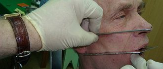
Changes in structure, various inflammatory and tumor processes lead to severe diseases of the jaw. The most common pathologies are osteitis, periostitis, osteomyelitis. At the first signs, such as swelling, redness, pain, fever, you should contact a dentist or surgeon.
ANATOMY OF THE JAW
UPPER JAW
The upper jaw is a fixed paired bone of the facial skeleton. It has an air cavity (sinus), 4 surfaces (orbital, anterior, posterior, internal), as well as 4 processes (frontal, zygomatic, palatine and alveolar).
The dentition of the upper jaw is deviated slightly forward and outward. The crowns of the teeth increase in size from the incisor to the first molar, but the second molar is smaller than the first, the third is smaller than the second. The height of the crowns decreases successively from the incisor to the third molar, with the exception of the canines. The large roots of the teeth provide significant stability to the dentition of the upper jaw and to each tooth individually.
LOWER JAW
The lower jaw is a movable horseshoe-shaped bone of the facial skeleton. A large number of muscles are attached to it. The dentition of the lower jaw has a parabolic shape.
The teeth of the lower jaw are characterized by the fact that the incisors and canines are located perpendicular to the alveolar process, the chewing teeth are slightly inclined towards the tongue. The pattern of changes in the size and height of tooth crowns on the lower jaw is the same as on the upper jaw.
JAW DEVELOPMENT
The growth and formation of the jaw is closely related to the development of tooth germs and alveolar process. This process begins as early as the 7th week of intrauterine life. It is influenced by various endogenous factors (maternal toxicosis, hormonal imbalances, levels of vitamins and minerals), and after the birth of a child - also exogenous (external).
The jaw of a newborn is not yet completely ossified and fused; the lower part is shifted back in relation to the upper.
When a permanent dentition is formed (from the age of 6), intensive growth occurs due to the eruption of molars and incisors. Also, growth spurts are observed at 11–13 years of age; in boys, this usually occurs later. By the age of 18, the formation of bone tissue is completely completed.
CHANGES IN THE SHAPE OF THE LOWER JAW WITH AGE
| Jaw of a newborn |
| Jaw of a 4 year old child |
| Jaw of a 6 year old child |
| Jaw of an adult |
| Elderly man's jaw |
For patients who want to get a free consultation with a dentist in Krasnodar , we inform you that pre-registration for an appointment is being made at Dentistry on Gagarin. Call the numbers indicated on the website and sign up for a free consultation with a dentist with a diagnosis (consultation duration up to 15 minutes).
Upper jaw
Most of the facial skeleton consists of the maxillary bones. Although they are called maxillae, the size and function of the maxillae include much more than just an addition to the mandible. They form the middle and lower part of the orbit. Between them there is a hole for the nose, under the lower edges of the small nasal bones.
The infraorbital foramen, the entrance to the floor of the orbit, is the end of the canal through which passes the infraorbital branch of the maxillary nerve, the second division of the fifth cranial nerve. It is located slightly below the lower edge of the orbit.
The alveolar margin, containing the alveoli or sockets in which all the upper teeth are located, forms the lower part of the upper jaw, while the lateral projection forms the zygomatic process, forming a connection with the zygomatic bone (cheekbone).
Facial contouring
V-shaped jaw line
Specially developed by clinics, the V-Line surgery technique has been practiced for more than 16 years and gives outstanding results. Clinic surgeons use a variety of surgical variations to create an attractive, natural jaw curve.
Resection of the lower jaw
This surgery creates a beautiful jawline by cutting or resecting the lower jawbone. The operation is considered safe, but the recovery period is longer due to swelling. The result is a beautiful natural look.
An incision is made in the oral cavity from the second molar to the canine. The desired shape is cut along a line from the back of the jaw to the chin hole. By removing only the outer layer of the jaw, the width of the face can be noticeably reduced. The volume of the cut depends on the width of the jaw.
| Price | $9000-12000 |
| Duration | 1 hour |
| Cut location | Intraoral |
| Recovery | Minimum 10-14 days |
| Anesthesia | General anesthesia |
| Removing stitches | In 1-2 weeks |
Chin reduction
This procedure makes the wide chin and jaw lines smaller and sharper, through T-Osteotomy or direct resection. The chin can be moved slightly forward or inward to create a beautiful, correct facial profile.
T-osteotomy
An incision is made inside the mouth from canine to canine, below the gum line. One horizontal and two vertical incisions are made down to the jaw bones. Another horizontal cut can be made to further reduce the length of the chin. The central bone fragment is removed and the remaining bone is fixed with a titanium plate and screws. Soluble material is also available. The lower jawline is then sanded down to create a smooth line. This procedure is often used in mandibular resection.
| Price | $5000-7000 |
| Duration | 1 hour |
| Cut location | Intraoral |
| Recovery | 1 Week |
| Anesthesia | General anesthesia |
| Removing stitches | In 1-2 weeks |
Direct resection (cut)
This procedure is suitable for people who do not need to correct their chin position, but only want to reduce the width of their chin and jaw. Also used to correct minor chin asymmetry.
| Price | $4000-5000 |
| Duration | 1 hour |
| Cut location | Intraoral |
| Recovery | 1 Week |
| Anesthesia | General anesthesia |
| Removing stitches | In 1-2 weeks |
Cheekbone reduction
Cheekbone reduction is a very effective way to make your face appear smaller and smoother. Large cheekbones can visually increase the size of the face. This procedure will help soften facial features. Also, often the presence of large prominent cheekbones makes the face wide and creates an irregular face line. In the clinics, our specialized surgeons can create a smooth and natural-looking face shape.
An incision is made in the upper part of the mouth (~4 cm) above the gum line. Incisions are made in the front and back of the cheekbone. To reduce the size of the cheeks, the cheekbone is moved inside the face. To prevent any movement, the position of the bone is fixed tightly using titanium plates. We can also use soluble materials that will be invisible under X-rays. The details of the procedure depend on the needs and preferences of our patients.
| Price | $5000-7000 |
| Duration | 1 hour |
| Cut location | Intraoral |
| Recovery | Minimum 10-14 days |
| Anesthesia | General anesthesia |
| Removing stitches | In 1-2 weeks |
Jaw surgery
Jaw surgery involves moving the upper and lower jaw, including the teeth, to create a beautifully balanced facial line.
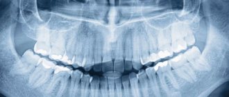
We use different approaches depending on the position and line of the patient's teeth. At the JK Clinic, 3D CT, X-rays and 3D virtual surgery are used to determine the best approaches.
a. Traditional approach 1-2 years before jaw surgery, teeth are optimally aligned through orthodontics. Then, after straightening the teeth, surgery can be done. You may then undergo minor orthodontic treatment to achieve optimal teeth alignment.
b. Surgery - First Approach This approach is used in cases where the position of the teeth is optimal for surgery. The total treatment time is much shorter than for the traditional approach due to the unnecessary preoperative orthodontic procedure. The jaw is corrected through surgery before orthodontic treatment is necessary.
c. Modified Surgery - First Approach This approach combines the traditional approach and surgery for cases where the teeth have minor line or orientation problems. Preoperative orthodontic treatment can be done for a short period (3-4 months) and then continued with jaw surgery with postoperative orthodontic treatment.
Upper jaw (Lefort I Osteotomy)
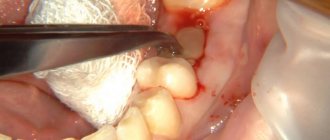
Lefort I osteotomy is the most commonly used procedure for cutting bones in the upper jaw. This procedure is performed under general anesthesia. The operation is performed entirely in the intraoral cavity. A U-shaped incision is made on the gums of the upper teeth to gain access to the jawbone. The gum is then lifted away from the bone. The upper jaw incisions are made using highly specialized instruments that allow the tissues to be clearly separated. If the upper jaw is to be reduced as much as possible, additional bone is removed. Bone may be grafted if it is necessary to lower or lengthen the jaw (bone graft). The upper jawbone is then inserted into its final position (planned prior to your surgery) and secured into place with tiny titanium plates and screws that remain in place forever. The gums are sutured using soluble ties, which dissolve within 3-4 weeks. After the bones heal, the jaw regains its strength.
| Price | 6000-8000 |
| Duration | 1 hour |
| Cut location | Intraoral |
| Recovery | Minimum 10-14 days |
| Anesthesia | General anesthesia |
| Removing stitches | In 1-2 weeks |
Lower jaw
1) Sagittal ramus osteotomy (SSRO)
This procedure is used to correct mandibular retrusion and mandibular prognathism (underbite and jaw protrusion). First, a horizontal incision is made on the inside of the ramus of the mandible, expanding access to the anterior aspect of the ascending ramus. Then, an incision is made downwards on the ascending ramus to the descending ramus, extending to the lateral border of the lower jaw in the area between the first and second molars. At the same time, a vertical incision is made, expanding access to the jaw, to the lower border of the jaw. All cuts are made to the middle of the bone, where the bone marrow is located. A medical chisel is then inserted into the pre-existing incisions and gently pressed into all areas to separate the mandible into left and right sides. From this position the lower jaw can be moved forward or backward. If retraction occurs, the additional segment must be trimmed to make room for the mandible to retract.
| Price | $6000-20000 |
| Duration | 1 hour |
| Cut location | Intraoral |
| Recovery | Minimum 10-14 days |
| Anesthesia | General anesthesia |
| Removing stitches | In 1-2 weeks |
Vertical ramus osteotomy (VRO)
Vertical osteotomy is a procedure in which the entire thickness of the mandibular rami is dissected. It is carried out on both sides, the lower jaw is divided into 2 small segments with the condyles and into a large segment consisting of the anterior part of the lower jaw and the part that includes the teeth and chin. The incisions are made behind the inferior alveolar canals, and therefore the chance of damage to the alveolar nerve is minimized. This surgery can be used to reposition, change the angle of the mandible, and also shorten the back of the mandible. However, this surgery cannot be used to lengthen the lower jaw.
| Price | $6000-10000 |
| Duration | 1 hour |
| Cut location | Intraoral |
| Recovery | Minimum 10-14 days |
| Anesthesia | General anesthesia |
| Removing stitches | In 1-2 weeks |
ASO (Anterior Segment Osteotomy)
The upper and lower first molars are removed from both sides of the mouth, along with the bone on which they are located. The front of the upper and lower mouth is moved back and attached to the jaw using titanium plates to secure it firmly. Typically done for people with protruding mouths (bimaxillary protrusion).
| Price | $6000-10000 |
| Duration | 1 hour |
| Cut location | Intraoral |
| Recovery | Minimum 10-14 days |
| Anesthesia | General anesthesia |
| Removing stitches | In 1-2 weeks |
Sunken chin
A sunken chin can also be considered a chin with an overbite, where the lower jaw is underdeveloped. This is very noticeable when looking in profile and full face.
a. Small chin: small chin but normal jaw size
Implant installation The operation is performed under local anesthesia, and the patient will not feel pain or discomfort. An incision is made through the lining of the mouth to the lower jaw, where a silicone or Gore-Tex implant is inserted. This simple procedure takes about 20 minutes.
| Price | $3000-4000 |
| Duration | 30 minutes |
| Cut location | Intraoral |
| Recovery | 7 days |
| Anesthesia | Local anesthesia |
| Removing stitches | In 1-2 weeks |
Chin forward
Sliding genioplasty This procedure is performed when the jaw position and development are normal, but the chin is sunken. A horizontal cut is made on the chin, and only the chin is moved forward. Titanium plates and screws are used to hold the chin in place.
| Price | $4000-6000 |
| Duration | 1 hour |
| Cut location | Intraoral |
| Recovery | 7-10 days |
| Anesthesia | General anesthesia |
| Removing stitches | In 1-2 weeks |
Moving the lower jaw forward
An incision is made into the mouth and the chin bone is cut out and moved forward and positioned using the SSRO (Sagittal Ramus Osteotomy) technique. Using titanium plates, screws and/or wires, the jaw is secured into place. The jaw should remain closed until the bones have healed. The operation itself takes about 1.5-2 hours, and there are no problems in going to work or light vigorous activity. Depending on the condition of the upper jaw bone, two operations may be necessary.
| Price | $4000-6000 |
| Duration | 1 hour |
| Cut location | Intraoral |
| Recovery | Minimum 10-14 days |
| Anesthesia | General anesthesia |
| Removing stitches | In 1-2 weeks |
Paranasal concavities
The nasolabial groove on the sides of the nose has a sunken appearance. This often makes you look tired and also older than your age. This problem also gives the effect of a “protruding” mouth. This procedure is popular among both young and old people who have this problem.
An incision is made inside the mouth about 1.5 to 2 cm long. The implant is inserted into the cavity of the maxillary bone. The implant can be made of silicone or Goretex. The surgeon can determine the size and shape of the implant before the procedure.
| Price | $2000-2500 |
| Duration | 30 minutes |
| Cut location | Intraoral |
| Recovery | 4-7 days |
| Anesthesia | Local anesthesia |
| Removing stitches | 7 days |
Forehead surgery
This procedure is for people who have flat, depressed or small foreheads. For a more rounded, beautiful forehead shape, an implant or filler is used. Particular care is taken to ensure a smooth, round appearance from all angles.
Implant preparation
Implants that are inserted into the forehead are custom-made and pre-checked for shape and size before surgery.
Implant installation
An incision is made along the hairline in the scalp where the implant will be inserted. The surgeon inserts the implant. This procedure is done under local anesthesia, where there is no pain or discomfort.
With. Lipofilling (Fat injections using your own adipose tissue)
Fat tissue is taken from your lower abdomen/hips and then a small (1cm) incision is made at your hairline. The fat is injected through the top of the forehead down to the eyebrows between the muscle.
| Price | $2500-3500 |
| Duration | 20 minutes |
| Cut location | Inside the hair |
| Recovery | |
| Anesthesia | Local anesthesia |
| Removing stitches | 7 days |
Revision surgery
Due to the complexity of facial contouring surgery, it is very important that the initial surgery is done correctly. Unlike other parts of the body, revision surgery for facial contours may be limited due to conditions of the jaw and facial bones.
Square jaw
You have serious asymmetry. There are no changes before or after the operation. You may have a depression below the ear. You may have part of your jaw protruding due to improper cutting (secondary angle). You have a functional jaw problem.
Jaw
You feel unevenness in the operated area. There are no changes before or after the operation.
Cheekbones
You have serious asymmetry. There are no changes before or after the operation. You have obvious sagging. Your cheeks are not the same width.
Frontal, nasolabial and chin implants
There are no changes before or after the operation. You can see the outline of the implant. Your implant is moving. The implant causes you severe discomfort or pain. You often have fluid build up around the implant.
"Back
Temporomandibular joint
The temporomandibular joint consists of articulations between three surfaces; the mandibular fossa, the articular tubercle (from the flat part of the temporal bone) and the head of the mandible.
This joint has a unique mechanism; The articular surfaces of the bones never touch each other - they are separated by the articular disc. The presence of such a disc divides the joint into two synovial articular cavities, each of which is lined with a synovial membrane. The articular surface of the bones is covered with fibrous cartilage.
Purpose of the dental system
The biological mechanics of the dental system and its understanding lead to timely recognition of pathologies of teeth and soft tissues of the oral cavity. The normal functioning of the temporomandibular joint largely depends on the structure and normal functioning of the periodontium - the soft tissues surrounding the tooth cavity.
The anatomy and physiology of the structure of the periodontium determines the biomechanics of the structure and the characteristics of the periodontium. In addition, periodontal biomechanics helps in the functioning of other organs.
Knowledge of the laws of biological mechanics of the jaw and joints helps to design and use features, as fundamental knowledge on the topic of prosthetics and dental implants. Knowledge of the anatomical features of the jaw is necessary in the manufacture of structures and dental materials.
Devices have been developed that almost completely repeat the movements of the jaw and make it possible to reproduce anatomically similar structures to the human jaw. Popular devices include the occluder, facebow, and articulator. The latter design plays an important role in the fitting and creation of individual prostheses.
If disorders of the lower jaw occur, disruptions in articulation, speech, nutrition and swallowing occur. Today, a system of articulation of movements has been developed, led by such authors as Ganau, Gisi, Monson. These authors most accurately described the biomechanics of mandibular movements.
The authors claim that dysfunction of the lower jaw, as well as sore neck muscles, necessarily lead to breathing problems. Different angles of inclination of the lower jaw determine human health.
Occlusion is a condition characterized by full contact of both jaws, which ensures complete chewing of food. Therefore, the contact of teeth is a determining factor for the characteristics of chewing mechanisms.
The work and action of the lower dentition during chewing is due to the synchronous work of all muscles and joints. The work occurs under the influence of the central nervous system. In this case, displacements that occur spontaneously occur under the influence of the neuromuscular structure.
Conscious movements of the dentition include the moment food enters the mouth and swallowing food. The remaining movements that occur after eating food are the result of unconscious motor functions of the body.
The lower part of the human masticatory apparatus is the only moving bone in the human body. However, the connecting and muscle tissue, which sets the mechanism in motion, is of great importance in the structure of the masticatory row and in the normal functioning of the muscles.
In the structure of the lower dentition, there are several muscle groups that are responsible for various movements of the jaw.
Reliable fixation using mini-implantation
Mini-implants are threaded titanium pins that are smaller in diameter than standard implants and are used primarily for more secure fixation of removable dentures. The threaded part serves to secure the implant in the bone, its head serves to fix the removable denture.
The advantages of using mini-implants include the following:
- Reliable fixation of a removable denture and, as a result, a reduction in the plastic base of the removable structure itself;
- Does not require bone grafting;
- Low trauma and short rehabilitation period after installation of a mini-implant;
- Short period of time within which it is possible to install prostheses after the operation.
- A small number of contraindications compared to the use of classical implants.
Sometimes mini-implants are used to install fixed small prostheses. For example, when the alveolar process of the lower jaw is too narrow, and the patient categorically refuses to undergo bone grafting to thicken it, which prevents the installation of classical implants.
It is important to remember that any type of implantation occurs through surgery. During the periods of preparation for surgery and rehabilitation, it is necessary to strictly follow all the doctor’s instructions, follow diet and oral hygiene, and give up bad habits.
Prosthesis on implants
The most important advantage of implantation is reliable fixation of the prosthesis. With this method of prosthetics, it again becomes possible to eat foods of any consistency, chew them thoroughly, thereby improving the functioning of the gastrointestinal tract. Diction improves, it becomes possible to smile widely and lead an active social life.
Most people who are hesitant about installing implants in the oral cavity are worried about whether the foreign body will take root in the gum, how long the rehabilitation process after surgery will take, and whether there is really a need for a rather expensive procedure, the cost of which depends on the number of implants and the condition of the bone. fabrics.
For an approximate calculation, it is worth taking the following indicators: when prosthetics of the lower jaw using one solid-cast bridge prosthesis, the installation of from 4 to 80 implants is required.