Fibrosing alveolitis
This type of pathology is notable for the fact that connective tissue develops to replace the necrotic residue of the hole left after tooth extraction. In principle, many dentists propose to consider fibrosing alveolitis not as a separate type of this nosology, but as one of the stages of its course, which is characterized by a slight decrease in the intensity of the manifestations of the inflammatory reaction and partial stabilization of the general condition of the patient. If you take a closer look at the condition of the oral cavity, you will notice a slight proliferation of soft tissues adjacent to the hole in which the pathological process occurs. The formation of a gap between the soft tissues and the hard, bone wall is also typical in this situation.
Symptoms of alveolitis and dry socket
The diagnosis of “alveolitis” is made quite simply. With alveolitis, the following symptoms are observed:
- Outwardly, it may be empty, with a yellowish coating on the walls of the hole and traces of food debris, and festering blood clots are also visible. The gum next to the hole is usually inflamed, red, swollen, and hurts when touched.
- The pain associated with this disease varies and can be both acute and mild. Some people also experience pain in the head when the socket becomes inflamed.
- When a blood clot festers, it always begins to smell unpleasant, and the hole that is inflamed also has an unpleasant odor. It can be described as the smell of rotting, decay. A clot that festers leads to intoxication of the body, which is expressed in the person’s poor condition, as well as weakness and fever.
- In most cases, alveolitis occurs without swelling of the soft facial tissues due to the fact that the infection and pus come out through the sore hole. But there are cases when this does not happen, the facial tissues and gums swell, all this is accompanied by high fever and acute pain.
Allergic alveolitis
When compounds that cause a pathological reaction in the form of an allergy enter the human body, not only the respiratory tract and the mucous membrane of the eyes react - swelling and soreness of the dental alveoli with the subsequent formation of an inflammatory process can sometimes also be noted. Accordingly, all these changes can safely be called allergic alveolitis, because their pathogenesis is based on the same antigen-antibody reaction.
This type of pathology is manifested by constant aching pain, which intensifies significantly during food consumption. Quite rightly, allergic alveolitis is recognized as one of the “mildest” forms of this disease, because it is not accompanied by a pronounced clinical picture. Usually the patient's general condition is relatively satisfactory, there is no fever. Upon careful examination of the hole left after tooth extraction, one can note the almost complete absence or insufficient amount of a protective blood clot. Remnants of food are usually visible here. It may also be that allergic alveolitis forms after 2-3 days from the moment of tooth extraction. And if timely treatment is not started, the prognostically favorable form of the disease can lead to serious complications.
Text of the book “Fundamentals of clinical dental morphology: a textbook”
5.3.1.2. Age-related changes in the periodontium In the periodontium, throughout the life of an individual, plastic processes constantly occur in the renewal of its constituent elements: fibroblasts and other cells, collagen fibers and intercellular substance. The intensity of collagen renewal in the periodontium is 2 times higher than in the gums, and 4 times higher than in the skin. The high rate of collagen renewal is evidence of the important role of continuous periodontal restructuring in the constantly ongoing processes of adaptation of the supporting apparatus of the tooth to changing loads.
With age, the plastic capabilities of periodontal cells decrease, which is reflected in a decrease in the number of active cells (fibroblasts, osteoblasts, cementoblasts), a reduction in the synthesis of collagen fibers, and a deterioration in the blood supply to pericemental tissues. There is a decrease in the width of the periodontal fissure. These changes undoubtedly cause a weakening of the fixing apparatus of the tooth root, its barrier properties, and an increase in tooth mobility, which inevitably leads to the need for medical intervention.
5.3.2. Dental alveolus
Dental alveoli (alveoli dentales)
located in the alveolar process of the upper jaw and the alveolar part of the lower jaw. These sections of the jaws form as teeth develop and erupt and almost completely disappear with their loss.
Each alveolus is a bone socket in which a tooth is located. The edges of the alveoli do not reach the neck of the tooth (enamel-cement border), therefore the depth of the dental alveoli is slightly less than the length of the root of the corresponding tooth and the latter protrudes slightly from the jaw bones. This part of the tooth root is normally covered by the gum. The deepest is the canine alveolus. At the bottom of the alveoli there are usually several openings for blood vessels and nerves leading to the tooth.
In the alveolar process of the upper jaw there are 16 dental alveoli for the roots of the teeth. The alveoli are divided into vestibular (labial or buccal) and lingual (palatal) walls. The alveoli are separated from each other by bony interalveolar septa (septa interalveolaria). The alveoli of multi-rooted teeth also contain interradicular septa (septa interradicularia), which separate the roots of the tooth from each other.
The shape and size of the alveoli correspond to the shape and size of the tooth roots. The alveoli for the roots of the incisors are cone-shaped, with rounded outlines, the alveoli for the canine and small molars are oval and compressed in the mesial-distal direction. The alveolus of the first premolar of the maxilla is often divided by an interradicular septum into lingual and vestibular root chambers. The last three alveoli of the upper jaw, the largest in size, contain the roots of large molars. Each of them is divided by interradicular septa into three root chambers, two of which face the vestibular and the third face the palatal surface of the alveolar process.
The vestibular chambers are somewhat compressed laterally, so their size in the anteroposterior direction is smaller than in the palatobuccal direction.
The palatal (lingual) chambers of the alveoli have a rounded shape. Due to the variable number and shape of the roots of the third molar, its alveolus is variable in shape and can be single-chambered or divided into 2-3 or more parts.
The alveolar part of the mandible also contains 16 dental alveoli for the roots of the teeth, separated from each other by interalveolar septa. The walls of the alveoli facing the lips and cheeks are called vestibular, and the walls facing the tongue are called lingual.
The shape, size, and thickness of the walls of the alveoli are different for teeth of different groups. The alveoli of the incisors are compressed from the sides, and the thickness of the lingual wall of the alveoli is greater than the vestibular one.
The shape of the alveoli of the canine and especially the premolars is rounded, the lingual wall is also thicker than the vestibular one. The deepest is the alveolus of the canine and second premolar. The thickness of the walls here is greater than that of the alveoli of the incisors.
The alveoli of the molars contain interradicular septa: in the first two molars, one septum divides it into anterior and posterior chambers for the corresponding roots, and in the area of the third molar there may be one or more septa. Often the alveolus of this tooth is conical in shape without septa. The walls of the alveoli of the molars are thicker than those of the alveoli of the remaining teeth of the lower jaw.
In the alveolar process of the upper jaw and the alveolar part of the lower jaw, two parts are distinguished: the alveolar bone of the alveolar wall itself and the supporting alveolar bone. The first is a thin (0.1–0.3 mm) bone plate surrounding the tooth root, to which the fibers of the periodontal ligament are attached. It consists of lamellar bone tissue that forms osteons, is penetrated by a large number of perforating (Sharpey's) periodontal fibers and contains many holes through which blood and lymphatic vessels and nerves penetrate into the periodontal space.
The supporting alveolar bone includes:
- compact bone
forming the outer (buccal or labial) and inner (lingual or oral) walls of the alveolar process, also called cortical plates;
– spongy bone,
which is located between the walls of the alveoli and the cortical plates.
The cortical plates of the alveolar process, without a sharp border, pass into the bone of the jaw body. The thickness of the plates is not the same in different parts of the alveolar process and the alveolar part: they are much thinner in the upper jaw than in the lower jaw. They reach their greatest thickness in the area of premolars and molars of the lower jaw, as well as on the lingual side.
Spongy bone fills all the spaces between the walls of the dental alveoli and the cortical plates, and also forms interradicular and interdental septa.
The crossbars (trabeculae) of the cancellous bone distribute the forces acting on the walls of the alveoli to the cortical plates, so their location usually corresponds to the direction of the forces acting on the alveoli during chewing movements. Thus, in the area of the lateral walls of the alveoli they are located predominantly horizontally, and at their bottom they have a vertical course.
The spaces between the trabeculae of the cancellous bone are filled with red bone marrow in childhood and adolescence, and with yellow bone marrow in adults. Individual areas of red bone marrow may persist throughout life [67].
5.3.2.1. Age-related changes in the dental alveoli
During the life of an individual, the bone tissue of the wall of the dental alveolus, as well as the supporting bone of the alveolar process, is in a state of constant restructuring. this ensures adaptation of the alveolar bone tissue to changing functional loads, which is clearly manifested during physiological and orthodontic tooth movements.
With age, teeth gradually wear down on the occlusal and proximal surfaces. The abrasion of the tooth is compensated by its gradual advancement from the bone alveolus. Important mechanisms of this process are, first of all, the deposition of cement in the area of the root apex, as well as the restructuring of the alveolar wall, at the bottom of which and in the area of the interradicular septa, new bone tissue is formed.
When the approximal surfaces are worn away, the teeth become less convex, but the contact between them is not disrupted, since at the same time the interdental septa become thinner (Fig. 116).
Rice. 116. Restructuring of the walls of the dental alveoli with age.
1 – enamel; 2—dentin; 3 – interdental gum (papilla); 4 – walls of the bony alveoli.
This compensatory process is known as approximal, or medial, displacement of the teeth, the driving factors of which are occlusal forces, as well as transseptal periodontal fibers that bring the teeth together. In this case, a number of changes occur in the wall of the alveoli and periodontium, which can be designated as a physiological restructuring of the supporting apparatus of the tooth.
On the medial side of the alveoli (in the direction of tooth movement), narrowing of the periodontal space and bone resorption occur. On its lateral side, the periodontal space expands, the fibers of the pericementum are stretched, and on the wall of the alveoli, new formation and deposition of coarse-fiber bone tissue occurs, which is later replaced by lamellar tissue.
This ability of the alveolar bone tissue to restructure is widely used in orthodontic interventions aimed at moving a tooth to a new location.
At the same time, thanks to the use of special devices, it is possible to provide such an effect on the alveolar wall, which leads to resorption of bone tissue in the area of pressure and the formation of new bone tissue in the area of tension, i.e., to correction of the position of the tooth in the dentition in case of some anomalies of the dentofacial system . 5.3.3.
Desna Desna
is divided into three parts: attached, free and interdental (Fig. 117).
Rice. 117. Diagram of the structure of the gums.
I – gingival sulcus; 2 – gingival epithelium; 3 – free part of the gum; 4 – gingival groove; 5 – attached part of the gum; 6 – lamina propria of the gum mucosa; 7 – bone wall of the tooth alveolus; 8 – cement; 9 – periodontal ligament; 10 – attachment epithelium;
II – enamel.
Attached part
The gums are firmly fused with the periosteum of the alveolar process of the upper jaw and the alveolar part of the lower jaw.
Free part
Gums are the edge of the gum, which is loosely adjacent to the surface of the tooth crown, separated from it only by a narrow gap - the gingival sulcus. The gum does not have a strong connection with the periosteum and has some mobility. The border between the free and attached parts of the gum is a shallow groove - a gingival groove, located parallel to the gingival margin at a distance of 0.5–1.5 mm from it, approximately at the level of the bottom of the gingival groove or slightly above it.
Interdental part
forms a triangular interdental papilla, filling the gap between adjacent teeth. There are two types of interdental papillae - vestibular and lingual, connected to each other through the interpapillary ligament.
Due to constant mechanical stress during the process of chewing food, the epithelium and connective tissue of the gums have structural features of an adaptive nature. The gum is lined with multilayered flat, partially keratinized epithelium with an average thickness of 220–250 microns, which loses the stratum corneum in the area of the gingival sulcus (Fig. 118). The gingival surface covered by non-keratinizing epithelium is about 8-10%, keratinizing by orthokeratosis - 12-15%, by parakeratosis - 70-75%.
Rice. 118. The structure of the gingival epithelium.
1 – epithelium; 2 – basal (germ) layer; 3 – lamina propria of the gum mucosa.
The cells of the basal layer of the gum epithelium are characterized by high mitotic activity, so the rate of epithelial renewal here is higher than in other areas of the oral mucosa. The basal layer contains numerous melanocytes that produce melanin, which accumulates in significant quantities in epithelial cells, determining the pigmentation of the gums.
The border between the epithelium and the lamina propria of the mucous membrane has a scalloped character due to the introduction of high connective tissue papillae of the lamina propria into it.
The gingival epithelium is directly continuous with the sulcal epithelium and the attachment epithelium. The sulcal epithelium forms the lateral wall of the gingival sulcus. At the apex of the gingival papilla it passes into the gingival epithelium, and towards the neck of the tooth into the attachment epithelium.
Gingival sulcus
(sulcus gingivalis) - a narrow slit-like space between the tooth and the gum, extending from the edge of the free part of the gum to the attachment epithelium. The depth of the groove is from 0.5 to 3 mm, on average about 2 mm. An increase in the depth of the gingival sulcus with age of more than 3 mm turns it into a gingival pocket between the gingival margin and the surface of the tooth. The bottom of the gum pocket can reach the level of the cervical part of the tooth enamel.
The gingival sulcus contains fluid in which there are desquamated epithelial cells, bacteria, and granular leukocytes that have migrated into the sulcus through the epithelium. Electrolytes, immunoglobulins, and antibacterial substances are transported from the blood into the gingival fluid.
Chemical analysis showed the presence of amino acids, peptides, carbohydrates, urea, bacterial toxins, enzymes (lysozyme, lactoperoxidase), and antibodies in the gingival fluid. Some antibiotics (in particular, the tetracycline series) can accumulate here in concentrations 4–8 times higher than their level in the blood serum. The volume of gingival fluid under physiological conditions is negligible, but it increases sharply during inflammation.
The epithelium of the sulcus is similar to the epithelium of the gums, but is thinner and does not undergo keratinization. Epithelial cells are small in size and contain a significant amount of tonofilaments in the cytoplasm. The border between the epithelium and the lamina propria of the mucous membrane is smooth due to the absence of connective tissue papillae. In the epithelium and underlying connective tissue there is a significant number of granular leukocytes, macrophages and lymphocytes.
The attachment epithelium is a continuation of the sulcus epithelium, lining its bottom and forming a cuff around the tooth, which is firmly connected to the enamel surface covered with the primary cuticle. In structure, it is a multilayered squamous epithelium, the thickness of which is greatest in the area of the bottom of the gingival sulcus (15–30 layers of cells) and decreases towards the neck to 1–3 layers. Its cells, regardless of their location in the layer, have a flattened shape and are oriented parallel to the surface of the tooth.
The most interesting morphological feature of the structure of the attachment epithelium is the arrangement of epithelial cells between two basement membranes. Epithelial cells are located on the basement membrane, connecting it with the lamina propria of the gingival mucosa. The surface cells of this epithelium provide attachment of the gum to the tooth surface using hemidesmosomes associated with a second (internal) basement membrane located between the tooth and the attachment epithelium.
Both basement membranes have a similar structure and consist of two zones:
– electronically transparent zone,
located at the base of the epithelial cells of the basal layer and called the transparent plate;
– electron-dense zone,
underlying transparent plate.
The plates contain type IV collagen, laminin, anchorin, and glycoproteins.
Epithelial cells connected to the inner basement membrane do not undergo desquamation, which is not typical for stratified squamous epithelial cells.
Desquamation is experienced by cells lying under the surface layer of the attachment epithelium, which are displaced towards the gingival sulcus and then desquamated into its lumen. The intensity of this process in the attachment epithelium is 50-100 times greater than desquamation in the gingival epithelium.
The loss of cells is compensated by their intensive new formation in the basal layer of the epithelium, the cells of which are characterized by very high mitotic activity. From the basal layer, cells move simultaneously towards the enamel and gingival sulcus.
Typically, the rate of renewal of the attachment epithelium in humans is 5–8 days, and if damaged, its complete recovery occurs within 5–6 days.
In terms of their ultrastructure, the cells of the basal layer of the attachment epithelium are somewhat different from the epithelial cells of the rest of the gum.
The cytoplasm of cells contains a well-developed granular endoplasmic reticulum and a pronounced Golgi complex, while tonofilaments occupy a significantly smaller volume in it.
The composition of cytokeratin intermediate filaments of the cytoplasm, as well as surface membrane carbohydrates, characteristic of poorly differentiated cells, indicates the presence of attachment of this class of cells in the epithelium. In this regard, a number of researchers suggest that maintaining attachment epithelial cells in a relatively undifferentiated state is important to preserve their ability to form hemidesmosomes, which ensure the connection of the epithelium with the tooth surface [66, 85].
The number of desmosomes connecting cells of the attachment epithelium is 3–4 times less than in the furrow epithelium. In this regard, the intercellular spaces are usually expanded, they contain cells such as granular leukocytes, monocytes and lymphocytes migrating to the surface from the lamina propria. At the same time, many substances are transferred through wide intercellular spaces in the opposite direction from saliva and the oral cavity (bacteria, bacterial toxins, other antigens), which may be necessary for adequate stimulation of the immune system.
Own record
The gum consists of papillary and reticular layers.
The papillary layer is formed by loose, unformed connective tissue containing a large number of blood vessels and nerve fibers with numerous nerve endings. The high papillae of this layer are smoothed in the area of the gingival sulcus.
The reticular layer is represented by dense connective tissue with a high content of collagen fibers, bundles of which firmly attach the gum over its greater extent to the periosteum of the alveolar process (attached gum). On the other hand [21], the supra-alveolar zone of the lamina propria of the gum contains functionally oriented bundles of collagen fibers that form fibrous ligaments in the gum (Fig. 119):
– vestibular-oral fibers (fibrae gingivales vestibulo-orales) are located along the midline, moving in the interdental spaces from the vestibular surface of the gum to the oral one;
– dentogingival fibers (fibrae dentogingivales) start from the cementum of the root at the bottom of the gingival sulcus and spread fan-shaped into the connective tissue of the gums. The bundles are well expressed on the vestibular and oral surfaces, weaker on the contact surfaces;
– spiral interdental fibers (fibrae interdentales spirales) begin in the gum at the bottom of the gingival groove on the medial surface of the tooth, surround it in the form of a spiral and are woven partly into the cement of the same tooth on the distal surface, partly on the medial surface of the adjacent tooth;
– interdental fibers (fibrae interdentales) form bundles 1.0–1.5 mm thick, running from the cement of the contact surface of one tooth through the interdental septum to the cement of the adjacent tooth. They continue to the roots (interroot fibers). this group of fascicles plays a special role in maintaining the continuity of the dentition. They are involved in the distribution of chewing pressure within the dental arch;
– dentoperiosteal fibers (fibrae dentoperiostales) pass from the neck of the tooth to the periosteum of the jaws. In the area of incisors and canines, these fibers are better expressed on the contact surfaces, and in premolars and molars they are well expressed on all sides.
Some of these ligaments are part of the periodontal membrane.
The lamina propria of the mucous membrane in the area of the dental-gingival junction is characterized by the following:
– relative paucity of collagen fibers and fibroblasts;
– an extensive network of blood vessels;
– high content of leukocyte cells.
Rice. 119. Gingival ligaments (scheme according to S.S. Mikhailov).
I – medial root of the second molar: II – distal root of the second molar; III – root of the third molar
1 – dentogingival fibers; 2 – spiral interdental fibers; 3 – interdental fibers; 4 – vestibular-oral fibers; 5 – circular fibers; 6 – interroot fibers.
Granulocytes (mainly neutrophils), lymphocytes and monocytes are continuously evicted from the lumen of numerous vessels, which move through the intercellular substance of the connective tissue towards the epithelium and ultimately enter the gingival sulcus and then into saliva.
Thus, the gums are one of the main sources of leukocytes found in saliva and transformed into salivary corpuscles. The number of leukocytes migrating through this route into the oral cavity is normally about 3000–5000 per minute. Most of them (70–90%) have high functional activity in the initial period after migration.
With pathology, the number of migrating leukocytes increases significantly. It is believed that the rate of movement of leukocytes is influenced by chemotactic factors secreted by bacteria that are located in or near the gingival sulcus.
Thus, such a high content of leukocytes is necessary to protect the relatively thin and non-keratinizing epithelium of the gingival sulcus and attachment epithelium, as well as the underlying tissues, from the penetration of microorganisms from the oral cavity.
Idiopathic alveolitis
If alveolitis has already developed, then you need to rush to take action, and not find out the reason why this happened. Yes, this is one of the diseases in which understanding the pathology of the process is not of primary importance, because regardless of what exactly caused the pathological process, treatment regimens will differ little from each other. The tactics of patient management are determined not by the origin of alveolitis, but by the severity of clinical symptoms.
It is not a fact that alveolitis arose due to a medical error - individual anatomical features could be the only factor that provoked the occurrence of such a situation. Take, for example, the same alveolitis after the extraction of a wisdom tooth - due to the difficulties with removing the “eights” itself, the likelihood of complications occurring is quite high. By the way, it is precisely the anatomy of the root system of wisdom teeth that causes the removal of a dental cyst most often to be carried out on the eighth molars.
What are the features of the upper alveoli of the teeth?
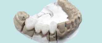
Photo: alveolitis
Regardless of which jaw the alveoli are located on, there are no significant differences in their structure. The only peculiarity of the upper alveoli of the teeth is that their structure affects diction and intelligibility of speech , which is due to the close location of the alveolar process and the palate.
The alveoli are susceptible to a number of dental diseases, the most dangerous of which is alveolitis. The disease can cause relaxation of the alveolar tissue, which can cause the tooth to shift, become loose, or even fall out. If you suspect that your teeth have begun to shift, you should immediately contact the dentist.
Exogenous alveolitis
A persistent febrile condition is characteristic; throughout the entire disease there is pronounced pallor of the skin. The patient's quality of life deteriorates significantly - he cannot eat properly, because it becomes very difficult to eat.
Lymphangitis and lymphadenitis are characteristic signs of exogenous alveolitis, because the infection quickly spreads to nearby areas. Upon visual examination, a gray coating and severe hyperemia are noted, and upon palpation the patient notes sharp pain.
Pushing the root into the maxillary sinus
Pushing the root into the maxillary sinus occurs when the forceps or direct elevator are incorrectly advanced, when the root of the tooth being removed is separated from the bottom of the sinus by a thin bone plate or it is thinned or completely absent as a result of a pathological process. The cheek of the instrument must be inserted between the root and the wall of the socket, and if the forceps are applied incorrectly, the cheek presses on the root of the tooth and thereby moves it into the maxillary sinus. In some cases, when the handles of the forceps are brought together and the cheeks are not applied deeply enough, the root slips out and gets into the sinus. To diagnose this complication, radiographs of the paranasal cavities and intraoral photographs are taken. X-ray examination helps to clarify the location of the tooth. Endoscopy is also used to detect a root or tooth in the sinus. A rhinofibroscope or endoscope is inserted into the defect in the bottom of the maxillary sinus through the socket of the extracted tooth and examined.
When the root is pushed into the maxillary sinus, perforation of the sinus bottom occurs, so this complication is accompanied by a clinic characteristic of perforation. 1. When you exhale sharply through the nose, you can observe the appearance of blood and small bubbles from the hole. 2. The appearance of blood in the nose on the side of the damaged sinus. 3. The appearance of a nasal voice. 4. The patient’s complaints about the passage of air through the socket, a feeling of pressure in the projection of the sinus. A root that has entered the maxillary sinus must be done as quickly as possible, otherwise the mucous membrane of the maxillary sinus may become infected and sinusitis may develop. The tooth or part of it cannot be removed through the socket, because this only worsens the conditions for subsequent closure of the sinus defect. The root is extracted in a hospital. A burr hole is made in the anterior outer wall of the maxillary sinus. In case of developed sinusitis, all stages of radical surgery of the maxillary sinus are performed. At the same time, plastic closure of the sinus floor defect is performed. Using an endoscope inserted through the formed hole in the lower nasal meatus, the location of the root is recorded and it is removed with special endoscopic instruments.
Post-extraction alveolitis
In this case, the name of the pathology indicates its direct origin - it is easy to guess that post-extraction alveolitis develops as a result of unsuccessful (complicated) tooth extraction. This variant of alveolitis involves inflammatory tissue damage not only to the socket itself, but also to the adjacent gum. The statistics of complications is impressive - even taking into account the potential of modern medicine, the incidence of post-extraction alveolitis is about 40% of cases, and most of them are associated with interventions performed on the lower molars.
The clinical picture of post-extraction alveolitis occurs 2-3 days after tooth extraction. Symptoms can be conditionally classified into two variants of manifestations - symptoms of general and local significance. Alveolitis necessarily has symptoms from both the first and second categories.
The first category includes:
- Low-grade or febrile fever.
- Lymphangitis and lymphadenitis (regional);
- Halitosis.
Manifestations related to the second category are:
- Local hyperemia and swelling.
- Lack of protective blood clot.
- The presence of a gray coating on the hole.
- Separation of pus.
- Local pain syndrome.
In this case, alveolitis treatment requires primarily symptomatic treatment.
An important point that must be taken into account when diagnosing post-extraction alveolitis - for the reason that the possibility of provoking the development of the disease by the remaining piece of tooth cannot be excluded, the patient must be referred for an x-ray. It may be that even if alveolitis develops, the symptoms of this disease do not indicate the presence of pathology for a certain period of time.
Many patients consider the occurrence of alveolitis to be solely the fault of the doctor, because there are articles that write that the socket can become inflamed as a result of improperly carried out preventive measures after tooth extraction. Yes, such cases happen, but not in the Academy Dent clinic - the use of modern techniques, coupled with the highest professionalism of our doctors, allows pain relief and removal without any consequences for the patient.
The worst option is when the tooth is not completely removed and some part of its root remains in the socket. This situation, without a doubt, is the fault of the doctor, because he was not convinced that the intervention was performed correctly and did not study the anatomy of the root system of the tooth using an x-ray. Severe purulent inflammation develops - body temperature rises, purulent discharge with an unpleasant odor is noted. The patient complains of a specific sensation of something sweet in the mouth - but in fact, the inflammatory process is to blame. Therefore, it is very important to ensure that after tooth extraction there are no foreign bodies left in the socket - in this case, their imperceptible resorption by cells of the immune system is a priori impossible.
The process in question, which occurs in one place, can easily inflame several adjacent holes, as a result of which eliminating the problem will become much more difficult. It will be necessary to drain the purulent lesion and use strong antibiotics administered orally. Alveolitis often requires surgical treatment, with dissection of the adjacent subcutaneous fat.
The structure of the alveoli in the mouth
Alveola is translated as cell. The term is used in pulmonology, dentistry and other medical fields. The alveoli have a spongy structure penetrated by:
- a network of nerve endings that ensure their sensitivity;
- a network of blood vessels that supply the alveolar processes with nutrients.
The walls of the dental alveoli are divided into internal, located closer to the throat, external, located on the side of the lips, and interdental. From the inside, the sockets are separated by thin bone partitions, the location of which depends on the structure of the roots of the teeth. Their inside is covered with a spongy plate, the size of which exactly matches the size of the tooth located in the socket. The outer edge is covered by a cortical jaw plate.
Photo: alveoli in the mouth
Elastic fibers predominate in the composition of alveolar tissue. The main function of the remaining cells is the constant restoration and renewal of bone tissue. The balance between the processes of its destruction and growth depends on them.
Bone tissue contains both organic and inorganic particles. Its main components are:
- proteoglycans;
- osteoclasts;
- collagen;
- osteocytes;
- osteoblasts.
If a tooth falls out or is removed, the hole heals quite quickly. A few weeks after extraction, the gums heal, and after a few months, a new gum cover is completely formed.
Treatment of alveolitis
Treatment of alveolitis, regardless of its etiology, is a very difficult task, the solution of which clearly requires professional medical care. In no case should you act independently and at home, because with an illiterate approach there is a high chance of causing significant harm.
Conditionally, the treatment of alveolitis can be classified into stages - depending on how timely the alveolitis was diagnosed, the treatment and general management tactics are determined.
Early treatment
If alveolitis was diagnosed in the initial stages, then to get rid of the disease it is sufficient to perform the following measures:
- The implementation of drug anesthesia is primarily because, despite its relative simplicity, the process of getting rid of alveolitis is quite painful.
- Treatment of the hole in which there is an inflammatory process is washed with an antiseptic solution.
- Removing necrotic debris from the socket.
- Repeated treatment of the hole with an antiseptic followed by drying with gauze.
- Placement on top of fabric soaked in antiseptic and drugs that provide an anesthetic effect.
- Apply applications with antiseptics daily (for a week) in order to eliminate the possibility of a relapse of the inflammatory reaction.
Soft tissue injuries
Damage to soft tissue is a common occurrence in the practice of a dental surgeon. For example, careless or incomplete detachment of the gum when inserting the cheeks of the forceps under the gum and further advancement will lead to stretching of the tissue with subsequent rupture. When forceps are applied to the edges of the alveolus and the gum is captured, crushing and even tearing off the marginal part of the gum occurs. Insufficient detachment of the circular ligament leads to rupture of the gums during removal of the tooth from the socket. Damage to the tongue, tissues of the floor of the mouth, cheeks, and palate occurs when the elevator slips during tooth dislocation.
Prevention of soft tissue damage
Prevention of damage to soft tissues is quite simple and consists, firstly, in careful exfoliation of periodontal tissues and careful handling of instruments in the oral cavity, secondly, in peeling off the gums to a sufficient depth, and thirdly, in using instruments for their intended purpose. When inserting the elevator blade and during tooth dislocation, it is necessary to block the direction of possible sliding of the tool with the finger of your free hand.
Treatment of soft tissue damage
If the soft tissue damage is not accompanied by bleeding, the tooth extraction should be completed, and then the edges of the wound should be brought together with sutures. If there is bleeding, it is necessary to stop it by ligating the vessel in the wound, bringing the edges of the wound together with sutures. In case of a blind wound, sutures are placed on the bottom of the mouth and tongue, after which the wound must be drained for 12-24 hours by inserting a rubber or polyethylene strip into it. When the gum edge is torn off exposing the alveolar process, it is necessary to cover the exposed bone surface. To do this, two vertical incisions are made in the gums to the arch of the vestibule of the mouth, and the mucoperiosteal flap is peeled off. In order to mobilize it, the periosteum is crossed. After this, the mucoperiosteal flap is lowered and fixed with sutures so that it covers the exposed area of the alveolar process. The second option is to form a mucoperiosteal flap in the area of the vault of the oral vestibule and move it to the area of the gum defect.