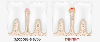
The periodontium unites a complex of tissues that surround the tooth and have genetic and functional commonality: periodontium, alveolar bone, gums, tooth root cement. This complex of tissues ensures reliable fixation of the tooth in the jaw bones.
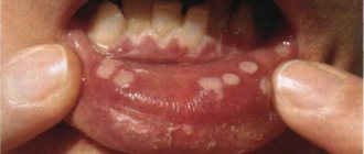
Periodontal diseases in children are widespread. According to WHO, 80% of children have some form of periodontal disease. They can be inflammatory, dystrophic and tumoral in nature. The largest group of periodontal diseases are inflammatory (gingivitis, periodontitis). They account for 94–96% of all periodontal diseases. Gingivitis (catarrhal or hypertrophic, edematous form) and periodontitis have the same causative factor. These are two interrelated forms of the disease. With gingivitis, the inflammatory process is limited only to the gums. The spread of inflammation to other periodontal tissues (periodontium, root cementum, alveolar bone) leads to the development of periodontitis. The prevalence of gingivitis in childhood is 80%, periodontitis is 3–5%. Most often, periodontal diseases are detected at the age of 9–10 years.
Gingivitis and periodontitis are divided into:
- according to the clinical course: acute, chronic, aggravated;
— by localization: localized, generalized;
- by severity: initial, mild, moderate, severe.
Depending on the age of the child, periodontitis is divided into:
— for prepubertal periodontitis — up to 11–12 years;
- puberty (juvenile) - from 12 to 17 years;
- post-pubertal - from 17 to 21 years.
Features of the development of periodontal diseases in children are associated with the fact that, firstly, the pathological process develops in the growing, constantly remodeling tissues that make up the periodontium, in tissues that are morphologically and functionally immature, capable of reacting inadequately even to minor damaging factors. On the other hand, periodontal pathology can develop against the background of disproportion in the growth and maturation of tissue structures both within a system that has common functions (tooth, periodontium, alveolar bone, etc.), and in structures and systems that provide the entire body and adapt it to changes in the external environment (nervous, humoral, endocrine, etc.), which causes the occurrence of periodontal diseases in the juvenile period. In addition, the condition of the periodontium may be influenced by the lack of synchrony between the rate of eruption of permanent teeth and the rate of construction of the alveolar bone, which leads to a decrease in the zone of attached (alveolar) gum, lengthening of the clinical crown of the teeth by 2–5 mm, and a decrease in the depth of the vestibule. Therefore, when assessing the clinical and radiological signs of periodontal diseases, it is necessary to take into account the structural features of the periodontium in childhood. The gingival groove in children is deeper, up to 3 mm, and during tooth eruption up to 4 mm; the periodontal gap in the cervical region during the period of incomplete root formation is 2 times wider than in adults, which must be taken into account when analyzing radiographs; mineralization of the tips of the interalveolar septa and the compact plate is completed simultaneously with the end of root formation, that is, in the frontal area at the age of 8–9 years, and in the lateral areas at 14–15 years.
The etiological factors of inflammatory periodontal diseases are divided into local and general. This division is conditional, since etiological factors can be closely related to each other and to the child’s body. Both local and general factors have different effects on immature periodontal tissues in childhood.
According to WHO, the leading role in the development of periodontal diseases belongs to the microflora of dental plaque, dental plaque, which is represented mainly by gram-negative and gram-positive cocci, obligate and facultative anaerobes, actinomycetes, protozoa, fusobacteria, yeast fungi, spirilla, spirochetes, bacteroides, etc.
The formation of dental plaque in large quantities is observed, on the one hand, with poor oral hygiene or lack thereof. On the other hand, the formation of abundant plaque and dental plaque is associated with a violation of the mechanisms of natural self-cleaning, which can be caused by a number of factors that occur in the child’s oral cavity: a) hyposalivation or viscous saliva; b) traumatic occlusion, which is observed with crowding of teeth, malocclusion in different planes, incorrect orthodontic treatment, early removal of primary molars, which leads to overload of permanent incisors; c) anomalies in the structure and attachment of the frenulum of the lips and tongue, small vestibule of the oral cavity; d) dysfunction of chewing (lazy or chewing on one side), swallowing (infantile type), breathing (oral, mixed type); e) bad habits; f) chronic periodontal trauma from decayed teeth, incorrectly applied fillings, parts of orthodontic appliances, self-inflicted periodontal injuries by adolescents with nails, pens, hairpins, etc.; g) insufficient load on the masticatory apparatus, associated with the predominance of carefully processed soft foods in the diet.
All of the above factors make it difficult to wash out microbes with saliva, which leads to an increase in the number of pathogenic microorganisms in the oral cavity and disruption of the dynamic balance between normal and pathogenic flora of the oral cavity. Self-cleaning is understood as the constant ability of the oral cavity to cleanse its organs of detritus, food debris, and microflora. The main role in this is played by the salivary glands, which provide an adequate volume of secretion, flow and quality of saliva necessary for the formation of a food bolus that is convenient for chewing and swallowing. The movements of the lower jaw, tongue, lips, cheeks, as well as the normal structure of the dental system, the correct functioning of the functions of chewing, swallowing, breathing, speech, the full load of the masticatory apparatus, the nature of nutrition (the predominance of coarse, hard food) are important. The dental pulp also takes part in self-cleaning due to the release of dental cerebrospinal fluid onto the tooth surface. It is known that teeth that are pulpless and located outside the dental arch are difficult to clean. Through the pulp, its vascular network, and connective tissue structures, the influence of the general condition of the child’s body on the ability of the tooth surface to self-clean is realized.
Until the 60s of the 20th century, the development of inflammatory periodontal diseases was associated, on the one hand, with systemic diseases of the body (however, the mechanism of changes in the periodontium was not clear), and on the other, with occlusal trauma. But overloading of the teeth leads to destructive processes in the periodontal bone tissue, and not to inflammation, and it does not occur in all patients. It was only in the 60–70s of the last century that dentists began to associate periodontal diseases with dental plaque.
It has been clinically and experimentally established that without dental plaque there is no periodontitis. All causative factors were divided into primary and secondary. The primary set of causes included dental plaque and the inflammatory reactions caused by it. The secondary complex of causes covered local and systemic factors that allowed the primary complex to be realized. At the same time, inflammatory periodontal diseases were considered as a consequence of nonspecific infection of the periodontium by microbes of dental plaque and dental plaque. And since the late 80s of the last century, the hypothesis about the existence of a specific microflora of dental plaque has come to the fore. New microorganisms from the bacteroid group were discovered: Actinobacylus actinomycetemcomintans, Prevotella itermedia, Porphyromonas gingivalis, Bacteroides melanogenicus, etc. The existence of periodontopathogenic bacteria was recognized. If in a healthy periodontium gram-positive aerobic microorganisms predominate, and the proportion of gram-negative microorganisms is 10–15%, then with periodontitis this ratio becomes the opposite.
Currently, inflammatory periodontal diseases are considered as an opportunistic infection that adapts to existence in the oral cavity and displaces other, less pathogenic microorganisms. This infection depends not only on the presence of pathogenic specific bacteria, but also on the environment conducive to their reproduction - local changes in pH, anaerobic niche (gingival grooves, pockets), as well as changes in the body's resistance.
The pathogenesis of inflammatory periodontal diseases (gingivitis, periodontitis) is based on an immunologically determined inflammatory reaction in periodontal tissues under the influence of specific microflora. The reaction involves systems of nonspecific and specific immunity (cellular and humoral immunity), inflammatory mediators.
The resulting inflammatory mediators (histamine, serotonin, bradykinin) increase vascular permeability, cause hyperemia, swelling of the gums, periodontium, alveolar bone, as well as gum pain. Initially, symptoms of gingivitis appear (catarrhal or hypertrophic, edematous form). During its long-term course in the absence of treatment, loosening and destruction of the periodontal epithelial attachment occurs, germination of the epithelium in the apical direction with subsequent bone resorption, due to both the cytotoxic effect of microbial endotoxins and an acidic environment, and the activation of osteoclastic resorption under the influence of inflammatory mediators (lymphokines, leukotrienes, interleukins, prostaglandin E2). Systemic diseases (endocrine, diseases of the cardiovascular system, blood, gastrointestinal tract, hypovitaminosis, dysfunction of the gonads, immunodeficiency states, etc.) lead to changes in the immunobiological reactivity of the body, to a decrease in protective and adaptive reactions that ensure the resistance of the body in general and periodontal disease in particular. There are numerous studies that indicate a significant weakening of nonspecific and specific immunity factors in patients with periodontitis. In this regard, conditions are created for the implementation of the primary set of causal factors. Differences in the course of periodontal diseases are determined by the different state of immunity in patients. Long-term contact of dental plaque microflora with periodontal tissues can lead to the development of autoimmune processes.
Systemic diseases certainly affect the condition of the periodontium, but this effect consists of aggravating the course of an already existing process, possibly increasing the risk of its occurrence, but not being the direct cause of the disease.
Pathomorphologically, the following occurs in the gum: swelling, lympholeukocyte infiltration, disintegration of mucopolyscharides, fragmentation and lysis of collagen, then destruction of the epithelial attachment, collagen fibers of the circular ligament of the tooth. Subsequently, proliferation and submerged growth of the epithelium, destruction of periodontal fibers, inflammatory osteoclastic bone resorption are noted, lymphoid and plasma cells are detected, cement resorption with the formation of depressions and bays is observed.
Periodontitis in children: a mouth full of worries
Periodontitis is a disease in which all periodontal tissues become inflamed. Inflammation of the gums occurs, destruction of muscle ligaments, pathological periodontal (so-called periodontal) pockets are formed, the process affects the bone, then teeth begin to loosen and fall out.
Periodontitis in children is most often detected at the age of 9 - 10 years. And although this is not the most common dental disease, it occurs in only 3 - 5% of children, nothing guarantees that your child will not be affected by this problem.
The peculiarity of the course of periodontitis in children is that the inflammatory process affects functionally immature, growing, constantly remodeling tissues, which sometimes react inadequately even to minor damage.
In addition to the standard division according to prevalence into local and generalized, as well as into chronic and acute - according to the nature of its course, childhood periodontitis is divided into prepubertal and pubertal.
Classification
Periodontitis in children occurs:
- local and generalized (widespread);
- acute and chronic;
- pubertal and prepubertal.
If everything is clear with the first two points, then we will dwell on the last in more detail.
The prepubertal form of the disease occurs when the mammary units erupt or immediately after this process is completed. Then the teeth cannot reliably connect to the gums, and the structure of the bone tissue changes. If the disease is not noticed in time, the child will become toothless very early. Afterwards, destructive changes can move on to the rudiments of permanent units.
It is believed that the prepubertal type of pathology is caused by:
- decreased immunity;
- the presence in the mouth of specific microorganisms that trigger the process of gum destruction.
Pubertal periodontitis manifests itself in adolescence. Its appearance is determined by factors such as:
- changes regarding sex hormone levels;
- poor oral hygiene;
- untreated dental curvatures.
In the pubertal form, adolescents complain of bad breath, itching of the gums and bleeding, changes in the consistency of saliva (it becomes very viscous), and local swelling.
Prepubertal periodontitis
It begins during the eruption of baby teeth or immediately upon its completion. It is characterized by disruption of the connection between the gum and tooth and serious destruction (change in structure) of the bone. Prepubertal periodontitis already at a very early age leads to the loss of a large number of baby teeth, after which the inflammatory process spreads to the rudiments of permanent teeth. The causes of the development of prepubertal periodontitis are most often weakened immunity, as well as the presence in the child’s oral cavity of specific types of microorganisms, the original carriers of which in most cases are the baby’s parents.
Periodontitis
In children, periodontitis is more common, localized in the area of 1–2 or groups of teeth in the frontal area of the jaws with crowded teeth or dental anomalies, with anomalies in the structure and attachment of the frenulum of the tongue, lips, and the small vestibule of the oral cavity. The following signs are characteristic of periodontitis:
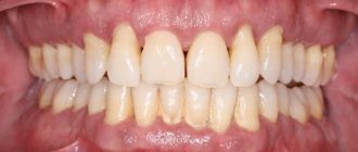
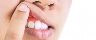
- complaints of bleeding gums, sometimes pain in them during exacerbation, bad breath; with initial changes, complaints may be absent;
— symptomatic gingivitis (catarrhal, hypertrophic);
— deposition of supra- and subgingival soft plaque and tartar;
— destruction of the periodontal epithelial attachment;
- formation of periodontal pockets with serous or purulent contents;
— destruction (destruction) of the edge of the alveoli (interalveolar septa), determined on an x-ray;
- exposure of the necks and roots of the teeth;
- tooth mobility and traumatic occlusion.
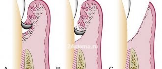
In the initial form of periodontitis, there may be no complaints; mildly expressed symptoms of catarrhal gingivitis, plaque deposition, false periodontal pockets up to 2–2.5 mm are clinically determined, the teeth are stable, the necks of the teeth are not exposed. X-rays reveal vagueness or destruction of the cortical plate at the apexes of the interalveolar septa and slight osteoporosis of their spongy substance.
In mild forms of periodontitis, patients complain of bleeding gums when brushing their teeth; symptomatic catarrhal gingivitis, small amounts of plaque and tartar deposits, periodontal pockets up to 3.5 mm deep, and slightly or 1st degree tooth mobility are clinically determined. The radiograph reveals the absence of a cortical plate at the apexes and lateral sections of the interalveolar septa, expansion of the periodontal fissure in the cervical region, and subsequently destruction of the interalveolar septa up to 1/3 of the root length; foci of osteoporosis of the interalveolar septa. In the chronic course, osteoporosis is not clearly expressed, and during exacerbation it is diffuse in nature.
In the moderate form of periodontitis, complaints of bleeding gums appear during brushing teeth and eating, symptomatic gingivitis, deposition of tartar and plaque, periodontal pockets up to 5-6 mm with serous or purulent discharge are clinically determined; mobility of teeth of the 1st–2nd degree, exposure of the roots of the teeth occurs. The X-ray shows destruction of the interdental septa from 1/3 to 1/2 of the root length, expansion of the periodontal fissure, osteoporosis is insignificant in the chronic course, and diffuse in case of exacerbation.
Severe periodontitis is characterized by complaints of bleeding and sore gums when brushing teeth and eating. Clinically, symptoms of catarrhal or hypertrophic gingivitis are detected, significant deposits of supra- and subgingival calculus and soft plaque, pockets more than 5-6 mm deep, filled with granulations and purulent contents. The radiograph shows destruction of the interalveolar septa within 2/3 of the root length, diffuse osteoporosis of the remaining bone tissue of the septa, mobility of teeth of the 2nd–3rd degree, exposure of the roots, displacement of teeth and traumatic occlusion.
Diagnosis of periodontitis in children
Examination of a child with periodontitis begins with a study of complaints, medical history and life history. Anamnestic data includes information about the physical and intellectual development of the child, common and infectious diseases suffered and existing at the time of examination. When examining a child, pay attention to his posture, since it is a factor in the full development of the child and a risk factor for anomalies in the development of the dental system.
Examination of the face in front and profile at rest and when closing teeth, during conversation and facial movements allows us to determine functional deviations in accordance with facial characteristics. Thus, the “thimble” symptom in the form of pinpoint retraction of the skin on the chin is a sign of impaired swallowing function. Pay attention to the state of tone of the facial and chewing muscles at rest and during function. The type of breathing (nasal, oral, mixed), the position of the lips (closed, open, tense or atonic), the state of the red border of the lips (color, volume, turgor, architectonics, etc.) are determined, which makes it possible to establish a violation of the basic functions of the dental system and bad habits. The condition of the temporomandibular joint and the lymphoid apparatus of the maxillofacial skeleton is assessed.
A study of the vestibule of the oral cavity allows us to judge its depth, severity and nature of attachment of the labial frenulum to the alveolar process, the state of the mucous membrane covering it, the degree of teething, their position in the dentition and the relationship of the jaws. The depth of the vestibule of the oral cavity is determined by horizontal abduction of the lower lip by the distance from the edge of the marginal gum to the level of the transition of the mucous membrane to the lip. The vestibule is considered shallow if the specified distance does not exceed 5 mm, medium with a depth of 5 to 10 mm, and deep over 10 mm. The area of the attached (alveolar) gum is measured in millimeters, as well as the depth of probing of periodontal pockets using a graduated blunt probe. In addition, the localization of the gingival margin relative to the enamel-cementum boundary (degree of root exposure) and the presence of bleeding upon light probing must be determined.
When examining the oral cavity, the condition of the salivary glands, the position, size of the tongue, the structure and nature of the attachment of the frenulum of the tongue, the shape and relationship of the dental arches, and the presence of crowding of teeth in the frontal area are assessed. The teeth are examined to identify carious lesions, and the quality of the applied fillings is assessed. The mobility of teeth and their occlusal and interdental relationships, the presence of plaque and tartar are determined.
To objectively identify the symptoms of gingivitis and its prevalence, the Schiller-Pisarev test is used, and the degree of gum inflammation is determined using the PMA index. The degree of destructive changes in the periodontium is determined by the periodontal index. Assessment of the hygienic state of the oral cavity is carried out using the Fedorov-Volodkina, Green and Wermillion hygienic indices. It is mandatory to determine the uniformity of the occlusal load using the fingerprint method (occlusiogram), as well as radiographic examination of the periodontium. If X-histiocytosis is suspected, an X-ray examination of the jaws and skull bones is performed.
In addition, a study of the child's developmental history is necessary; conducting clinical blood tests (general, for glucose levels to exclude diabetes), as well as determination of immunoglobulin levels to exclude immunoglobulinemia, a general urine test, and if X-histiocytosis is suspected, for neutral fat. According to indications, cytological and microbiological methods are used to examine periodontal pockets. The child must be examined by a pediatrician, hematologist, endocrinologist, immunologist, pediatric gynecologist, psychoneurologist, etc. in order to identify and treat systemic diseases that may affect the course and outcome of treatment of gingivitis and periodontitis.
Differential diagnosis
Periodontitis must be differentiated from chronic gingivitis and changes in the periodontium with X-histiocytosis, diabetes mellitus, constant and cyclic neutropenia, etc. Some difficulties arise in the differential diagnosis of the initial forms of periodontitis with gingivitis. X-ray data are decisive in this case. With chronic gingivitis, as a rule, there are no changes in the alveolar bone on the radiograph; only in rare cases, with long-term and untreated gingivitis, osteoporosis of the interalveolar septa is observed. When analyzing radiographs, it is necessary to take into account the variability of the shapes of the interalveolar septa in children, as well as the fact that the periodontal gap in the area of the necks of the teeth before the formation of the permanent dentition is wider than in other areas; during the period of teething, the interalveolar septa are not formed, the cortical plate at their apexes is not determined.
When differentially diagnosing periodontitis with changes in the periodontium with X-histiocytosis, one must keep in mind that with X-histiocytosis, along with changes in the alveolar bone, there are foci of bone tissue destruction in the body and branches of the lower jaw. In diabetes mellitus, vertical resorption of the interdental septa with funnel-shaped, crater-shaped pockets predominates, which does not extend to the body of the jaw. With constant and cyclic neutropenia, the process is limited to the interalveolar septa and has clear contours.
With cyclic neutropenia, exacerbation of the process occurs at strictly defined intervals, characteristic of a particular child (usually 21 days) and lasts 4-5 days.
Prepubertal periodontitis
Occurs in children under 11 years of age. The most common form is a generalized one. The disease begins during or shortly after the eruption of baby teeth and is manifested by a violation of the attachment of the gums to the teeth and severe destruction of the alveolar bone, leading to premature tooth loss in children from the age of three. The early and aggressive course is due to the fact that such patients have immune defects - there are few monocytes and polymorphonuclear leukocytes in the blood. All children from birth have decreased resistance to infections, furunculosis, pustular skin diseases, otitis, and pneumonia. The children are pale and have low nutrition. This disease is associated with microorganisms of the bacteroid type: Porphyromonas gingivalis, Actinobacylus actinomycetemcomintans. Infection occurs from parents, the window of infection is from 1 to 3 years. It begins with the primary destruction of the periodontium, while at first there are no symptoms of gum inflammation. Subsequently, the architectonics of the gingival margin changes, the gums seem to recede, the necks of the teeth are exposed, deep periodontal pockets form, and then inflammation develops. X-ray reveals vertical destruction of the alveolar bone. Subsequently, mobility, movement and loss of teeth occur. There are no symptoms of bleeding or pain. There is no tartar, the teeth are covered with a soft plaque. Children first lose their milk teeth, then the process spreads to the rudiments of permanent teeth, they erupt early, similar changes occur with them, and by the age of 14–15 all teeth fall out. However, the pathological process does not extend beyond the alveolar bone. With the loss of permanent teeth, the destructive process in the jaws stops.
The same changes in the periodontium, combined with palmoplantar hyper- or dyskeratosis in the form of cracks on the palms, feet, forearms, and increased sweating, are also observed in Papillon-Lefevre syndrome. There is evidence in the literature that taking tetracycline antibiotics for Papillon-Lefevre syndrome during the primary dentition period prevents the development of generalized periodontitis in the permanent dentition and the loss of permanent teeth.
Pubertal (teenage) periodontitis
There are 2 forms: localized and generalized. Localized pubertal periodontitis is observed in practically healthy children and adolescents without systemic diseases. It is characterized by rapid and severe destruction of the alveolar bone in the area of the first permanent molars, and sometimes the incisors. Clinically, mild inflammation (which may be absent) and a small amount of plaque or tartar are observed. Bone tissue destruction progresses 3–4 times faster than with periodontitis in adults. Currently, this disease is associated with a bacteroid of the Actinobacylus actinomycetemcomintans type. In most cases, it occurs in children whose parents are carriers of this microorganism. It enters the periodontal tissue soon after teeth erupt. The process occurs with minimal inflammatory reaction. The rapid spread of the process is due to the fact that specific microflora not only populates the groove and subsequently the periodontal pocket, but also penetrates deep into the periodontal tissues, including bone tissue, where it is quite persistently preserved. On the other hand, this microorganism has the ability to suppress the chemotaxis of leukocytes, and antibodies in such cases do not have time to form. The periodontium is rarely damaged after the eruption of permanent teeth, since by this time specific antibodies have time to form and exert their protective effect. Previously, this disease was described as local desmodontosis, local monoalveolysis according to Orban.
Generalized pubertal (teenage) periodontitis is rare. In this case, the process often involves the periodontium of both jaws. It is characterized by an aggressive course, rapid and severe destruction of the alveolar bone. It is commonly called aggressive, or rapidly progressive, periodontitis. According to foreign and domestic literature, a large number of facultative anaerobic gram-negative rods, mainly Porphyromonas gingivalis, accumulate in the area of the affected teeth. The most aggressive course is associated with the combination of this microorganism with Actinobacylus actinomycetemcomintans and Prevotella itermedia. It is with this combination that microorganisms exhibit a sharply positive mutual influence.
Local factors
Insufficient level of oral hygiene
The inability to properly brush your teeth with a special children's toothbrush and the reluctance to regularly carry out hygiene procedures lead to the accumulation of microorganisms in the periodontal pockets, and the toxins they release trigger the inflammatory process.
Short frenulum of the tongue or lip
Insufficiently long frenulums provoke atrophy and inflammation of the adjacent gum area due to constant tension on the frenulum fibers. The inflammatory process is localized in the area of the lower central teeth.
Chewing food on one side
The habit of chewing food on one side is fraught with the accumulation of plaque on teeth that are not involved in chewing. It is necessary to explain to the child that food should be chewed on both the left and right sides.
Anomalies and deformations of the dental system
The development of the inflammatory process in periodontal tissues can be caused by both functional overload of certain teeth and insufficient load on them. The reason for this is an incorrect bite or an incomplete set of teeth (the permanent teeth have not yet grown in, and some of the baby teeth have already fallen out).
Gum injuries
This group of factors that provoke the occurrence and development of periodontitis includes chronic trauma to the gums from decayed teeth or parts of braces, as well as filling defects and damage to periodontal tissue by nails, pens, and hair clips.
Nutritional nature
Too soft food does not contribute to a sufficient load on the child’s chewing apparatus and self-cleaning of the teeth, which results in the accumulation of plaque at their base, and as a result, the appearance of tartar in children.
Reasons why periodontitis develops in children
In addition to the above-mentioned causes of the disease, dentists also focus on the following:
- Untimely removal of soft and hard plaque. If a child is not able to clean his teeth independently, then he must undergo a professional hygiene procedure twice a year. It is absolutely painless. It is performed by a dentist using special equipment. Using a device that supplies a pressurized stream of air with small abrasive particles, the doctor treats one tooth after another. This simple measure is the best prevention of gum disease.
- Shortened frenulum of the lip or tongue. If this feature is not detected, the area of the gum adjacent to the frenulum will be under constant tension. As a result, an inflammatory reaction will occur.
- The habit of chewing food with only one side of the jaw. Then plaque will begin to accumulate in large quantities on the opposite side. Over time, it will transform into stones and will irritate the mucous membranes. That is why it is important to teach your baby to chew on both sides of the jaw.
- Severe anomalies in the structure of the jaws and the location of the teeth. Due to an altered bite, individual units may either suffer from overload or not participate in the chewing process at all. Both the first and second options are dangerous due to adverse consequences. Therefore, if a child has obvious disorders, it is necessary to consult with an orthodontist about their elimination. The doctor may prescribe wearing plates, mouthguards or braces.
- Traumatic damage to the gums. Children can damage the soft tissues of the oral cavity when they chew on foreign objects, fall, or hit something. Chronic injuries can also be caused by damaged teeth and inaccurately fixed parts of braces.
- Refusal to eat hard foods. If the baby eats pureed food all the time, then the necessary load is not placed on the chewing apparatus. Then plaque begins to accumulate intensively on the teeth - tartar appears. As it grows, it penetrates under the gums.
It has also been proven that the risk of periodontitis in children increases if they have diabetes or congenital heart disease.
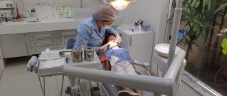
Treatment of periodontitis in children: the work of professionals
Non-surgical methods for treating the inflammatory process of periodontal tissue are the same as in adults.
This includes professional oral hygiene and various physiotherapeutic procedures. Surgical interventions, such as lip frenulum correction, may also be required. In any case, before prescribing certain procedures, a child must visit pediatric dentistry for consultation with three specialists - a therapist, a surgeon and an orthodontist. They will determine the true causes of periodontitis and develop an individual treatment plan. It is also important to exclude systemic diseases that can provoke periodontitis. Their treatment must be carried out with the participation of a specialist of the appropriate profile.
Diagnostics
If plaque and deposits begin to appear on your child’s teeth, you need to visit the pediatric dentist ahead of time.
Generalized periodontitis is more expressive than localized periodontitis, so it can be diagnosed visually. To determine the gap between the tooth and the gum, a special instrument is used - a periodontal test. Normally, the depth of the gap should be 2-3 mm. A value above 5 mm is a sign of pathology.
To obtain a clearer picture of the disease, the doctor may order an x-ray examination.
How to treat pathology in children
Treatment of periodontitis in children is carried out comprehensively. All therapeutic measures (depending on the severity of the case, they can be combined) are aimed at stopping the destructive process, preventing its further development, and eliminating provoking factors. The main thing that the doctor needs to do is to help the little patient save his teeth, prevent the pathological process from affecting the permanent bite and the rudiments of permanent teeth, and normalize the function of complete and painless chewing of food.
During therapy, the following measures must be taken.
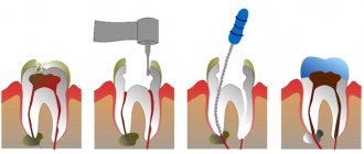
1. Removal of tartar and sanitation of the oral cavity
Bacterial plaque is a source of infection. Without its removal, all further therapy simply will not make sense. This is where you need to start treating periodontitis, gingivitis and periodontal disease not only for children, but also for adults.
Gentle techniques are used for children: using ultrasound, hand tools, Air Flow treatment and a professional brush with paste, the doctor will carefully remove plaque from the teeth and from under the gums. You can use an effective and safe laser, which, among other things, helps relieve the inflammatory process. Afterwards, the doctor will apply a strengthening fluoridated compound to the enamel, which will make it more resistant to bacterial attack.
Important! In severe conditions, if plaque sits deep in periodontal pockets and cannot be completely removed using non-invasive methods, then surgical intervention is indicated - gingivectomy (dissection of the periodontal pocket with its subsequent curettage) or curettage.
After removing plaque, it is necessary to cure diseased teeth from caries, pulpitis, periodontitis, if such dental problems occur.
Use of medications
To stop the inflammatory process, removing plaque is not enough. After visiting the dentist, parents will have to make every effort to ensure that the little patient follows all the doctor’s recommendations. Drug therapy may include the following:
- anesthetics – reduce discomfort and pain,
- antiseptics – have an antimicrobial effect,
- anti-inflammatory drugs – eliminate redness, swelling, stop the inflammatory process,
- enzyme proteolytic preparations - used if there is purulent discharge from the periodontal pocket,
- antibiotics – indicated for moderate and severe forms of the disease.
Medicines are used in the form of tablets, irrigations, applications, rinses and mouth baths.
Application of vitamins
The most useful and necessary vitamin for gums is vitamin C. It will help strengthen the mucous membrane, normalize blood flow in it, and provide it with the necessary nutrients. Vitamin C is contained in rose hips, so children are often prescribed its decoction or syrup. More specialized medications are prescribed by the doctor based on the clinical picture, so do not try to buy vitamins and medications based on reviews on the Internet. Remember that the same vitamin C in different dosages can cause an allergic reaction, you need to be careful with it.
It is also necessary to strengthen your teeth. Vitamin complexes containing calcium, fluorine, and phosphorus are suitable for this.
In some cases, the doctor prescribes physiotherapeutic procedures: electrophoresis, laser therapy, UHF, ultraviolet irradiation.
On a note! You need to strengthen your gums from the outside as well. For example, using sea buckthorn oil or vitamin E will help regenerate the mucous membrane and promote its speedy healing.
Splinting teeth
If the teeth become mobile, this causes severe discomfort and makes it impossible to chew food without pain. To solve the problem, doctors use splinting structures. However, their use will be appropriate only in the early stages of pathology development. If the disease is severe and advanced, then the splints will not be able to save the situation; they may fall out along with the diseased teeth.
Read on the topic: splinting teeth for periodontitis and periodontal disease.
Also, during and after treatment, it is necessary to strengthen oral hygiene, so a dental specialist will tell you and your child how to do this correctly and what personal hygiene products are suitable in each clinical situation.
It will be necessary to adjust your diet. So, during the period of treatment you should not eat hard and irritating mucous foods, you should not eat too hot or too cold foods and drinks. After treatment, you will need to gradually introduce solid vegetables and fruits into your diet, which help remove plaque.
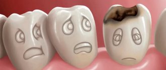
Caries, pulpitis and periodontitis of primary teeth in children
Milk teeth have thinner enamel and dentin layers than permanent teeth, they have a larger pulp chamber and, therefore, infection spreads faster to the pulp. They are more often susceptible to carious lesions - and not least because of night feeding or the habit of drinking drinks from a bottle at night (mostly sweet or sweetened - tea, juices).
In addition, caries is often provoked by plaque on baby teeth, which forms faster than in adults, and which not all parents are in a hurry to remove. Many people begin brushing their children’s teeth at a more or less conscious age, while a child should be taught hygiene and regularly remove plaque from the moment the first teeth erupt, that is, from about six months of age. Also, the cause of diseases of the dental system can be an insufficient amount of solid food in the baby’s diet, since chewing solid food helps to clean the dental surfaces themselves.
The child's nutritional habits, lack of hygiene, and too thin enamel layer provoke a phenomenon called circular caries of baby teeth - carious lesions spread over the entire tooth surface, as if encircling the crown.
Caries quickly turns into pulpitis - an inflammatory lesion of the dental pulp. Most often, a doctor is consulted precisely at this stage of the infectious process, ignoring the initial stages of damage to dental tissues. Pulpitis, in turn, provokes periodontitis of primary teeth in children - inflammation of the periodontal tissues near the tips of the tooth roots. This condition is usually accompanied by severe pain, severe swelling of the affected area, and often an increase in body temperature.
Removal of baby teeth in children
Removal of baby teeth in children is carried out only for specific indications or in the case when the tooth is ready to fall out on its own and just needs a little “help”. If there is a choice - to remove or treat, a competent dentist will always advise treatment, since the early loss of baby teeth leads to a number of serious disorders of the dental system, including the appearance of permanent bite defects.
Extraction operations should be carried out exclusively in the dentist’s office - attempts to remove teeth at home can lead to injury to soft tissues, infection of the socket, injury to the rudiment of a permanent dental unit, etc. The doctor will perform the extraction under local anesthesia quickly, competently and completely painlessly.
How to protect baby teeth from caries?
Prevention is the best way to cope with any disease. Therefore, those parents who care about the health of their children should definitely think about caries prevention. How to protect baby teeth from caries? The recommendations of experts in this matter are as follows:
- The correct diet for the mother during pregnancy is with a sufficient amount of foods containing calcium, fluorine and phosphorus: cottage cheese, beef and beef liver, sea fish, nuts, fruits and vegetables, cereal porridges.
- Compliance with hygiene standards: daily brushing from the moment the first teeth appear, first with a cotton or gauze swab with boiled water, then with special baby brushes and pastes. Plaque on baby teeth should be removed promptly and thoroughly.
- A healthy diet for a child: as little fast carbohydrates, carbonated drinks, sweets as possible, more fruits and vegetables, cereals, and dairy products.
- Timely visits to the dentist for preventive examinations (at least once every six months).
It is important that parents develop a conscious attitude towards the health of their children's teeth and oral cavity in general. A timely visit to the dentist in case of any identified problems, as well as for regular medical examinations, will help maintain the health of the child’s dental system – and therefore the digestive system too.