- What are root canals?
- What are roots, how many roots do teeth have?
- How many canals in a tooth
- Indications for dental canal treatment
- Canal diseases
- Pus in the dental canal
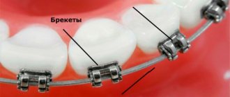
- Endodontics
- Stages of root canal treatment
- Canal filling methods
- Canal treatment under a microscope
- Possible complications after endodontic treatment
- Prevention of dental canal diseases
- Choosing a paste for prevention
- Tooth canal treatment: price
What are roots, how many roots do teeth have?
The roots of the teeth are located in the jaw under the gum, bone formation, 70% of the entire tooth, thanks to them the teeth are firmly held in the jaw and cope with chewing everyday loads.
The number of roots does not equal the number of tooth crowns. The total number of roots is different for each person, it is individual. It differs depending on the location in the oral cavity, race, age, and load taken.
Number of roots in different teeth:
- 1 root has incisors and fangs,
- 1-2 roots have premolars,
- 3-4 roots at molars,
- Wisdom teeth can have up to 5 roots.
Roots are also present in baby teeth; they are the attachment of the teeth to the jaw; one tooth can have three roots. During the change of teeth, they dissolve, giving way to growing permanent ones, so the fallen baby teeth do not have “roots”.
Features of the structure of the root system
Human teeth have a purely individual structure, so the root system of the same element may differ from person to person, including the number of canals in it and their branches. The structure of the roots is directly related to the immediate purpose of the tooth:
- incisors of the upper and lower jaws - help to bite off food,
- canines and premolars - perform the primary chewing function,
- molars - take on the main chewing load and help crush solid food as thoroughly as possible.
The photo shows teeth with roots.
The last ones in the row, sixes and sevens, and sometimes eights, require more nutrients to remain strong and wear-resistant throughout a person’s life. Therefore, premolars and molars usually have a developed canal system. But before we move on to a more detailed consideration of the structural features of the root system, let’s look at what a tooth actually consists of.
Indications for dental canal treatment
Root canal treatment
- a necessary procedure in case of inflammation, helps to save the tooth. Since tooth canals are treated endodontically, a number of indications and contraindications must be taken into account. You should immediately contact a dentist for treatment when the tooth hurts when pressing on it, if there is pain in the cheek, swelling of the gums, and the tooth reacts to cold and hot.
You should consult a doctor if you have symptoms:
- Inflammation, there is a risk of pulp damage
- Deep caries, there is a risk of developing pulpitis, periodontitis, and abscess.
- Pulp chamber injury
- Toothache and swelling of the gums.
- After dental treatment, after a few days, painful sensations arise and do not go away.
Contraindications:
inflammation of the tooth root and maxillary sinus, root fracture, narrowing of the canals, destruction of the alveolar process.
If it is impossible to pass through the canal, it is removed or treated with mummifying paste.
Implantation of 6 teeth
The implant on the sixth tooth is installed more often than others, since it is a “running” tooth, that is, it actively participates in chewing, and therefore deteriorates faster than others. As a rule, the restoration of the sixth tooth is performed using the classical (two-stage) method: first, an implant is installed, and after 4-6 months, when the implant has completely taken root, a crown is installed.
A long-term absence of sixes can lead to displacement of neighboring units and, accordingly, to a change in the bite. The teeth located on the opposite jaw will move forward, which threatens to expose the roots. Therefore, to maintain healthy teeth and bite, it is recommended to install an implant.
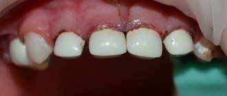
Canal diseases
Canal diseases make themselves felt by inflammation and pain, so you need to see a dentist. If there is internal inflammation, but there is a possibility of soft tissue necrosis.
- Pulpitis
- inflammation of the dental pulp. With pulpitis, blood vessels collapse and die.
- Periodontitis
- inflammation of connective tissues resulting from complications of pulpitis.
- Abscess
- the presence of pus in the gums, which arose as a complication of pulpitis
- Advanced caries
leads to inflammation of the tooth canals.
In what cases are the nerve and pulp removed?
- Pulpitis and spread of carious destruction;
- Periodontitis;
- Several carious cavities at once or one, but large carious cavity;
- Mechanical trauma to the tooth (impact, chipped tooth, bleeding from the roots).
Of course, if there is an opportunity to keep the tooth “alive” and leave the nerves and pulp, the doctor will certainly take advantage of it. However, with pulpitis or severe damage, there is one difficulty: there are no 100% methods that would cure pulp inflammation. That is, if once it becomes inflamed, then with a high probability it will be an irreversible process. Of course, if you looked into a tooth with a microscope, you could accurately determine whether the pulp can be cured, or whether it is easier to remove it - but the problem is that you cannot look at a living patient under a microscope, and dentists are not pathologists after all. Therefore, to avoid risk, the pulp is almost always removed.
Also, in some cases, the pulp is removed before prosthetics.
- especially if the original, natural tooth has been severely damaged. In addition, when using metal-ceramic prosthetics, most of the tooth is still filed down, and during this process, the nerve can be damaged - so it is also easier and more reliable to remove.
Pus in the dental canal
Pus can accumulate in the dental canal in case of improper therapeutic treatment, or as a complication of this treatment, tooth trauma. Pus in the dental canal
appears as a consequence of deep caries, when the carious process has reached the canal and pathogenic microorganisms began to develop, causing pain.
Pus accumulates due to infection and the development of bacteria in the dental canal, and has two exits: through the carious cavity of the tooth or through the gum. It is very important to seek help in time, since the presence of pus can lead to the following complications: spread to the tissues of the oral cavity, spread to nearby organs. The presence of pus over time can lead to a fracture of the jaw and dissolution of bone tissue.
Treatment boils down to removing the pus and rinsing the canal cavity through the tooth tissue or gum. The canal is washed daily for several days and done in the dentist's office. The patient is taking antibiotics to prevent the infection from spreading. It is important to see a doctor in a timely manner because of the danger of an acute condition becoming chronic.
Dystopic and impacted wisdom teeth –
From the word “retention” - delay in eruption. Thus, an impacted wisdom tooth is the eighth tooth that cannot erupt for a long time. This may be due to lack of space in the dentition (for example, due to insufficient length of the lower jaw), or due to the complete or partially horizontal position of the tooth in the jaw. Impacted eighth teeth pose quite a danger to the bite, because... if there is insufficient space for eruption, they will push adjacent teeth, moving them towards the center. Accordingly, this will lead to crowding of the front teeth.
What impacted wisdom teeth look like -
In turn, a wisdom tooth is called dystopic when it has fully erupted, but not quite correctly. For example, it may be in an incorrect position, located not along the arch of the dentition, but, for example, closer to the cheek (or the tooth may have a strong buccal or lingual inclination). The decision to preserve such teeth is made by the dentist on an individual basis. For example, if, when closing the teeth, it injures the mucous membrane of the cheek, the patient is sent for removal.
What do dystopic wisdom teeth look like?
Endodontics
Endodontics
is a branch of dentistry that studies the methods and techniques of prevention and treatment in the root canals of human teeth. Here we can draw a non-trivial analogy: a tooth is like a tree - if the roots are deep and powerful, then both the stem and crown will be strong and beautiful. Similarly, the tooth - its aesthetic beauty and health largely depend on the condition of the root canals!
Root canal treatment
necessary for complications of caries disease, namely pulpitis and periodontitis. This is the so-called primary root canal treatment. When re-treating a tooth that has been manipulated - re-canal treatment, or re-treatment.
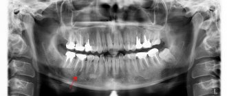
Why do we need to know the structural features of roots?
X-ray diagnostics and computed tomography in particular allow us to carefully study the root system and at the same time identify the narrowest and most intricate branches. And if the length of the canal is determined instrumentally, then the exact number of internal passages will be clearly visible on the x-ray image. Information regarding their cross-country ability is also extremely important:
- curvature up to 25 degrees – tool processing available,
- within the range from 25 to 50 degrees – the cavity is considered impassable,
- from 50 degrees - it is impossible to carry out instrumental treatment, but if the corner is in close proximity to the mouth, a specialist can try to improve patency.
An X-ray examination allows you to examine the canals and roots of the teeth.
Due to severe narrowing or even overgrowth as a result of an advanced pathological process, the doctor may not find the canal at all. Among other reasons for this situation, experts in the field of endodontics identify age-related changes and previous medical errors.
The complex anatomy of teeth makes endodontic treatment a complex and risky undertaking. But with the advent of modern technologies in the arsenal of specialists, the likelihood of medical errors has noticeably decreased. We are talking about treatment under a microscope, when during work the doctor has a clear picture of what is happening in real time before his eyes. The use of optics can significantly reduce the risk of poor-quality filling or root perforation, which means that treatment can be carried out efficiently and without complications
- Dmitrienko CB, Krayushkin A.I. Anatomy of human teeth, 2003.
Stages of root canal treatment
- The preparatory stage is when a diagnosis is made, a treatment plan and a method of pain relief are thought out. According to X-ray
- Preparing a carious cavity, it is opened, the dentin is removed, and access to working with the canals is opened.
- If the pulp is inflamed, a devitalizing paste is applied and a temporary filling is placed.
- The tooth cavity is opened and the pulp chamber arch is removed.
- The pulp of the crown is removed.
- Pulp extraction. The endodontist treats the canal up to the apical foramen.
- Treatment of the canal using instruments and medications.
Canal filling methods
Canal filling
helps prevent the development of dental canal diseases. Canal treatment is a painstaking process, complicated by the fact that the dental canals themselves are narrow, the shape can be curved, which requires painstaking work to fill the entire canal. Today, dentistry has several methods for filling canals.
1. Heated gutta-percha
Gutta-percha is a hard material that becomes elastic when heated and ideally fills the canal cavity. Several methods are used to treat a tooth canal using gutta-percha:
1) liquid injectable gutta-percha;
2) continuous wave;
3) vertical condensation;
4) syringe administration of gutta-percha.
2. Lateral condensation - cold gutta-percha
A gutta-percha pin is inserted into a canal filled with sealer paste, compacted, and sealed.
3. Thermofil - volumetric filling with hot gutta-percha
A plastic rod is inserted into the canal and the canal cavity is filled with hot gutta-percha, penetrating into all branches, leaving no free space.
4. Depophoresis technology
It is used in cases of difficult access to a curved canal that was previously filled, as well as in cases where the canal contains a part of an instrument broken during treatment.
All methods are painless for the patient. After treatment, pain is possible for two weeks if the root is removed.
Teething
The emergence of permanent teeth from the place of origin and development into the oral cavity, provided that the child develops correctly, coincides with the period of loss of milk teeth. As a rule, replacement occurs in the same sequence as eruption during the formation of a temporary bite.
The first permanent teeth appear between 5 and 6 years of age. These are the first permanent molars that have no temporary predecessors. They are the first to undergo the process of calcification of hard tissues after the birth of a child. After this, premolars, second molars, central and lateral incisors, and canines appear.
If there are problems with resorption of temporary roots and other difficulties that prevent normal eruption, a baby tooth is removed and other dental care is provided. This takes into account the fact of genetic predisposition and other features of the child’s development.
According to WHO, the standard terms of development and eruption are:
- Central incisors. Mineralizes at 4-5 years of life. They appear in the mouth at 6-8 years of age, while root formation is completed only by 10 years of age.
- Lateral incisors . They erupt at the age of 8-9 years. Complete mineralization of the enamel occurs at 4-5 years of a child’s life, and the root system continues to develop up to 10 years;
- Fangs . Enamel mineralizes at 6-7 years of age. They emerge in place of temporary ones at 10-11 years of age and complete full formation at 13 years of age.
- First premolars. They undergo complete mineralization at the age of 5-6 years and appear in the mouth at 9-10 years. The root system of these teeth is formed by the age of 12.
- Second premolars . Mineralizes by 6-7 years. They usually erupt at the age of 11 years and complete development by 12 years.
- First molars . They are the very first permanent teeth. They mineralize at 2-3 years, erupt at 5-6 years and are fully formed by 10 years.
- Second molars . They appear in children at the age of 12-13 and complete development by the age of 15. Moreover, their intramaxillary growth and mineralization takes place in the period of 7-8 years.
The last permanent teeth to appear between the ages of 18 and 25 are the third molars. Their final development can last up to 30 years.
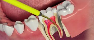
Canal treatment under a microscope
In endodontics, the method of treating tooth canals under observation of the process through a microscope is widely used. This method allows you to see much more and provide better treatment. Root canal treatment under a microscope is a convenient method for the patient and the doctor. A magnification of 30-40 times under a microscope makes it possible to see all the branches of the canals, clean the canal perfectly and seal it.
The microscope allows you to see cracks in the canal, find all the branches of the canal, remove foreign objects, precisely treat hard-to-reach areas of inflammation, and remove the nerve. A microscope helps the dentist determine the condition of the filling, avoid damaging healthy tissue, and fill all the voids in the canal, leaving no room for infection to develop.
Complications resulting from the growth of “wise teeth”
- Malocclusion and crowded dentition.
In this case, the eighth tooth simply does not have room for normal eruption and placement. - Pressure and damage to the adjacent tooth.
If there is not enough room for the eight to grow, it begins to push against its neighbors. - Pericoronitis.
When the eruption of the figure eight is difficult, the soft tissues in this area become inflamed due to the activity of pathogenic bacteria. A so-called gum hood forms over the tooth, in which food debris accumulates, which provokes infection. The main syndrome is severe aching pain, inflammation, and sometimes purulent discharge. - Inflammation of the trigeminal nerve.
Incorrect position of the wisdom tooth can affect the trigeminal nerve, which manifests itself in the form of pain and uncontrollable twitching of the facial muscles. - Osteomyelitis and dental cyst.
Possible complications after endodontic treatment
- The walls of the tooth cavity and the bottom are perforated, subject to the presence of dentin, if the instrument penetrates too deeply.
- The contents of the root canal are not completely removed in cases of obstruction, lateral branches, denticles or bleeding.
- The lumen is clogged with dentin filings, pulp residues, or an instrument that has broken in the canal.
- If the canal is bent, perforation of the root walls is possible
- The canal was not sealed thoroughly enough.
- The canal lumen was not expanded correctly.
- Filling falling out.
Complications may not appear immediately. After root canal treatment, the patient has slight sensitivity, pain, and discomfort in the area of the treated tooth and gums. If these sensations do not go away after two weeks and the pain intensifies, you should contact your doctor for an examination to determine the cause.
Recovery methods
You can restore sevens by:
- fixation of a dental bridge;
- installation of a removable denture;
- implantation
A bridge in the absence of one tooth consists of three crowns. The outer ones are fixed on the supporting teeth, and the middle one compensates for the lost unit. Since the seventh tooth is usually the end tooth, there is nothing to attach the permanent bridge to. In this case, removable structures are used.
Removable dentures are fixed on the teeth on one side, and glued to the gums with a special gel on the other. This is an inexpensive option, but unreliable - the fixation of the prosthesis is unstable and does not provide functional qualities.
Implants completely imitate the structure of the tooth and are the best solution to the problem. A titanium root is screwed deep into the jaw, and a crown is placed on top. Neighboring units of the dentition are not involved in any way. An artificial tooth is no different from a natural one and takes the same chewing loads.
- Dental bridge
- Removable denture
- Implantation
Prevention of dental canal diseases
Prevention of dental canal diseases begins with the prevention of diseases that precede them. It is necessary to take care of the condition of the teeth, preventing the progression of carious processes. Regular dental hygiene: brushing with a toothbrush with the right toothpaste, using dental floss, and rinsing your mouth reduces the number of bacteria and slows down the development of caries and inflammation.
Regular preventive visits to the dentist will allow you to detect the carious process in the initial stage. Do not wait until the tooth makes itself known with painful sensations. Compliance with preventive measures will not only preserve teeth for many years, but also significantly save on treatment and avoid complications caused by inflammatory processes in the dental canals.
Choosing a paste for prevention
Dental health should be taken care of from early childhood. The most effective way is prevention and regular teeth cleaning.
For daily use you need to choose a toothpaste. With a variety of options to choose from, your dentist can recommend the right one for your teeth type. To avoid the addictive effect, it is recommended to periodically change the toothpaste.
Hygienic toothpastes do not have medicinal properties and are intended for everyday use. Daily use of hygienic pastes ensures cleansing of the oral cavity from food debris after eating, removes surface plaque, and has a short-term refreshing effect.
Medicinal pastes should be used as prescribed by a doctor, since they contain high concentrations of medicinal substances, and it is not recommended to use them constantly. The use of medicinal pastes helps fight the development of caries and inflammation in the gums.
Therapeutic and prophylactic are suitable for regular use. They contain active medicinal substances and natural ingredients, but in smaller quantities.
A small amount of toothpaste (about the size of a pea) is enough to effectively brush your teeth and prevent large amounts of toothpaste from ending up in your stomach. It is important to remember that to get results from toothpaste, you must use your toothbrush correctly and change it every three months. The toothbrush is selected depending on the condition of the teeth and oral cavity.
What is the difference between implantation on the upper and lower jaw?
Features of the operation from below
Implantation of the 6th tooth from below is performed using a two-stage method with delayed loading. The implant is implanted using the patchwork method. The crown is installed on the abutment after the artificial root has healed (after 2-4 months). Features of implantation are associated with the anatomical structure of the mandibular structures:
- high jaw bone density - the implant takes root faster, its good primary stability is achieved;
- bone tissue is stronger, therefore it decreases more slowly than in the upper jaw;
- the volume of the mandibular bone is anatomically higher - there are more opportunities to replace the lower tooth without tissue expansion;
- absence of sinuses - there is no risk of injuring the membrane of the maxillary sinus during implantation.
But the trigeminal nerve passes through the base of the lower jaw; if the bone atrophies, it can be damaged (if the technology of the implantation protocol is violated).
Installation of the implant from above
Implantation of the first molars of the upper jaw also has features associated with the anatomy of the jaw structures. The upper teeth are located close to the sinuses (maxillary sinuses), damage to which is accompanied by serious consequences. The maxillary bone is looser and thinner, so it decreases very quickly, and implants take 1-2 months longer to take root than in the lower jaw.
- The choice of method for implanting first molars is a two-stage protocol with a break for osseointegration of the titanium root. The average healing time for upper jaw implants is 4.5 months.
- Since the classical protocol requires sufficient volume and good quality of bone tissue, treatment is often complemented by osteoplastic surgery (sinus lift).
- When making prosthetics, the increased mesiodistal space and the distribution of occlusal forces are taken into account.
Tooth canal treatment: price
Tooth canal treatment price
differs depending on how many roots are in the canal, whether access to them is difficult or not. The price is influenced by the technique used in the treatment process and the equipment involved (microscope).
Dental canal treatment cost
consists of many components: consultation, examination, opening access to the canal, treatment with medications. The patient needs to expect an amount of 5,000 rubles above. On our portal you can navigate prices, find great deals and trusted clinics near your home.
Guarantees
The Center for Private Dentistry “Doctor Levin” has a guarantee program. The patient of the clinic will receive a guarantee for:
- implant;
- surgery;
- sinus lifting and osteoplasty;
- prosthetics.
Only Lifetime on Nobel Biocare issues a lifetime warranty on the implant; other companies usually provide 25 years.
The warranty provides a year of free service for implantation and two years for all types of treatment. The contract is valid subject to systematic medical examinations and proper care.