From this article you will learn:
- methods of treating pulpitis in dentistry,
- how to remove a nerve from a tooth - video, stages of therapy,
- Is it painful to remove a nerve from a tooth?
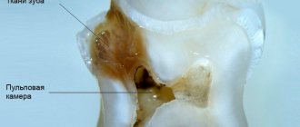
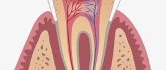
Pulpitis is an inflammation of the neurovascular bundle located inside the tooth, which in dentistry is called the term “dental pulp.” The neurovascular bundle is located in the center of the tooth (in the cavity that dentists call the pulp chamber), as well as in the root canals. Through the tips of the root canals, the vessels and nerves of the pulp anastomose with the vessels and nerves outside the tooth. The name of such a disease as pulpitis arose from a combination of the word “pulp” and the ending “it” (the latter denotes the presence of inflammation).
In an article devoted to the reasons for the development of pulpitis (you can read them at the link above), we have already written that the main reason is long-term dental caries. As a result of the carious process in the hard tissues of the tooth, a carious cavity is formed, the walls of which contain a large amount of infection. When the thickness of healthy tooth tissue separating the pulp from the carious cavity becomes thin, the infection will begin to penetrate into the tooth cavity, causing inflammation of the pulp.
Inflammation of the pulp produces characteristic symptoms – pain. In most cases, treatment will require removal of the inflamed pulp from the tooth cavity and root canals, but in some cases conservative therapy is also possible. The latter is possible only in the early stages of inflammation and in certain groups of patients (we will also discuss this below).
Symptoms of tooth pulpitis -
Pain with pulpitis can be of varying degrees of severity - from minor pain, which is provoked by thermal irritants, to acute paroxysmal spontaneous pain, which makes you want to climb the wall. Given the difference in symptoms, it is customary to distinguish 2 forms of this disease. Below we have described what pulpitis symptoms and treatment will have in each of these cases.
- Acute form of pulpitis – this form is characterized by acute paroxysmal pain that occurs especially at night.
It is typical that the pain increases, while pain-free intervals become shorter and shorter. As a rule, pain occurs spontaneously, i.e. without the participation, for example, of thermal stimuli. However, during the pain-free interval, in some cases it can be provoked by cold or hot water. With pulpitis, it is typical that after the removal of the irritant, the pain persists for about 10-15 minutes (this makes it possible to distinguish pain with pulpitis from pain with deep caries). With the latter, the pain stops immediately after the cessation of exposure to the stimulus. Very often, patients cannot even indicate which tooth exactly hurts, which is due to the irradiation of pain along the nerve trunks. The pain increases due to the gradual transition of inflammation from serous to purulent. With the development of purulent inflammation in the pulp, the pain becomes pulsating, shooting, and pain-free intervals almost completely disappear.
- Chronic form of pulpitis - in this form the inflammation is unexpressed. Patients usually complain of slight aching pain, most often arising from exposure to heat and cold irritants. Sometimes, by the way, with this form of pain there may be no pain at all. Keep in mind that the chronic form of pulpitis can periodically worsen, and during periods of exacerbation of inflammation, the symptoms will be exactly the same as in the acute form.
Clinical manifestations
Treatment of chronic pulpitis does not always begin on time, since this disease is very insidious. Acute pain is always the reason for seeking professional help, but with chronic pulpitis, pain symptoms are not pronounced and the patient prefers to postpone his visit to the dentist. In this case, the disease develops quite quickly and, at best, will lead to an exacerbation of chronic pulpitis with severe pain symptoms or to the occurrence of periodontitis, in which it can be very difficult to save the tooth from removal. No less pleasant consequences may include the formation of cysts, ulcers, phlegmons and even sepsis. This is why timely diagnosis of chronic pulpitis is so important!
The main manifestation is an acute reaction to cold and hot food. However, it occurs late (sometimes 5-10 minutes after consuming food or liquid). In a calm state, a person experiences heaviness inside the tooth.
The hypertrophied form is not accompanied by severe pain. Overgrown tissue appears that ruptures when eating food. Pain may occur when eating hard foods or when pressing on the inflamed area.
Gangrenous pulpitis causes severe pain when eating hot food, which does not go away for a long time. The pulp begins to rot, which causes bad breath. The color of the tooth enamel changes (it becomes dark gray).
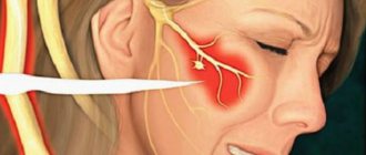
An exacerbation is accompanied by severe pain even without mechanical and thermal effects on the affected area. There are positive symptoms of vasoparesis, swelling of the gums, and pain in the trigeminal nerve.
Treatment of tooth pulpitis: methods
Treatment of pulpitis is most often carried out by depulping the tooth. This method involves completely removing the nerve in the tooth, after which the doctor mechanically expands and then fills the root canals. In young patients (provided they apply at an early stage of inflammation), it is possible to carry out treatment while preserving the living pulp of the tooth.
Of course, it is best to leave the nerve alive, because pulpless teeth become more fragile and also change their color to a grayer color. However, in most cases, the use of biological methods for treating pulpitis is impossible, because Patients extremely rarely apply at the very beginning of pulp inflammation (when the first symptoms have just appeared). Age also plays a decisive role in the choice of this method - this method is indicated for use only in people under 25-27 years of age.
Below we will talk in detail about the traditional treatment of pulpitis (read about the conservative method in the link above). By the way, according to official statistics, treatment of pulpitis is carried out poorly in 60-70% of cases, which requires subsequent retreatment of the tooth. This is due to poor-quality root canal filling.
Causes of pulp inflammation
The main cause of pulpitis is the penetration of infection into the pulp cavity, in which a bundle of blood vessels and nerves is located.
The causative agents of the disease can be both pathogenic (Staphylococcus aureus) and conditionally pathogenic (lactobacillus) microorganisms. They can penetrate into the tooth in several ways:
- through the dentinal tubules in case of poor oral hygiene, accumulation of bacterial plaque and stone in the gum pockets;
- through cracks in the enamel caused by tooth trauma;
- through cracks due to a fracture of the dental crown;
- through the apical opening of the root canal against the background of periodontitis, osteitis and other chronic inflammatory processes of the oral cavity.
- Inflammation can also occur after treatment of deep caries
How to remove a nerve from a tooth - video, stages
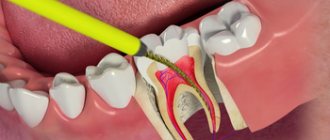
Removal of the tooth nerve is a classic method of treating pulpitis. Its essence lies in the following steps:
- drilling out all tissues affected by caries (Fig. 2),
- removal of dental pulp (carried out using a special instrument),
- mechanical expansion of channels (Fig. 3),
- filling of tooth root canals (Fig. 4),
- filling the coronal part of the cube (Fig. 5).
Treatment of pulpitis: stages of tooth depulpation
Below we will describe in more detail each stage of treatment for pulpitis; perhaps this information will help you identify a would-be dentist and prevent poor-quality treatment and its complications.
Treatment of pulpitis: video of nerve removal from a tooth
Video 1 clearly shows how the pulp is removed (time - 1 minute 5 seconds), video 2 shows how the canals are mechanically treated with a special endodontic tip, and then they are filled.
Possible consequences
If pulpitis is advanced and nerve removal is already required, then after 3-4 months the pathological process will make itself felt by darkening of the teeth. In addition, they will become brittle and dull. If the enamel has acquired a bluish tint, the dentist may have made a mistake when filling. This happens if there were traces of blood in the canal at that moment.
It is worth remembering that it is impossible in principle to cure pulpitis using traditional methods. Moreover, there is no single medicine that a doctor can prescribe to a patient for this diagnosis. Each case is individual, so treatment methods are selected taking into account the characteristics of the clinical picture: the degree of tooth damage, the presence of caries, purulent contents, diseases of the oral cavity, etc. Therefore, if signs of this disease occur, be sure to consult a doctor.
Algorithm for the treatment of pulpitis using a specific example -
If you have pulpitis, treatment of a single-rooted tooth with one canal is usually carried out in two visits (at the second visit a permanent filling is already placed). In multi-rooted teeth, which have a significantly larger number of canals (from 2 to 4), pulpitis treatment is carried out in 3 visits.
The rule is categorical - a permanent filling on a tooth is not placed in the same visit as the root canal filling, i.e. The filling material in the root canals must first harden (moisture evaporates). Only after this can a permanent filling be placed. But to save time, some dentists neglect this. Below we will look at the algorithm for treating pulpitis of a multi-canal tooth in three visits.
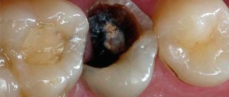
Elimination of tissue damaged by caries
Most often, tooth pulpitis is a consequence of advanced deep caries, and therefore, when treating inflammation in the pulp, a mandatory procedure is the elimination of the enamel layer and dentin with carious lesions. Damaged tissue is removed with a special tool using a drill.
However, when treating pulpitis, intact tissue will be partially removed; this is necessary to gain access to the pulp chamber and canal stomata that need to be treated.
First visit:
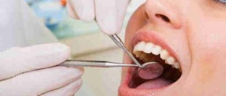
Anesthesia or is it painful to remove a nerve from a tooth?
How painful is it to treat pulpitis: It is definitely very painful if you decide to do it without anesthesia. Fortunately, modern anesthetics can completely solve this problem. If you still feel pain after anesthesia, this may be due to the anesthetic not being strong enough or the anesthesia technique being used incorrectly. The latter usually happens when the doctor tries to anesthetize large molars in the lower jaw (mandibular anesthesia, which is complex in technique, is performed there).
An example of anesthesia (video) –
Drilling out all carious tissues with a drill -
Firstly, at this stage all carious tissue is removed. Secondly, healthy tooth tissues are also partially removed, namely all tooth tissues above the pulp chamber and the mouths of the root canals. This is necessary to ensure visualization of the root canal orifices and ease of their processing with instruments. In Fig. 6-7 you can see the boundaries of excision of hard tooth tissues in the treatment of pulpitis. Figure 8 shows a view of the root canal mouths after they have drilled into the required amount of tooth tissue.
Tooth isolation from saliva –
This is done using a rubber dam. Isolation is necessary to prevent infection from the oral cavity from getting into the root canals along with saliva. This is standard international practice, but in Russia a rubber dam can often be seen only when a doctor fills a tooth.
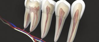
Removal of pulp from the tooth crown and root canals –
It is carried out with special tools designed to work in canals. In Fig. 9 you can see tooth pulp wound around such a tool. By the way, video 1, which we posted above, shows the process of pulp removal.
Measuring the length of root canals in a tooth –
This is one of the most important stages, because... if the length of each channel is determined incorrectly, it will cause -
- or underfilling of the canals, which will lead to complications after the end of treatment,
- or refilling the canals, which can lead to long-term pain and injury to the mandibular nerve.
Measuring the length of the canals is ideally carried out using a combination of the x-ray method and the use of an “apex locator”. In this case, first, special K-file instruments are introduced into each root canal in turn (Fig. 10), which are connected to the apex locator using a thin electrode (Fig. 12). The K-files are gradually advanced deeper into the root canal until there is a signal on the apex locator screen that the tip of the instrument has reached the apex of the tooth root.
It is necessary to measure each channel in turn, because The length of each channel is unique and there are no exact standards. After the measurements are completed and the data are recorded, K-files are simultaneously inserted into all channels (each to its own depth), and a control x-ray is taken (Fig. 11). The apex locator sometimes makes mistakes, so the x-ray will show how accurately the length of the canal was measured and whether adjustments are needed.
Mechanical processing of channels –
Usually carried out with manual files (K-files or reamers). In Fig. 13 you can see the K-file in the root canal. The dentist rotates this instrument by the handle with his fingertips, and the cutting edges of the instrument excise chips from the walls of the canal, expanding it. The purpose of mechanical treatment is to widen the canal so that later it can be properly filled.
Mechanical processing of each channel is carried out to the depth determined at the previous stage. This is necessary to ensure that each root canal is filled exactly to the root apex. During the expansion process, it is very important to constantly rinse the canals with antiseptics, which is necessary for disinfection, but first of all, to wash out the shavings from the canal (24stoma.ru).
Placing a temporary filling –
After the canals are washed and dried to remove excess moisture, turundas soaked in antiseptic are left in them, and a temporary filling is applied to the tooth. The cost of treatment is calculated based on the number of root canals in the tooth.
Complications
In the absence of adequate treatment, pulp inflammation spreads beyond the boundaries of the root canal, to the periodontium and periosteum. In addition to the formation of a purulent focus (abscess) that opens onto the mucous membranes of the oral cavity next to the affected tooth, there is a possibility of developing more serious complications:
- flux or inflammation of the periosteum, which leads to the loss of a diseased tooth, and sometimes healthy teeth located nearby;
- osteomyelitis or purulent inflammation of the bone, which requires complex, expensive therapy;
- phlegmon - lesions of the soft tissues of the face, their “melting” with purulent exudate.
Each of these complications can, with a certain degree of probability, lead to life-threatening conditions, for example, sepsis.
Second visit:
By the way, it is preferable to fill root canals without anesthesia, but this is not necessary. This is due to the fact that if a slight pain occurs when filling the canals, the doctor immediately understands that he has moved the gutta-percha pin beyond the apex of the root. Accordingly, the doctor can change the filling depth in time.
- Removing a temporary filling.
- Flushing the canals with antiseptics.
- Filling canals using gutta-percha and sealer - after the root canals have been washed and dried, it is necessary to seal them tightly.
This is done using gutta-percha pins of different sizes (Fig. 16) and a sealer (this is something like a paste). The pins are inserted into the root canals and compacted there. In Fig. 14-15 you can see the mouths of the root canals Before and After the canals were filled with gutta-percha. Read more about this stage in our article: → “Mechanical treatment and filling of canals for pulpitis”
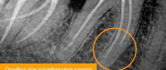
- X-ray control of the filling ( required!!! ) if everything is OK on the x-ray, we proceed to the next stage.
But, if we see that the canal is not filled up to the apex, or the gutta-percha pins extend beyond the root into the surrounding tissues, it is necessary to remove all the gutta-percha pins and start filling the canals all over again. In Fig. 17-19 you can see well-sealed root canals (all root canals are sealed to the apex of the root). Unfortunately, it is worth noting that the vast majority of dentists, even if they see that the root canals are filled, do not redo the work. This is precisely what is connected with the percentage of poor-quality treatment of pulpitis that we announced at the beginning of the article (in 60-70% of cases). Type of high-quality filled root canals –
At the end of the visit, a temporary filling is placed, and the patient is warned that the tooth may begin to hurt after undergoing anesthesia. Good tablet analgesics will help relieve pain. A little pain is normal because... during instrumental work in canals, K-files slightly injure the tissue in the area of the root apex.
Introduction of an anesthetic drug
It is impossible to do without anesthesia when treating pulpitis. When the pulp is damaged, the nervous tissue becomes inflamed and therefore any therapeutic manipulations will be unbearably painful for the patient. Therefore, before starting treatment of pulpitis, the doctor will administer an anesthetic, the action of which will remove the pain for the time necessary for carrying out therapeutic actions.
Sometimes, even after the administration of pain medication, the patient may feel pain. This suggests that either the amount of the drug was calculated incorrectly, or that the doctor made a mistake when giving the injection. Do you want to cure pulpitis painlessly and efficiently? Contact the specialists of the Vanstom dental clinic in Moscow! Our dentistry works for you at the address: Moscow, Baumanskaya metro station, st. Bakuninskaya, 17/28.