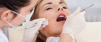
Making the correct diagnosis during a clinical examination of a dental patient is a necessary condition for successful treatment of the patient.
Examination methods such as history taking, examination, electroodontodiagnostics, temperature diagnostics, and x-ray examination are used in dentistry. Laboratory tests and tests are also carried out (general clinical blood test, allergological and cytological, etc.).
Inspection process
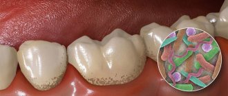
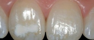
If the patient comes to the clinic without any complaints about his health, the doctor conducts an examination according to the classic scenario. First of all, a visual assessment is necessary: hard and soft tissues, existing crowns and implants, mucous membranes, palate, etc. are carefully examined. This allows you to detect excessive accumulation of plaque, tartar and other deposits. If a stone appears, this may indicate that a person does not pay enough attention and time to oral hygiene.
Professional cleaning using an effective method such as Air Flow may be required to remove stone and hard deposits. The doctor must also examine the enamel, determine the presence of changes in its color, density, and identify the presence of chips and cracks on the surface. When tissue demineralization occurs, strengthening therapy using fluoride-containing drugs may be prescribed.
After this, you can move on to studying the gums: you should check the tissues for the presence of compactions, foci of inflammation, ulcers, etc. The appearance of the tongue, palate, and cheeks is also important. If there are signs of mechanical damage or injury, the doctor may recommend changing your toothbrush and other hygiene devices to more gentle ones.
Probing
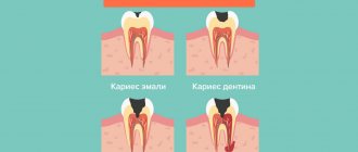
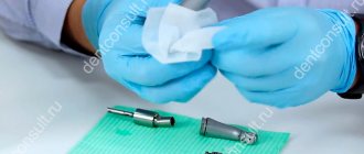
This research method can be carried out to evaluate the reliability and density of the enamel of real teeth and artificial crowns. To do this, the dentist uses an instrument called a probe. With its help, it is possible to determine the presence of carious cavities and the stage of tissue damage. The method is also actively used for the diagnosis of other non-carious diseases, for example, hypoplasia, erosion, fluorosis, etc.
Probing is an absolutely painless and safe process. To check your teeth, the doctor simply needs to run the blunt end of his instrument over the surface of the enamel. This allows you to find irregularities and defects, determine density and hardness. This device can also be used to detect the presence of gum pockets.
Additionally, the dentist can check for gaps between the gum and the crown (or between healthy areas of enamel and the filling material). If there is a severe gap, the filling will need to be reinstalled.
Percussion - tapping on the tooth - is used to determine the condition of the periodontium. Using tweezers or a probe handle, tap the cutting edge or chewing surface of the tooth. If there is no focus of inflammation in periodontitis, percussion is painless; in the presence of an inflammatory process, a painful sensation occurs. The blows should be light and even. Percussion should begin with obviously healthy teeth, so as not to cause severe pain and to allow the patient to compare the sensations in healthy and affected teeth.
There are vertical percussion, in which the direction of the blows coincides with the axis of the tooth, and horizontal, when the blows have a lateral direction.
Palpation - feeling - is used to determine swelling, swelling, compaction, mobility of organs or tissues of the oral cavity. The palpation technique depends on the location and size of the lesion.
In some cases, palpation is performed with one index finger, in others (when palpating the tissues of the cheek) with the index fingers of the right and left hands, with one finger located outside and the other on the side of the oral cavity, in third cases the mucous membrane is taken into the fold with two fingers.
It is recommended to begin palpation from an undamaged area of the mucous membrane, gradually approaching the lesion: in this way the border of pain and compaction is more accurately determined.
When palpating ulcers of the oral mucosa, determining the density of the edges and their soreness is of great diagnostic importance. The absence of pain on palpation of ulcers with dense edges should raise suspicion of its malignant quality or the presence of a syphilitic ulcer.
The mobility of the teeth is determined with tweezers by rocking them. The tooth has physiological mobility, which is normally almost invisible. When the periodontium is damaged and there is exudate in it, severe tooth mobility occurs. There are three degrees of mobility:
- I degree - confusion in the vestibular-oral direction;
- II degree - displacement in the vestibular-oral and lateral directions;
- III degree - displacement in the vestibular-oral, lateral and axial (vertical direction).
Currently, objective methods for measuring the amount of tooth deviation from the axis have been proposed, but they have not yet been put into practice.
Temperature diagnostics. Determining the tooth's response to temperature stimuli is one of the oldest physical research methods; it is used to identify the condition of the pulp. Ether is used as an irritant, but more often - cold or hot water, which is a stronger irritant due to its greater heat capacity.
The easiest way is to irrigate the teeth with water from a syringe, but sometimes it is difficult to determine which tooth reacts to the irritant. In such cases, a swab moistened with cold or warm water is inserted into the carious cavity or applied to the surface of the tooth.
A study of the pulp's response to stimuli showed that a tooth with a normal pulp reacts to significant temperature deviations. The indifferent zone (zone of no reaction) for incisors is 30 °C (50-52 °C - reaction to heat, 17-22 °C - to cooling).
Teeth have both cold and heat sensitivity. An adequate reaction (if heating and cooling cause the appropriate sensation) indicates the normal condition of the pulp. When it becomes inflamed, the indifferent zone narrows and even with minor deviations from body temperature (by 5-7 °C), a response occurs in the form of prolonged intense or aching pain. In addition, during inflammation, an inadequate reaction is noted: pain occurs from cold or warm.
Teeth with necrotic pulp do not respond to temperature stimuli.
Did you like the article? Share with friends
0
Similar articles
Next articles
- Electroodontodiagnosis
- X-ray examination of teeth
- Luminescent diagnosis of caries
- Functional tests
- Functional research methods
Previous articles
- Dental examination
- Examination of the oral cavity itself
- Oral examination
- External examination of a dental patient
- Anesthesia technique
Add a comment
Palpation and percussion
This technique is used to assess the health of periodontal tissues. To do this, the specialist carefully moves the tooth to the right and left using medical tweezers, checking its mobility and ability to move. If the coronal part moves easily, this indicates the presence of serious inflammation or indicates tissue swelling.
Such consequences can occur due to severe injuries to the jaw, as well as diseases such as periodontitis, periodontitis or periodontal disease. Using palpation, the doctor assesses the degree of pain of the procedure for the patient, because pain can be of varying intensity depending on the complexity of the pathology and its location.
Percussion is a method in which the doctor gently taps the surface of the tooth crowns with a probe or other device. If unpleasant sensations occur with horizontal impacts, this indicates the development of periodontitis; if with vertical impacts, then this indicates an inflammatory process in the pulp chamber. Based on the data obtained, the specialist can make a preliminary diagnosis and send the patient for further examination. Sometimes, during the general examination, an x-ray is prescribed; this is necessary to confirm the diagnosis.
Palpation examination
Using this research method, inflammatory processes are identified, as well as the nature and stage of development of periodontal destruction. To more accurately understand the condition of the tooth, they begin to move it in different directions using tweezers, this makes it possible to understand how it moves in the alveolus:
- In three directions
- In five together with vertical
- In one
- In two.
If a tooth gives in very easily with a little force, this means that there are deviations from the norm, which may be accompanied by tissue swelling and an inflammatory process. These symptoms may indicate the presence of traumatic tooth lesions, such as periodontal disease, periodontitis and periodontitis. It is by palpating the cheeks and gums that the “One to One” doctor receives information about the presence of purulent or bloody discharge, pain, compaction and swelling. A big plus that can be given to the method is that it allows you to instantly obtain the research result upon examination. The only negative that unfortunately exists is the inability to make a definitive diagnosis.
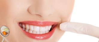
When should you undergo a general examination by a dentist?
Experienced dentists and orthodontists strongly recommend visiting a dental clinic for a preventive examination at least once every 6 months. This should be done even if there are no complaints. If you have any unpleasant sensations, you should not delay your visit to a medical facility for even a minute.
There are several main causes for concern: increased sensitivity of the enamel, pain in the gums under the denture, swelling, redness and any other visible tissue changes. Also, one of the symptoms of developing pathology is a strong smell from the mouth, which does not disappear even after brushing.
Abundant accumulation of plaque and tartar, changes in the shade and transparency of the enamel, bleeding - all this is a good reason to visit your doctor. Also, consultation and examination are required before orthodontic treatment.
previous post
Why are my teeth numb?
next entry
Modern ideas about the classification of pulpitis
In modern dentistry, medical knowledge is constantly updated. As new information becomes available, there is a need to change treatment methods, equipment and drug use.
However, no technology or new materials can replace the tactile sensations and common sense of a doctor. The author hopes that dentists will continue to monitor modern high-tech devices and treatment methods that are not a substitute for clear, sound thinking. A few years ago, students performed routine endodontic procedures without thinking about quality standards. In recent times, dental school graduates have become better at performing almost all phases of routine endodontic treatment. As complication-free endodontic treatment becomes an integral part of modern dental care, its “mystery” is fading. In Russia and the republics of the former USSR, the classification of pulpitis by I. G. Lukomsky, developed back in 1936, is most widespread. The classification of I. G. Lukomsky attempted to establish a correlation between clinical manifestations and the characteristic histological picture of the pulp, and has undergone almost no changes to the present day.
The author does not agree with the currently accepted classification of pulpitis adopted in Russia, according to which pulpitis is acute, divided into serous focal, serous purulent diffuse, purulent diffuse; or chronic, divided into simple, having 2 stages, hypertrophic, gangrenous, granulomatous and exacerbation of chronic pulpitis.
The pulp may be infected or sterile. Clinically, acute pulp inflammation has certain symptoms, while chronic inflammation is asymptomatic. These opinions often do not correspond to histological observations. It is impossible to clinically determine the degree of pulp inflammation (partial or complete). According to modern concepts, any type of irreversible pulpitis requires endodontic treatment.
In the past, many attempts have been made to establish correlations between clinical manifestations and the characteristic histological appearance of the pulp. The futility of this approach to classification has long been proven all over the world (Garfunkel, A, Sela. J., and Ulmansky, M.: Dental pulp pathosis; clinicopathologic correlations based on 109 cases. Oral Surg. 35:110, 1973). Since it is believed that there is no reliable correlation in dentistry, histological and clinical data are usually studied separately. Therefore, after collecting clinical data, the doctor has no idea about the histological state of the pulp. However, by understanding the basics of the pathological process, you can make a fairly correct decision about the need for pulp treatment, and if it is indicated, then determine what kind of treatment should be carried out - preventive or endodontic. This is what is important for a doctor. Moreover, after endodontic treatment, the doctor must be able to make a prognosis and evaluate the results of treatment.
Since there is no exact correspondence between the clinical condition of the pulp and histological data, the modern classification is based on other principles. The most difficult thing for a modern doctor to understand is the fact that there is no correlation between the severity of pain and the degree of pulp damage. It is extremely important for the doctor to determine which treatment is indicated - endodontic or prophylactic. If the decision is made to perform endodontic treatment, the exact histological condition of the pulp is of academic interest only, since the treatment generally accepted throughout the world involves its complete removal. In other words, for various forms of pulpitis (irreversible), the same type of treatment will be performed, which, regardless of classification, aims to completely remove the infection from the canal and periapical tissues and ends with a complete, dense and hermetically sealed filling of the root canal system with non-irritating materials. In this regard, the above classification used in Russia loses its meaning.
This clinical classification especially complicates treatment planning and prognosis for combined endodontic and periodontal pathologies; therefore, the author did not aim to dwell on it in detail, since it is quite fully described in the publicly available domestic literature.
In modern dental practice in the USA, England, Israel, Australia and other developed countries, the condition of the pulp is divided into: - normal, - reversible pulpitis, - irreversible pulpitis, - pulp necrosis.
Periapical diseases are divided into: - acute apical periodontitis, - acute apical abscess, - chronic apical periodontitis, - phoenix abscess, - radicular cyst, - condensing osteitis (periapical osteosclerosis). To understand the pathology of the pulp, you need to thoroughly know what is considered normal.