A mouth smear is not the most common test in dermatovenerological practice.
However, venereologists sometimes suggest that their patients be examined for sexually transmitted diseases (STDs) in this way.
It happens that such patients are referred to a dermatovenerologist by doctors of other specialties - dentists, therapists, otolaryngologists.
The reason is simple.
Many pathogens of STDs, in addition to the genitals, feel very good in the oral cavity or on the epithelium of the ENT organs.
Who needs a mouth swab and when, how and where it is done - we will tell you below.
What is it and why?
The oral cavity of every person is very densely populated with microorganisms.
Most of them are harmless saprophytes, among which there are cocci and bacilli.
There are even non-pathogenic spirochetes.
Our body has learned to coexist safely with them, and we simply do not notice them.
But such prosperity does not always happen.
Problems arise when foreign microorganisms enter the oral cavity.
For example, pathogens of sexually transmitted infections that thrive on the mucous membranes of the mouth and genitals.
The list of microbes that are capable of such a change in habitat is quite wide:
- Trichomonas
- treponema pallidum
- human papillomaviruses (HPV)
- herpetic group viruses
- gonococci
- chlamydia
In principle, this list can include almost all STD pathogens.
A smear helps to identify them in the mouth.
Diagnostic value of mouth swab
At its core, it is completely, 100% analogous to a urogenital smear.
The only difference is where the diagnostic material is taken from.
A mouth smear helps determine the spectrum of oral microflora in the person being examined, regardless of gender.
This method refers to laboratory-instrumental techniques and performs one task: obtaining diagnostic material for a detailed study of microflora.
Samples obtained using a mouth swab are examined in different ways:
- microscopy of native material and fixed, stained smear
- bacteriological examination (culture)
- immunological studies
- molecular genetic
The main thing is to correctly and accurately collect diagnostic samples.
But before you collect them, you need to determine who needs it.
How is scraping performed for bile acids?
Such a scraping also allows for analysis of bile acids. The quality of the analysis is ensured by high-tech thin layer chromatography.
For such a high-precision analysis, the material is taken in the morning. The patient should not have time to eat, drink or take medications.
Don't be afraid of this simple procedure. It consists only in the fact that in the laboratory, using an ordinary teaspoon, the plaque that has accumulated overnight is carefully removed from the tongue. Then the material is placed in a test tube, filled with ethyl alcohol and left for a while under a tightly closed lid. All components must dissolve.
Then the material is evaporated in a water bath. It should decrease in volume by half. And only one drop of the resulting extract is applied to a heated chromatographic plate. Then the plate is dried again, cooled and developed. If there are few bile acids, the line will be solid, but if their concentration is high, dense spots will form.
For treatment to be effective and quick, it is necessary to identify the disease in its early stages. If the doctor has prescribed a scraping, there is no need to postpone this procedure. It is absolutely painless and safe. Moreover, such an analysis is very informative.
Indications for performing an oral smear
The reasons for conducting such a study differ for doctors of different specialties.
As for venereology, here are the main symptoms for which dermatovenerologists take a swab from the mouth:
- burning and pain in the mouth
- painful rash on the mucous membranes of the mouth and cheeks
- enlarged tonsils without pronounced clinical signs
- complaints of a sore throat for a long time
- the appearance of painless lumps and wounds in the mouth, on and under the tongue
- unpleasant odor with the exception of dental caries
- sour taste, foamy saliva
In principle, everything is decided individually each time.
We have listed only a few signs that indicate the main STDs characteristic of lesions in the oral cavity.
4. Results of throat culture for microflora
Culture results will be ready within 1-2 days, in some cases testing is carried out within a week.
There is also a rapid test for streptococci.
The results of such a test for streptococcus will be ready in 10-15 minutes. This test is only for bacterial infections caused by streptococcal bacteria.
A rapid test for streptococcus can show two results:
- Negative test results for streptococcus.
Streptococcal bacteria were not detected. Microflora culture may be recommended. - Positive test results for streptococcus.
Streptococcal bacteria were detected. This means you have strep throat (strep throat). You must start taking antibiotics immediately.
Throat culture for microflora can also be negative or positive:
- Negative culture results for microflora. There are no bacterial or fungal infections and their growth is not observed. Negative results may mean that your infection is caused by a virus rather than bacteria or fungus. Throat infections can be caused by different viruses - enteroviruses, Epstein-Barr virus, herpes simplex virus, respiratory syncytial virus (RSV).
- Positive results of throat culture for microflora. Bacterial growth is observed. Examples of bacterial throat infections are strep throat, diphtheria (diphtheria corynebacterium), and whooping cough (whooping cough). Fungal growth is observed. The most common fungal infection in the throat is oral candidiasis, which is caused by yeast-like fungi of the genus Candida.
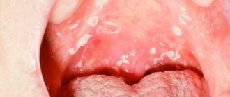
Typical signs of oral STDs
It often happens that people come to dermatovenerologists from dentists or laryngologists.
After the treatment from these doctors turned out to be ineffective.
Or after the patient has had a mouth smear examined for microflora.
And they found sexually transmitted infections in the material.
How they get there is sometimes quite difficult to figure out.
The most common method is oral sex.
It is also necessary to remember that more than 200 thousand germs are transmitted with a deep kiss.
And, if your partner had herpetic rashes in the mouth, infection cannot be avoided.
The same applies to atypical forms of primary syphilis, gonorrhea and other STDs.
Gonorrhea is transmitted through oral sex.
It manifests itself as a sore throat, hoarseness and other symptoms of a prolonged cold.
Syphilis is the same.
Characteristic signs are painless lumps and reddish ulcers on the tongue and red border of the lips.
May occur as chronic flaccid tonsillitis.
Trichomonas, chlamydia and mycoplasma when localized in the mouth do not cause specific symptoms.
They can be suspected only by a lingering cough or tonsillitis.
The patient's general well-being does not deteriorate.
Herpes is easy to detect: pain and burning, rashes on the mucous membranes, transforming into erosions.
Relapses up to 5-6 times a year.
Against the background of immunodeficiency, constant persistence of rashes in the mouth is possible.
Human papillomavirus lesions are painless and usually have the appearance of genital warts.
Having reached large sizes, they can make swallowing and speech difficult.
Infection through domestic means cannot be ruled out.
All infections can be transmitted through a toothbrush.
Because of this, children whose parents suffer from the oral form of one or another sexually transmitted infection often suffer.
Another reason for taking a swab from the mouth is the need to assess the prevalence of STDs when examining a patient with established damage to the reproductive organs.
Sowing the contents of the gingival pocket for anaerobic flora
A microbiological study aimed at isolating the pathogen and, if identified, determining its sensitivity to antibiotics in order to select the optimal therapy for periodontitis. When pathogenic and/or opportunistic microorganisms are identified, their sensitivity to antimicrobial drugs (antibiotics and bacteriophages) will be determined. When microorganisms that make up the normal microflora are detected, sensitivity to antibiotics and bacteriophages is not determined, because has no diagnostic value.
Synonyms Russian
Sowing for facultative anaerobic bacterial flora; microflora in diseases of periodontal tissues; culture for anaerobes; culture for anaerobic bacteria; anaerobic bacteria, culture, gingival pocket discharge.
Research method
Microbiological method.
What biomaterial can be used for research?
A smear from a gingival pocket.
How to properly prepare for research?
- Do not perform oral hygiene on the day of taking biomaterial for examination.
General information about the study
A gingival, or periodontal, pocket is a depression between the gum and tooth. If this depression pathologically increases, then we are talking about the appearance of a periodontal pocket. During the development of periodontitis, inflammation of the gums occurs due to exposure to bacterial agents. Changes associated with the occurrence of a pathological periodontal pocket are associated with changes in the microbial composition of dental plaque. Normally, gum tissue contains a small amount of bacteria: rods and cocci. During inflammation, the number of mobile rods and spirochetes increases. The presence of a periodontal pocket can be suspected when there are complaints of localized pain, thickening of the gums, gingival bleeding and suppuration, tooth mobility, and a blue-purple color of the gingival margin. The main diagnostic method is to probe the gingival margin along each tooth surface. The contents of the gingival pocket consist mainly of a mixture of microorganisms, leukocytes and their metabolic products, food debris, and epithelial cell debris. Changes in the periodontal attachment due to periodontitis can be of a constant progressive nature, during which periods of remission and exacerbation are distinguished. If during remission the inflammatory reaction is reduced or absent altogether, then during an exacerbation there is an increase in clinical manifestations with the presence of gingival exudate, in which a large number of mobile bacteria and spirochetes can be identified. Various methods are used in treatment: antibacterial therapy, curettage, surgical treatment, etc., including combinations of various methods.
Anaerobic microflora are microorganisms that do not require oxygen for their life and reproduction; for many of them, on the contrary, it is destructive. Anaerobes inhabit the human body normally (in the digestive tract, respiratory organs, genitourinary system). With a decrease in immunity or injuries, damage, activation of infection with the development of the inflammatory process is possible. The human body can then essentially become a source of infection for itself (endogenous infection). Less commonly, anaerobes enter the body from the outside (deep puncture wounds, infected abortion, wounds to the abdominal and thoracic cavity, insertion of needles and prostheses). Developing in the thickness of the skin, soft tissues and muscles, anaerobic organisms can cause cellulite, abscesses, and myositis. Symptoms that allow one to suspect an anaerobic infection of soft tissues: dense swelling, gas formation (a feeling that air bubbles under the skin are bursting when pressed), putrefactive inflammation, fetid odor.
The main treatment for anaerobic inflammation is surgical. In this case, it is necessary to eliminate the source of inflammation or open the wound, providing access to oxygen, which is harmful to anaerobes.
For differential diagnosis of anaerobic and aerobic infections, the biomaterial is cultured for flora, since the principles of treatment in one case or another will be different. Anaerobic microorganisms are sensitive to a rather narrow range of antibacterial drugs, so it is advisable to carry out drug therapy after determining sensitivity to antibiotics. Based on the grown culture, the type of microorganisms that participate in the formation of the inflammatory reaction is determined. Knowing the type of pathogen, you can select an antibacterial drug that can successfully influence these microorganisms and contribute to more effective sanitation of periodontal pockets.
After identifying a bacterial culture, it is advisable to determine their sensitivity to various antibiotics. Due to the fact that the development of antibiotic resistance in microorganisms is increasingly observed, the selection of antibiotics according to their spectrum of action on bacteria can lead to ineffective or completely unsuccessful treatment. The advantage of the method for determining sensitivity to antibiotics is the accurate determination of the antibacterial drug that has the highest effectiveness in a particular case.
List of tested antibiotics:
- Vancomycin / 5 mcg
- Clindomycin / 2 mcg
- Erythromycin / 15 mcg
- Amoxicillin clavulanate / 20/10 mcg
- Metronidazole / 50 mcg
- Kanamycin / 30 mcg
What is the research used for?
- For a comprehensive diagnosis of periodontitis and periodontal diseases;
- for differential diagnosis of anaerobic and aerobic infections, for selection of adequate therapeutic treatment taking into account the detected microflora;
- to monitor the therapy of periodontitis or inflammatory processes in the gum canal.
When is the study scheduled?
- In case of inflammatory processes in the area of periodontal pockets;
- with bleeding gums, sore gums, bad breath;
- when the position of the teeth changes, gaps appear between the teeth;
- when purulent-inflammatory processes appear in the area of teeth, abscesses.
What do the results mean?
Normally, anaerobes are not sown in diagnostically significant quantities.
The reason for identifying microorganisms is the presence of growth of colonies of this type of microorganisms on a nutrient medium.
Also recommended
- Sowing for aerobic and facultative anaerobic flora
- Sowing wound discharge into aerobic and facultative anaerobic flora
Who orders the study?
Dentist, periodontist, infectious disease specialist, therapist, general practitioner, otorhinolaryngologist, pediatrician.
Literature
- Fermin A Carranza, Paulo M. Camargo. The Periodontal Pocket. / Carranza's Clinical Periodontology, 2012, 127-139.
- Mirela Kolakovic, Ulrike Held, Patrick R Schmidlin, Philipp Sahrmann. An estimate of pocket closure and avoided needs of surgery after scaling and root planning with systemic antibiotics: a systematic review. / BMC Oral Health. 2014; 14:159.