Causes
The main causes of damage to the NAS are:
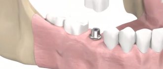
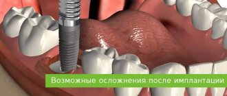
- implantation (doctor’s errors, lack of a full preliminary examination);
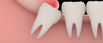
- removal of dystopic “eights” on the lower jaw;
- errors when performing conduction anesthesia;
- exit of the filling material beyond the root apex into the nerve canal;
- infectious lesion of the periapical region of the lower row.
But the most common reason is the first reason - damage as a result of implant installation, usually in the chewing area.
Experts' forecast
The prognosis for damage to the NAS is uncertain and depends on the degree of damage to the nerve structures. With neurotmesis, it is unfavorable; neurosensory changes most often cannot be stopped.
Axonotmesis - with adequate treatment, nerve sensitivity is partially restored within 2-4 months.
With neuropraxia, complete restoration of normal sensitivity occurs within a few days or weeks.
The main treatment for severe neuropathies is surgery, leading to success in 50-80% of cases
.
First signs of damage
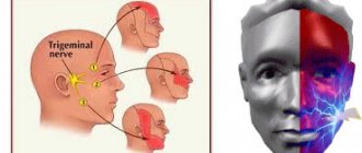
The first symptoms of nerve damage are discomfort in the gums, cheeks, and lower lip. Manifestations of the problem are:
- paresthesia, that is, a change in the level of sensitivity without pain;
- dysesthesia with pain in the affected area, a feeling of “pins and needles”, changes in the general sensitivity of the area;
- anesthesia - complete loss of sensation in a certain area.
In some cases, the lingual nerve, which runs from the side of the tongue into the gum tissue, may be affected. This is usually observed as a result of the removal of "eights" (in approximately 2.1% of all cases). When implanted, this nerve is affected less frequently. If this situation occurs, the following symptoms appear:
- salivation becomes profuse;
- involuntary biting of the tip of the tongue appears;
- diction disorders;
- burning sensation, numbness in the tongue;
- loss, change in taste;
- swallowing is impaired.
In 90% of cases, the problems go away on their own within seven to ten weeks; no special treatment is required.
Ways to assess case complexity
In diagnostic studies for neurological lesions, 2 types of testing are used:
- mechanoceptive;
- nociceptive.
Using the first, the reaction of tissues innervated by the NAS to mechanical stress is checked:
- The brush test involves lightly passing the brush across the lips and asking the patient to determine the direction of movement of the brush.
- The two-point stimulation method is based on the use of a probe with two legs, the distance between which varies in 2 mm increments. By touching the legs to the skin, it is determined at what distance between them the patient ceases to feel each point of contact individually.
Nociceptive studies can determine pain and temperature sensitivity. Pain sensation is tested using a pin, temperature sensitivity is tested using ice and the handle of a dental mirror heated to a temperature of 43 °C.
A study of taste sensations is also carried out using sugar and salt sprinkled on a cotton pad.
To compare the results, not only the affected area is checked, but also the opposite side of the face.
The essence of the method of compression dental implantation and the dental structures used.
In this article we will tell you what alternatives to dental implants exist.
Here https://zubovv.ru/implantatsiya/metodiki/vliyanie-stabilnosti-vnutrikostnyih.html we will consider ways to determine the stability of implants.
Classification
In accordance with Seddon's classification, the following types of NAS injuries are distinguished:
- Neuropraxia. This injury is reversible; the sheath of the nerve fibers does not suffer, that is, there is no development of degeneration. After treatment, the problem goes away, which usually takes a couple of weeks.
- Axonotmesis. A reversible type of lesion, for which long-term, intensive treatment is prescribed - from three to six months. The development of degeneration and fiber damage are observed.
- Neurotmesis. A serious disorder affecting all structures, including fibers and connective membranes. Scar tissue develops, and surgery is indicated to correct the problem.
In accordance with the generally accepted WHO classification, there are five categories of NAS injuries:
- compression, nerves are not damaged;
- swelling of tissues;
- partial rupture of fibers;
- complete ruptures of the NAS are observed;
- post-traumatic fibrosis develops.
Assessment methods
To determine the cause and extent of the disorder, diagnostics are required. For this purpose they prescribe:
- mechanoceptive methods, in which the response to mechanical irritation and stimulation is recorded;
- nociceptive, allowing to determine sensitivity to painful stimulation.
The first type of research includes tests of the following types:
- tests with a brush, during which the Patient’s lip is passed with a brush and asked to determine the direction of movement;
- two-point irritation using a probe.
The second diagnostic method includes:
- pin tests;
- Temperature tests for sensations of cold or heat.
Tests are also carried out for the presence of deficits in the sense of taste, and data from the right and left sides of the maxillofacial apparatus are compared. The accuracy of the research should be no more than 1 mm.
Treatment
The following regimens are used for therapy:
- observation of the Patient at intervals of 2, 3, 4, 6 months;
- drug therapy;
- unscrewing, removing the implant;
- microsurgical intervention.
There is no strict protocol; tactics are determined individually in each case. But there are limitations to surgical intervention - it will be effective only in the first year, after which this method is considered ineffective.
Drug therapy includes:
- taking painkillers;
- use of hydrogen pump blockers;
- prescription of anti-inflammatory drugs (glucocorticosteroids are prescribed);
- therapy for sensory impairment.
If symptoms appear in the first 1.-1.5 days after installation of the implant, its removal may be indicated. During this period, the prognosis is favorable, but over a longer period of time, removing the artificial root will no longer bring a noticeable improvement. The reason is irreversible changes in nerve fibers.
Surgery is indicated in cases where all previous measures have not brought the expected effect. To do this, the doctor must determine the location of the NAS and evaluate sensitivity disorders. The effectiveness of the operation is influenced by the following factors:
- time between injury and surgery;
- severity, type of lesion;
- blood supply;
- Preparation;
- tension zone;
- Patient's age, health status.
Predictions and prevention
Treatment has an uncertain prognosis; with microsurgical intervention, the positive result is in the range of 55-82%. If the damage is severe, complete recovery does not occur with any treatment method. According to some experts, complications such as pain persist even after the intervention. It is also worth noting that the success of treatment depends entirely on the extent of the lesion. It is impossible to guarantee a high result in any situation, that is, the forecasts are uncertain.
To avoid complications, prophylaxis is recommended. This is a mandatory full examination before the implantation procedure. The specialist will determine the exact location of the nerve, the topography of the nerve trunks and the maxillofacial system. This allows you to exclude the development of pathology and conduct conduction anesthesia correctly, without the risk of complications.
The main preventive measures include:
- competent selection of implantation system;
- diagnostics, including CT, production of a surgical template, computer modeling;
- planning the procedure for introducing an artificial root;
- determining the paths of nerve trunks, ensuring the distance between them and implants is not less than 2 mm;
- when drilling, it is necessary to avoid excessive force;
- When immersing a drill or implant, stoppers are used to limit the depth and ensure safety.
Treatment of damage to the mandibular nerve during implantation requires the doctor to have high qualifications and experience. But even under such conditions, the success rate does not exceed 82%. Some complications may remain, such as numbness or soreness.
Probability percentage
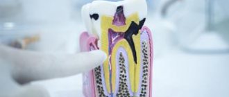
Statistics on damage to the NAS during dental implantation seem contradictory. According to some studies, nerve damage occurs in no more than 3% of cases. Other data suggests that this happens in 30-40%.
This variability is explained by the fact that the statistical results depend on many factors - the calculation method, the age group of the patients undergoing the examination, the time period during which the information was collected, and some others.
If we consider all cases of damage to the NAS during dental intervention, then the frequency of their occurrence, depending on the cause, is in the following order:
- conduction anesthesia;
- removal of third molars;
- entry of filling material into the canal with NAS;
- surgical intervention (periostitis, resection of the apex of the tooth root, etc.);
- implantation.
However, it has been established that in older age groups, implantation takes first place in the frequency of damage to the mandibular nerve.
There is one more factor that influences the statistical results. The later the data is taken, the lower the percentage of damage to the NAS during implantation, and this is quite understandable.
The implantation of artificial roots is becoming an increasingly popular technology, the qualifications of doctors are improving, safer technologies are being used, and high-tech diagnostic equipment is being used.
How to properly organize nutrition after dental implantation and a sample menu for the day.
Come here to find out how long does a tooth hurt after implantation.
At this address https://zubovv.ru/implantatsiya/metodiki/unikalnyie-vozmozhnosti-skulovoy.html we will consider the technique of zygomatic dental implantation.
Preventive actions
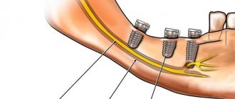
The main prevention of NAS lesions is adequate diagnosis before implantation. It is necessary to establish the exact location of the mandibular nerve, and if there is a risk of damage, take measures to eliminate complications.
The most important factors in preventing damage are the following:
- Accurate pre-implantation diagnosis , correct planning and competently carried out implantation.
- During diagnosis, the location of the mandibular nerve must be determined and ways to bypass it must be outlined. The use of CT scanning and a surgical guide virtually eliminates the risk of damage.
- During implantation , it is necessary to ensure at least a 2 mm gap between the nerve bundle and the instrument/implant.
- When creating a bed and fixing the implant, excessive force should be avoided .
An important factor in ensuring safety is the high qualifications of the doctor performing implantation.
The video shows a diagram of the movement of the mandibular nerve during dental implantation.
Reviews
Damage to the mandibular nerve is a very unpleasant complication of implantation, although it is relatively rare.
If you have experienced it yourself, please share your experience of overcoming it. You can do this by leaving comments at the bottom of the page.
If you find an error, please select a piece of text and press Ctrl+Enter.
Tags: implantation, complications of implantation
Did you like the article? stay tuned