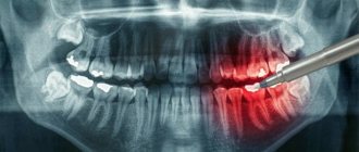
Radiovisiography is a modern diagnostic method that is used in leading dentistry around the world.
This is not only the most accurate way to detect pathologies, but also the most gentle method in terms of radiation (it is 10 times weaker than with x-rays). Note! This technology has been used worldwide since 1987. Moreover, the equipment is found not only in elite, but also in budget clinics. In the West, X-rays have not been used in dentistry for a long time due to the fact that the negative impact on the body is too strong and there is a high risk of developing cancer.
Why is the research being conducted?
A targeted photograph of the tooth is taken to identify inflammatory processes in the periodontal tissues and to search for hidden carious lesions. For this task, a radiovisiograph is used - a special device that takes high-quality images of the affected area and displays the image on the monitor screen. In this case, the doctor can minimize the risk, since the patient avoids strong radiation, such as with an x-ray.
Fact: radiation exposure from x-rays is 10 times greater than from radiovisiography. Because of this, dentists are abandoning the outdated method in favor of new technologies. Regular exposure to X-rays can lead to accelerated aging of the body and deterioration of blood composition.
This is a win-win option because targeted intraoral radiography is more than affordable. The results of the study are stored on a hard drive, so even after many years, specialists can access it to study the medical history.
Types of dental radiography
Depending on the purpose and method of implementation, there are different types of radiography. You can familiarize yourself with them and their features in our table below.
| Type of X-ray | Objectives |
| Sighting/intraoral | Provides an image of one or several (up to 4) teeth that are located nearby. Allows you to determine:
|
| Panoramic | Allows you to obtain accurate data on the condition of the jaw in general. Thanks to this diagnosis, it is possible to determine foci of infectious lesions, along with the condition of the maxillary sinuses and the ANS. It is indispensable when planning various types of dental operations, including implantation. |
| Digital | Allows you to obtain a model of the jaw in three-dimensional format and requires the use of a computed tomograph. Allows you to accurately and in detail determine the condition of the patient’s dental system, determine the scope of the upcoming surgical intervention and develop surgical tactics. |
Our doctors
18 years of experience
Baghdasaryan
Armen Evgenievich
Chief physician, dentist-orthopedist-therapist
Graduated from VSMA named after. N.N. Burdenko. Internship on the basis of MGMSU named after. A.E. Evdokimov in “General Dentistry”.
Clinical residency at the Moscow State Medical University named after. A.E. Evdokimov in “Orthopedics”.
More about the doctor...
5 years experience
Sadina
Ekaterina Vladislavovna
Dental therapist, surgeon
Penza State University Medical Institute, specialty “Dentistry”.
In 2021, she underwent professional retraining in the specialty “Therapeutic Dentistry” at the Moscow State Medical and Dental University named after A.I. Evdokimov.
More about the doctor...
Indications and contraindications for radiovisiography
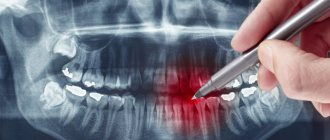
The procedure is prescribed in the following cases:
- Severe pain in the tooth area.
- Detection of periodontal lesions, pulpitis, caries, granulomas, neoplasms, periodontal disease.
- Canal filling.
- Assessing the condition of teeth after strong impacts or falls.
- Checking wisdom teeth (direction of growth, condition of roots, size).
The study is not recommended for patients for whom even minimal doses of radiation are contraindicated. For example, if a representative of the fair sex is in the first or third month of pregnancy. It is not advisable to perform radiovisiography if there is bleeding or autoimmune diseases are observed. You must also not exceed the maximum number of shots per year. A person can safely undergo the procedure a maximum of 50 times. After this, the annual radiation norm is exceeded.
Stages of the procedure
To carry out the procedure, a separate room, equipped accordingly, is required. The procedure itself does not cause virtually any discomfort and consists of several stages.
- The position of the person in the chair, the doctor’s clarification of the location of the problem.
- It is imperative that the patient wears an apron that protects against harmful radiation.
- Fix your head to get a good shot.
- Taking an image with the patient in a stationary position.
The entire procedure from taking a photo to receiving it in electronic or printed form takes a short period of time - about 20 minutes.
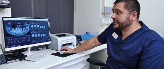
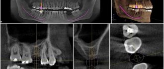
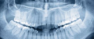
Analysis of the finished image
The dentist himself or a radiologist is responsible for describing the image. If the image quality does not meet the required quality, the procedure is repeated. The specialist examines the patient’s teeth, roots, and jaw bones in detail. If there are light and dark areas, this indicates the appearance of inflammation, cysts and other neoplasms.
In the pictures you can see inhomogeneities, damage to bone tissue, damage to the enamel, cement and root part, damage to the septum and other abnormalities that indicate the presence of a particular disease.
Features of performing a targeted shot for children
The younger generation needs this procedure more than adults, because caries develops more intensively in children. Another reason is to identify deviations and defects at an early stage of tooth growth. If any are detected, the necessary preventive and therapeutic measures are taken.
But it is not advisable to use the targeted photo procedure for children under 2 years of age. It is prescribed only in cases of birth or mechanical injuries, or to monitor the development of teeth.
Advantages and disadvantages
Each research endeavor has its pros and cons.
Advantages:
- Safety.
- High image quality.
- A large amount of information received.
- Convenient storage.
- Speed of execution.
Disadvantages for dentists include:
- It is impossible to detect cracks in the roots.
- Pictures of only one plane.
- Small viewing area.
- Narrow survey area.
- Limitations for use
The procedure is considered harmless and therefore has no visible contraindications for its implementation. Considering the minimum level of radiation (the permissible dose is 1000 microsieverts per year, i.e. about 100 images per year), a targeted image can be taken even during pregnancy. But, nevertheless, it is not recommended to resort to the procedure under the age of 2 years, in the presence of bleeding, reduced immunity, or in poor health.
A radiovisiograph provides a lower dose of radiation, an X-ray machine.
Price of the procedure
| Click to sign up for a FREE consultation |
Imaging services are provided in almost every hospital. Its cost will vary from 150 rubles and above. Sometimes the procedure is included in the cost of treatment.
In public clinics, under the compulsory medical insurance policy, you can take an x-ray and print the image for free.
Thus, a targeted image is a safe and effective procedure that can identify problems with the dental elements in the early stages if the patient seeks qualified help in a timely manner.
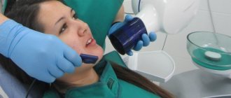
Stages of targeted dental imaging (intraoral x-ray)
It only takes a few minutes to take a targeted photograph of a tooth. The person is placed in a comfortable chair, after which the specialist covers him with a heavy lead apron to protect him from radiation. Next, the person bites the CCD sensor, which is previously placed in a sterile film (disposable covers are used). In just one second, a specialist takes a photo, after which you can remove the sensor and go to get the results.
Note! It is not uncommon for people to experience a gag reflex. This most often occurs when examining posterior teeth. For the same reason, you may not get an accurate image, so you will have to take it again (the same problem occurs if a person is sent for radiovisiography after treatment due to the effects of anesthesia).
Safety of X-ray diagnostics
Surely many people have a question: does X-ray photography harm my health?
X-rays are included in the spectrum of solar radiation, they are partially deflected by the Earth's magnetic field, some are attenuated in the earth's atmosphere, but a significant part reaches the surface of the planet. In addition, various radioactive elements that emit X-rays are present in the Earth's atmosphere, soil and rocks. This is not only uranium, plutonium and other substances used in the production of nuclear weapons and fuel for nuclear power plants. Some radioactive elements are widespread and ubiquitous. These are isotopes of carbon, iodine, hydrogen and others. The total amount of X-ray radiation is called natural background ionizing radiation and is equal to approximately 2.4 millisieverts per year. Compare this figure with the power of X-ray machines released during different types of shooting.
Radiation exposure for various types of X-ray examination in millisieverts (mSv)
| Type of study | Radiation dose | Number per year until the permissible dose is reached |
| Digital radiovisiography of teeth | 2 - 4 μSv | 330 shots |
| Digital panoramic image (OPTG) | 10 - 40 µSv | 40 shots |
| Digital teleradiography (TRG) | 15 - 50 µSv | 33 pictures |
| Computed tomography (CT) | 35 - 55 µSv | 25 shots |
| Film radiovisiography of teeth | 5 - 23 μSv | 30 shots |
| Film fluorography | 800 μsv | 1 photo |
| Travel by plane | 5 - 20 µSv per hour | 100 hours |
| Scanners (introscopes) at airports | 1 μsv | 1000 checks |
As can be seen from the table, the radiation exposure of modern digital X-ray machines is tenths of a percent of a millisievert. That is, the radiation received naturally in a year is equivalent to 300-400 x-rays. Even several dozen images add only a few percent to the natural radiation dose.
With this proportion, Western dentists consider it acceptable to take as many x-rays as necessary to ensure quality treatment. X-ray tubes in many clinics in the USA and Europe are located directly in treatment rooms; the images are taken by the doctors themselves and their assistants.
Russian legislation imposes increased safety requirements when operating ionizing apparatus. The Chief State Sanitary Doctor of Russia, by his Resolution No. 11 of April 21, 2006 “On limiting exposure of the population during X-ray medical examinations,” obliged medical organizations to ensure that patients’ X-ray loads are taken into account and to observe an annual effective dose of no more than 1 millisievert when conducting preventive medical X-ray examinations. This is equivalent to several hundred intraoral photographs or dozens of complex X-ray studies, such as teleradiography or computed tomography of the dentition.
Our legislation largely lags behind existing realities. Many of the requirements for the arrangement of X-ray rooms are excessive. The unit for accounting for radiation exposure is still 0.4 millisieverts, which is comparable to the radiation of an antediluvian film fluorographic apparatus. The technique used in Denta-Lux emits only hundredths of this “unit” in one shot.
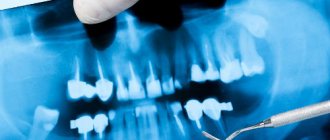
Analysis of the procedure performed
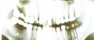
After receiving the results, the specialist must describe the image to make a diagnosis. In the picture you can clearly see the quality of the treatment performed if the following was performed:
- Removal of affected teeth.
- Canal filling.
- Fixation of implants.
If the procedure was performed to identify pathologies, then experts can detect neoplasms, caries, cysts, granulomas, pulpitis, and periodontitis. Additionally, the image shows dense elements, including fillings, dentures, and pins. You can also monitor the condition of bone and connective tissue.
Take a targeted photograph of a tooth in Moscow at a competitive price
Dentistry “Good Hands” has been working on the Moscow market for several years. Our team prefers an individual approach and performs work of any complexity - from professional oral care to root canal filling and implant installation. By contacting us, you receive the following benefits:
- Our dental clinic in Lyublino is equipped with new equipment from leading European manufacturers, so maximum safety and diagnostic accuracy is achieved.
- Only here you can take a targeted photograph of a tooth for only 300 rubles.
- All specialists are highly qualified and undergo certification annually.
- Diagnostics are carried out by appointment, so you don’t have to wait in line.
To make an appointment for diagnostics, contact our administrator by phone right now.
Intraoral scanner TRIOS TM
This model has all the advantages of intraoral systems. This thin flexible device penetrates even hard-to-reach areas of the oral cavity without causing any discomfort and does not require the use of anti-reflective powder. The color images obtained with its help are highly accurate even at the boundaries of the restoration.
The complex process of modeling an impression of various parts of the jaw now takes only a few minutes, since the scanner computer is connected to the milling department of the dental workshop.