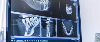
A photograph of the jaw is an important study at the preparatory stage for implantation, which involves installing an artificial titanium root into the jaw bone. To assess the quantity and quality of bone tissue, determine the condition of neighboring teeth, and identify hidden problems (cysts, granulomas), the ROOTT clinic uses modern diagnostic equipment of the latest generation. Before implantation, 3 types of images are taken - targeted X-ray, orthopantomogram (OPTG) and computed tomography (CT).
Orthopantomogram or OPTG is an important component of preparation for dental implantation
Article navigation
- Orthopantomogram - the essence of the study
- Indications for OPTG
- Advantages and disadvantages
- CT or panoramic image - differences
- What types of photographs are there and what is better to choose?
- Preparing for the examination
- How is the procedure performed?
- Are there any restrictions
- Acceptability during pregnancy
- Potential dangers and possible complications
- How to decrypt a photo
- How often can OPTG be performed?
- Cost of dental examination
Question for a specialist
An orthopantomogram is a separate type of x-ray examination that allows you to obtain a panoramic image of the teeth - more precisely, the entire dental system of the patient.
It reveals detailed information not only about the condition of the teeth of both jaws, but also about the maxillary sinuses and other important anatomical structures. This is a modern, affordable and effective way to conduct a diagnostic examination, making it possible to identify all problems at once. True, the detail of OPTG is always slightly lower than that of targeted images and computed tomography results. But today we will take a closer look at the orthopantomogram, find out what it is, how it is done and what it shows. Well, let’s answer the most important question – is it needed before implantation.
Safety
According to the established SanPiN requirements, the annual exposure rate should not exceed 1000 microsieverts. Modern X-ray equipment is safe - the table below shows the radiation exposure during one image and the permissible number of procedures per year.
| Radiation exposure per 1 procedure | Safe number of procedures per year | |
| Sight shot | 1-3 µSv | 500 |
| OPTG | 13-17 µSv | 80 |
| CT | 50-60 µSv | 20 |
| Type of diagnosis | Radiation exposure per 1 procedure | Safe number of procedures per year |
| Sight shot | 1-3 µSv | 500 |
| OPTG | 13-17 µSv | 80 |
| CT | 50-60 µSv | 20 |
X-ray diagnostics are not recommended during the first and last trimester of pregnancy.
Orthopantomogram - the essence of the study
An orthopantomogram is a complete panoramic x-ray of all teeth at once, which allows you to get a general picture of the condition of the dentofacial apparatus. Today, this technique is actively used in cosmetology and various fields of dentistry, including dental implantation and maxillofacial surgery. The procedure is carried out using a special device - an orthopantomograph or computed tomograph (this is more powerful and modern equipment that is installed in clinics that also offer computed tomography services).
Thus, the snapshot allows you to examine the status of the following elements:
- jaws and bite,
- teeth, their shape, position, roots, as well as the rudiments of future teeth, including eights, impacted elements, bone tissue,
- gingival tissue, vascular system, dental canals - examination allows us to identify possible pathological conditions, such as periodontitis or the presence of neoplasms,
- soft fabrics,
- fillings, crowns, dental bridges.
An orthopantomogram (OPTG) is a mandatory procedure in preparation for dental implantation. The examination allows you to identify hidden pathologies, eliminate possible contraindications to treatment and avoid many problems even at the preparatory stage. The diagnosis itself lasts about a minute. After this, the received data is loaded into a computer, where further image processing is carried out.
Important! In addition to OPTG of the dentition, computed tomography is often also prescribed, which allows you to obtain not just a flat, but a three-dimensional image - on it, of course, all elements can be viewed in 3D. This is especially important when all teeth are implanted, immediate loading methods are used, or complex osteoplastic surgery is required.
DENTAL PROSTHESIS WITH 4 OSSTEM IMPLANTS FOR 1 JAW - RUB 130,000.
Implantation even with bone tissue atrophy. Work guarantee! Save RUR 20,000.
on promotion >> Free consultation with an implantologist +7 (495) 215-52-31 or write to us
3D tomography after implant installation
The question often arises: is it possible to perform 3D tomography after installing implants, crowns, and pins?
Unlike MRI, in which artificial bodies “sound” and mislead the dentist, computed tomography after implant installation is allowed, and even recommended. It will be needed in cases where it is required:
- determine the engraftment of the artificial root;
- establish the reason for the instability of the structure or the shortening of its service life;
- check the quality of implant installation if the operation was performed without preliminary computer modeling;
- promptly detect possible complications invisible to the naked eye.
As for metal crowns, there is no need to rush to get a CT scan. First you need to consult a specialist. Conventional metal structures can complicate the examination, as they create optical effects on 3D images, which can lead to incorrect diagnosis.
A separate case is the presence of a pin in the oral cavity. If it is made of fiberglass or ceramic, then CT is acceptable. And if it is made of metal, then the choice of examination method is made based on the criteria:
- level of technical equipment of the clinic;
- qualification of an implantologist;
- the possibility of using alternative diagnostic methods.
Remember that only a specialist with many years of experience can correctly decipher the results of a tomogram that contains titanium or metal pins.
Indications for OPTG
The diagnostic technique under consideration is widely used in various fields of dentistry. Before moving on to the question of how and where a panoramic photograph of teeth is taken, it is worth understanding in which cases such a procedure is indicated. Among the mandatory indications for an orthopantomogram, experts identify the following cases:
- removal of teeth and roots in order to prevent possible complications,
- treatment of periodontal tissue diseases,
- diagnosis of jaw tumors,
- diagnosis of periostitis,
- orthodontic treatment – before installing plates or braces,
- diagnoses of unknown etiology,
- prosthetics, implantation.
Computed tomography or panoramic image - differences and which is better for implantation
What is the difference between an orthopantomogram and a CT scan and which is better to choose? When it comes to preparing for implantation, a two-dimensional panoramic image alone is not enough. In order to thoroughly examine the condition of hard and soft tissues from all sides (and not just one) for the subsequent selection of implants and places for their implantation, you should also undergo computed tomography (CT). Only CT provides the most complete, detailed information about the quality and volume of the jaw bone. And here the diagnostic result will be three-dimensional, volumetric, and not “flat”. Single-stage implantation protocols place even higher demands on preliminary X-ray examination. But OPTG can also be useful for monitoring treatment results.
It is interesting that modern CT machines have such capabilities that they can be used to perform computed tomography, orthopantomograms, and targeted x-rays. The laboratory technician or doctor only needs to change the settings in the device.
CT and especially MSCT, in comparison with conventional OPTG, allows one to achieve the highest degree of detail in images, which means giving an accurate assessment of the quality and condition of bone tissue, guaranteeing the exclusion of possible contraindications, and selecting the optimal models of implants and places for their implantation.
Today, many modern dental clinics offer X-ray examination services, but if we are talking about implantation (especially of all teeth), then preference should be given to a specialized diagnostic center.
The importance of X-ray diagnostics before implantation
The images allow you to identify obstacles to the installation of implants. They identify problems that can complicate the operation.
Using images, the implantologist determines:
- distance to the mandibular nerve and maxillary sinuses;
- density, volume of bone tissue to select the shape and size of the implant;
- the condition of adjacent teeth to prevent infection from entering the surgical area.
Lack of diagnosis can lead to:
- damage to the nerve and sinuses;
- unstable fixation of the artificial root;
- infection of the operation area;
- reducing the service life of the implant and replacing it with a new one;
- increased sensitivity of the gums due to incorrectly selected orthopedic design.
X-ray diagnostics makes it possible to exclude complications and install implants according to the correctly selected protocol in accordance with the patient’s clinical situation.
What types of photographs are there and what is better to choose?
As for the classification of OPTG, there are 2 types of panoramic X-rays of the jaw - film and digital orthopantomogram. Sometimes you can also hear about three-dimensional, but in fact this type of OPTG does not exist - all X-ray results here are only two-dimensional, “flat”.
- 1. film – this type of OPTG appeared first. Initially, models of devices were used that involved the use of a special film. It was placed on one side of the patient's head, while the X-ray tube was on the other side. After the procedure, the image was transferred to film, after which it was processed and developed,
- 2. digital – a modern version of the examination involves recording images on a medium in digital format. The obtained data is loaded into a special computer program, and the doctor can carefully study the already processed image from the computer screen.
What is better to choose – digital or film orthopantomogram? Of course, it is better to give preference to the digital option. It is more modern, accurate, and the image can be enlarged many times on a computer to examine the pathological area in detail.
Preparing for the examination
The procedure does not require any specific preparation; it can be carried out directly on the day of contacting the dentist or diagnostic center. However, before sending a person for OPTG, the specialist is obliged to conduct a visual examination of the oral cavity and collect a complete anamnesis, that is, take into account all complaints and possible pathological phenomena in the maxillofacial area. After the initial diagnosis, all that remains is to settle some formalities: the doctor will ask you to sign a consent to conduct the examination.
Advantages of the method
- The ability to rotate, enlarge, and examine images in any projection and section, which is impossible with conventional 2-dimensional scanning.
- The examination lasts only a few seconds (8-20 seconds).
- Complete diagnostic information.
- Maximum security.
- Digital information format.
- Detection of any pathological processes at an early stage.
- No prior preparation required.
- 3D reconstruction without distortion or artifacts.
- A wide range of purposes - from endodontic dental treatment and implantation to maxillofacial operations.
How is the procedure performed?
We have figured out why an orthopantomogram is done in dentistry, and now we will look at the progress of the procedure. Diagnostics is completely painless for the patient, and the operation of the device itself takes no more than a minute. In this case, the patient immediately receives the examination results. Immediately before the procedure, the specialist will ask you to remove all metal items and jewelry and put on a protective vest. The doctor will indicate the place where you need to stand during the diagnosis. After that, he will raise the working area of the device and fix it at head level.
“I took a panoramic photo before implantation - this is the most harmless procedure I have ever undergone in dentistry, especially compared to the operation itself! I don’t understand the reason for the excitement of some patients, after all, they probably underwent fluorography, everything is similar here, only they examine not the chest, but the jaw. Everything really takes no more than a few minutes. Moreover, the doctor immediately showed me the result on the computer screen, gave some comments, gave me the picture, and I also wrote it down on a flash drive, just in case. In general, don’t be afraid, there’s nothing scary there at all...”
Mila1989, Moscow, from correspondence on the woman.ru forum
After this, the person’s head is fixed using a special plate. At one end it is attached to the equipment, and its second free end is opposite the mouth. A sterile tip is fixed on it so that the patient can clamp it in his teeth - this helps ensure the immobility of the jaw apparatus during the examination and eliminate any distortions.
When all the equipment settings are ready, the specialist will definitely warn the patient about the start of the examination, and also remind him of the importance of remaining still. After turning on the tomograph, the plates will gradually rotate around the head, making two revolutions. Next, the doctor will be able to evaluate and show the patient the result on the computer screen, and will offer to print the image or record it on a digital medium.
Types of devices for performing CT scans of teeth
Several types of devices are used in performing dental CT scans. In this case, it is necessary to clarify which device will be used in your particular case, since not all models can guarantee clear images, while others have too intense radiation exposure on the body.
- Step-by-step computed tomography of teeth . Devices of this type have been used for quite a long time. Their distinctive features include poor clarity of bone tissue structures and low image quality. Moreover, when a CT scan of teeth is performed, the radiation dose (1500 μSv) applied to a person during one examination is one and a half times higher than the permissible values for a year;
- MSCT ( Spiral tomograph ). In this case, the person receives a smaller dose of radiation exposure, but the danger to the body still remains. Similar to the above model, there is insufficient structural clarity of bone tissue and low image quality. A similar device gives better results when examining lymph nodes, facial muscles and salivary glands;
- CBCT ( Cone beam computed tomography ). This is the latest type of equipment that provides maximum accuracy when examining root canals, teeth and bone structures. Moreover, the radiation dose when performing one specified study does not exceed 90 μSv, which makes it possible to carry out a secondary procedure.
It should be borne in mind that the degree of radiation exposure for devices of the latter type will have some differences, depending on the manufacturer of the tomograph, as well as the weight and size of the patient.
In addition, computed tomography systems may differ from each other in terms of the convenience of the programs used, which directly affects the usefulness of the final examination results.
The Implantmaster dental clinic uses CBCT, which is based on the use of the latest professional equipment, high professionalism of doctors, and the availability of software for most specialists.
Are there any restrictions
This examination option is considered safe and harmless, however, there are some contraindications. The procedure is not performed if the patient has malignant neoplasms in the head and neck area, as well as blood cancers - leukemia, lymphoma and others. In the first case, under the influence of radiation, the tumor can begin to grow and mutate, which will lead to even more severe complications. In the second case, even a slight effect of radiation on the bone marrow, a hematopoietic organ, can provoke and accelerate the development of the disease, including fatal consequences.
Also, performing OPTG is not always possible in a small child or in a person with serious mental disorders - when the patient is unable to remain still for even 1-2 minutes.
Acceptability during pregnancy
Panoramic tomography is extremely undesirable for patients during pregnancy - in such cases it is performed only if there is an urgent need. The procedure poses an increased risk during the first trimester. In later stages of pregnancy, X-ray radiation will not have such a severe effect, but can still cause certain disturbances in the functioning of internal systems. For example, the thyroid gland is especially sensitive to the effects of radiation.
Popular questions about dental CT
How often can a CT scan be performed?
During one examination procedure, the radiation dose reaches about 60 microsieverts, which is significantly lower than the radiation level during standard radiography. That is, even if you need to perform two CT sessions of the jaw joint, it is absolutely safe for the body.
Does the patient feel any discomfort during the examination?
This procedure is characterized by efficiency and ease of implementation, which eliminates the occurrence of any pain during the operation of the device.
Is such a diagnosis allowed during pregnancy?
A similar technique is allowed for pregnant women and women during breastfeeding. But since the effect of X-rays on a child has not been fully studied, the study is prescribed only in extreme situations and only when using a cone-beam tomograph. The lowest risk to the fetus is observed in the second trimester.
Author:
Potential dangers and possible complications
Many patients in dental clinics are interested in quite natural questions regarding whether an orthopantomogram is harmful and how often it can be done so as not to cause harm to health. Let's start with the fact that if you follow the technology of the procedure, you should not be afraid of any problems. One of the potential complications may be excessive radiation exposure, however, with modern equipment this situation is excluded, even though OPTG will have to be performed at least 2-3 times during implantation, as well as several more times during the year after it to monitor the results of treatment .
Numerous studies on the safety of using an orthopantomograph have proven that with a single procedure, the patient receives a radiation dose corresponding to values in the range from 10 to 40 μSv . This is an acceptable and completely harmless dosage that does not pose any threat to the human body. For comparison: with a single fluorography procedure, the radiation dose is up to 500 μSv.
Beam prosthesis RUB 180,000.
Preparation of the oral cavity, individual bar with installation, taking impressions, manufacturing, installation and fitting of dentures. The price is for 1 jaw. Work guarantee up to 2 years! Consultation with a doctor is free! Call now: +7 (495) 215-52-31
Initial examination
Before dental implantation, you need to visit the dentist's office for an initial consultation. During this procedure, the doctor will carefully examine the oral cavity, assess its condition and give a referral for x-ray diagnostics and tests. Already at this stage, the dentist can:
- determine indications and contraindications for implantation;
- inform the patient about the features of the upcoming operation.
In addition, during the initial examination, the patient must fill out a questionnaire (survey card), in which the presence of bad habits, chronic diseases and surgical interventions should be noted.
| We strongly recommend that you fill out the survey card honestly. Then our doctors will know better how your body will behave during implantation. |
How to decrypt a photo
In good clinics and specialized centers, the radiologist often provides a detailed description of the image, which identifies all the identified problems. However, such an approach to work is not found in all medical institutions today. Usually, the patient is given only an envelope with an image, and its detailed study and analysis is carried out by the attending physician.
If you try to “read” the results of an X-ray examination yourself, then, if you wish and have some skill, you can assess the position of your wisdom teeth, detect inflamed areas, and identify a decrease in the level of bone tissue. In addition, on the Internet today you can find a lot of sources with visual examples of dental problems shown in panoramic photographs of different patients - they can be used for comparison with your results.
Our clinic uses a safe device that meets international standards
The digital X-ray system with a minimum level of radiation SIEMENS SIRONA GALILEOS allows you to perform the most accurate studies for patients. The device is intended for the study of bone tissue, tooth roots, temporomandibular joint, nasopharynx, and upper spine. An analysis of local problems and the condition of a large volume of jaw tissue is carried out.
Levin Dmitry Valerievich
Chief physician, Ph.D.
Cost of dental examination
The cost of an orthopantomogram in clinics in Moscow and the regions starts from 700 rubles. But sometimes patients undergo it for free, for example, during turnkey implantation, when the total price of dental restoration already includes all the necessary diagnostic methods, consultations, implants, dentures and the work of specialists.
Chibisova M.A. Digital and film radiography in outpatient dentistry, 2004.
Author: Bespalov R. D. (Thank you for your help in writing the article and the information provided)