An x-ray is the dentist’s main tool in making the correct diagnosis. However, a conventional orthopantomogram or targeted photograph has limited diagnostic potential and does not provide complete data on the condition of the teeth and maxillofacial area. But technologies are constantly being modernized and today, more informative technology has come to the aid of conventional radiography - dental computed tomography (CT).
What does a 3D dental x-ray show?
3D dental tomography is a highly accurate diagnostic method that makes it possible to obtain a three-dimensional image of the dental system in different projections. Volumetric images obtained with CT allow the specialist to enlarge, rotate and examine the area of interest from all sides and at different depths:
- The entire maxillofacial apparatus.
- A dentition or an individual tooth.
- Paranasal sinuses.
- Bone and periodontal tissues.
Dental CT allows you to detect inflammation, assess the homogeneity of the filling material and check the quality of installation of a filling, crown or implant, see the number of dental roots and their fragments, identify neoplasms, assess the degree of curvature of teeth, determine the exact parameters of bone tissue (height, width, density, etc.). d.). The information obtained allows the doctor to optimize treatment measures and predict the result.
Rehabilitation after 3D computed tomography
As after a classic X-ray, there are no special recommendations for rehabilitation after a CT scan. This is a safe and easy way to find out about the presence of diseases, after which a person can immediately return to his usual lifestyle. In the rarest cases, a tomography with the introduction of a contrast agent may be prescribed. This is done extremely rarely for dental treatment; the recovery plan will be the same as after a CT scan of any other part of the body.
Experts strongly recommend drinking as much liquid as possible on the first day after the manipulations, and also giving preference to liquid foods, such as soups. You can also drink milk, but you can increase your usual portions. If the doctor has not prescribed antibiotics and other medications, you can drink a glass of red, dry wine (no more than 250 ml). Many doctors recommend eating natural honey, drinking freshly squeezed juices with pulp, and if there is a lack of hemoglobin, adding beets and pomegranates to the diet.
If you act correctly, the contrast is eliminated from the body within the next day (up to 90%). However, if there are problems with the functioning of the kidneys, the volume of fluid consumed should not be sharply increased. If a person remains in the hospital after a CT scan, he may be treated with saline and glucose intravenously. General recommendations after CT include eating plenty of nuts, fruits, parsley and beans. It is important to note that this procedure does not affect the possibility of conception in any way.
Why are dental x-rays prescribed?
A 3D photograph of teeth is performed if the following indications exist:
- Injuries of the maxillofacial area.
- Preparation for endodontic treatment (structure of root canals, pathological processes in the periodontium, degree of pulp damage, etc.).
- Diagnosis of neoplasms (cysts, abscesses, granulomas, tumors).
- Anomalies of development and deformation of the maxillofacial apparatus.
- Quality control of filling and implant installation.
- Planning of orthodontic treatment (identification of impacted and dystopic teeth, analysis of the condition of the tissues around each tooth, etc.).
- Detection of hidden periodontal cavities and pockets.
- Implantation planning (assessment of jaw bone parameters, indications for sinus lift or osteoplasty, modeling the result of implantation).
- Endogenous pathologies of the maxillary sinuses.
Three-dimensional x-ray examination is the gold standard when planning any complex dental procedure or surgery. CT allows you to quickly make an accurate diagnosis, competently plan treatment or dental prosthetics, and monitor the results.
Features of the use of CTCL in dentistry
In dental practice, cone beam tomography of the jaw is used to solve complex problems:
- removal of roots or “figure eights”;
- removal of a foreign body, fragment;
- canal inspection;
- exclusion of jaw neoplasms;
- detection of bone tissue destruction;
- assessment of periapical dentin tissues of teeth;
- detection of anomalies and birth defects;
- preparation for implant installation.
The examination remains the only diagnostics in dentistry that allows one to correctly assess the position of the canals, the condition of the pulp, and the presence of nerve endings. This is an important part of the examination before complex operations on the periosteum and implantation of an osteoprosthesis.
Due to its high effectiveness, the risk of metal pin rejection and other complications is reduced several times. Advantages of CBCT of the jaw over other techniques:
- Comfort for the patient, absence of unpleasant sensations, which is important for people who are afraid to go to the dentist.
- Minimum time spent on a quality examination, which reduces the number of visits to the dental office.
- To examine dental canals, the use of CBCT is the only way to measure the length. It is easier for the doctor to choose the ideal shape of a filling or pin.
In addition to studying the condition of teeth and bones, CBCT allows you to examine soft tissues. This helps to timely detect a cyst in the root of a molar, a fistula or an oncological tumor in the oral cavity, and select therapeutic agents. The data obtained is subsequently used in preparing the operation. Dentists can create a visual model and show the patient what the face will look like after all the procedures, and make sure that the crowns are in the optimal shape.
Study radiation dose
Many people naturally ask what is the radiation dose of cone beam computed tomography. This research method has a much lower x-ray load than when examining with spiral tomography. This is due to the high rotation speed of the tube. However, you should not prescribe this diagnosis yourself, since only a doctor can assess the actual need for it.
In addition, the following factors should be taken into account:
- Conducting conventional fluorography gives an exposure of 0.18 mSv;
- from the natural background of the Earth, each person receives radiation of about 1000 μSv;
- The maximum permissible dose at which no significant changes occur in the human body is 5000 μSv.
Due to the short examination time, the radiation dose of cone-beam computed tomography is in the range of 40–120 mSv. If we examine the skull with spiral computed tomography, the radiation exposure will increase from 400 to 600 mSv. In addition, testing on a cone-beam tomograph allows one to exclude further examination using other diagnostic techniques, which results in a low total radiation exposure to the body of the person being examined.
Where is the research applied?
The operating principle of cone beam computed tomography is based on visualization of the examined area. This device is not only distinguished by the ability to obtain a three-dimensional model of the problem area, but also by its compactness and safety. Research on it began for the first time in the USA, then came to Europe.
Modern devices are equipped with a robotic arm that allows you to select the desired trajectory for the task of sensor movements. Basically, they allow examination to be carried out on a small area, but, if necessary, the gluing function is used to expand the volume.
This type of tomography is widely used to identify problems in the following areas of dental research:
- Therapeutic dentistry allows us to identify acute inflammatory processes not only of the teeth, but also of the soft tissues surrounding them. Used to study canals, recognize the area of tooth root destruction, and control therapy.
- Surgical dentistry allows you to determine the location of the inflammation, its size, and the location of bone collection for the implant. It is used to detect pathologies formed as a result of low-quality therapy, surgical intervention, and allows you to detect parts of the tooth remaining after extraction.
- In orthopedics, it allows you to accurately draw up a treatment plan, assess the condition of the supporting tooth, and promptly identify complications that have developed due to the installation of the structure.
- In orthodontics, it is used for planning, allowing you to make the right decision about the need to remove teeth that interfere with the installation of the prosthesis.
A modern research method allows the doctor to receive results on his computer.
The use of cone beam computed tomography is widely known not only in dentistry, but also for solving problems:
- in implantology, it allows you to prepare the patient for implantation, assess the condition of the bones, and obtain accurate information about the place where the manipulation is supposed to be carried out;
- Maxillofacial surgery is used to assess bone injury, detect tumors, and treat inflammation;
- Otorhinolaryngology allows you to assess the condition of the nasal cavity and its sinuses, make the right decision on the advisability of operations, monitor and adjust treatment.
This is interesting: Pulpotec: composition, instructions for use, analogues, reviews, price
These directions have a small scanning area, usually assessing the conditions of the jaw bones, nasal septum, soft and bone tissues of the skull. Most often, this method is used to diagnose congenital pathologies of the palate, select implants, study diseases of the nasal sinuses, and abnormal positions of teeth, when other methods do not make it possible to make an accurate diagnosis.
Limitations of the study
Although the cone beam computed tomography method is considered an innovation that allows you to accurately determine the condition of the area under study, it must be borne in mind that it belongs to the category of radiation techniques and therefore requires some caution. First of all, it is prescribed in a limited manner:
- for children under 5 years of age. For this category of the population, this research method can be prescribed only if there are vital indications;
- persons suffering from renal failure;
- people who cannot remain still for 2–3 minutes;
- patients who have pronounced pain syndrome;
- pregnant women.
During pregnancy, any tomography or radiography is contraindicated. The only exception is a vital need to obtain urgent medical care, provided that the benefit from the examination for the mother is lower than the expected risk to the fetus. You should also talk to your doctor about preparing yourself to reduce the risk of harmful effects from x-rays.
Carrying out the procedure in the third or second trimester reduces the likelihood of developing pathologies in the fetus. Cone beam tomography requires the appointment of a doctor who can adequately assess all the risks to the body from this study.
Children's Study
There are cases where it is necessary to conduct this study for young children. Of course, a child’s body is more sensitive to radiation, but if there are serious indications, diagnosis should not be abandoned. If the child does not have an absolute contraindication to the study, then this method can be used even for babies in their first year of life.
Such restrictions include:
- birth injury;
- congenital anomalies;
- allergic reactions to drugs used to give anesthesia;
- heart disease.
Older children tolerate this examination quite easily
Before children are examined, they should not be fed for 2.5 hours before the intended procedure, otherwise aspiration pneumonia may form. When at the time of the study the child is already 4 years old, you should talk to him. At the same time, try to explain the procedure, be sure to emphasize that mom and dad will be nearby at all times.
For younger children, the examination is performed under anesthesia. Moreover, during the diagnosis, parents can be with the baby and wear lead aprons for protection.
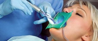
Description of the diagnostic procedure
To understand how computed tomography is done, it is worth considering a step-by-step description of the process:
- immediately before the procedure, the specialist will ask you to remove all metal jewelry to avoid equipment malfunctions,
- then the patient puts on a special protective vest to reduce the degree of radiation exposure to the body as a whole,
- the patient stands or sits with his back to the device, and his chin is fixed using a special stand - this is necessary to eliminate unnecessary movements and get the most accurate image possible,
- after turning on the device, a scanner with an emitting tube begins to rotate around the patient’s head - it is this that transmits the three-dimensional image to the computer.
The photo shows a tomography being performed
The procedure lasts less than a minute. The diagnosis is completely painless and does not require any serious preparation from the patient.
What equipment is used
To carry out 3D diagnostics, a three-dimensional computed tomograph SOREDEX Scanora 3D with advanced functionality is used. This is the latest generation equipment, which allows you to obtain three-dimensional images of the anatomical structures of the maxillofacial region in a few seconds, with the least radiation exposure for the patient.
The program analyzes the obtained multiplanar sections and builds them into a 3D model, thanks to which the specialist is able to accurately assess the condition of the dental system, detect all pathological processes occurring in this area and competently plan a treatment regimen.
A virtual 3-dimensional model of the scanned area can be recorded on any digital media (CD, flash drive), which allows the attending physician, if necessary, to view diagnostic data or involve related specialists in the analysis of the received information.
New teeth for patients protected by computer technology
As you know, medicine has undergone significant changes in recent years, which allows patients to receive all their guarantees regarding safety, efficiency, reliability, comfort and good treatment results. Such positive trends are also occurring in the dental industry, and in particular in dental implantation.
Today, everyone can safely decide to transform their smile with the help of 3D implantation, or rather, thanks to 3D technologies in dentistry. In the article below we will take a closer look at what “secrets” are available to professional doctors and how new teeth can now be obtained in just a few days.
Possible harm
Cone beam dental computed tomography is the safest and fastest diagnostic method. Thanks to the use of a conical X-ray beam, the radiation dose received during the study is 10 times less than when using spiral CT. And the pulsating mode of the X-ray beam further reduces the radiation dose. The three-dimensional computed tomograph SOREDEX Scanora 3D is one of the safest devices in terms of X-ray radiation dose - only 0.035 m3v.
However, despite the safety of the study, CT also has contraindications. If we just talk about dental tomography, it is not performed during pregnancy (in the 1st trimester). 3D dental x-rays with contrast are prohibited for pregnant and lactating women, patients with endocrine disorders (diabetes mellitus, thyroid pathologies), renal failure and intolerance to iodine-containing drugs.
Indications for this type of examination
A 3D image for dental examination is used in the diagnosis of complex injuries and diseases of the jaw. Indications for prescribing computed tomography include:
- incorrect position of the tooth in the gum;
- neoplasms in the maxillary sinuses or jaw;
- extensive inflammatory process;
- malocclusion;
- disorders of motor functions of the mandibular joint;
- difficulties with implantation and prosthetics;
- fractures of the lower jaw;
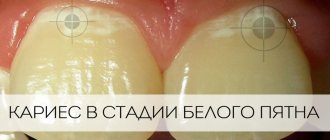
- diagnosis of caries complications;
- planning orthopedic treatment and evaluating its results;
- planning cephalometric changes;
- diagnosis of periodontitis.
Main contraindications
3D computed tomography of teeth is based on x-rays, so this procedure cannot be called completely safe. The radiation exposure during this examination ranges from 0.045 to 0.06 mSv. This is not a very high figure, given that the annual exposure limit is 5 mSv (according to the Russian Ministry of Health).
As for contraindications, they are standard for all types of x-ray examinations. The main limitation is the period of pregnancy (especially in the first trimester). In this case, the situation is considered individually, based on the ratio of harm to the child and benefit to the mother.
If a contrast agent is used (it is not used so often in CT scans of the teeth and jaw), then the following can be added to the contraindications:
- Patients suffering from thyroid diseases.
- Kidney failure.
- Allergy to drugs containing iodine.
The lactation period is not a contraindication, but after the procedure at least 48 hours must pass before the next breastfeeding.
Can this be done for children?
Many parents think that the use of 3D computed tomography is impossible in childhood due to the negative impact on the child’s body. This is not entirely true, because special attention to this study is given specifically in pediatrics.
If you follow all safety rules and do not exceed the frequency of procedures (for children – once a year), then the examination will not lead to any pathological changes in the child’s body.
3D CT in pediatric dentistry allows you to assess the condition of the sinuses, gums and see the rudiments of baby teeth. This method also allows you to identify malocclusion pathologies in a child.
Advantages of the method
- The ability to rotate, enlarge, and examine images in any projection and section, which is impossible with conventional 2-dimensional scanning.
- The examination lasts only a few seconds (8-20 seconds).
- Complete diagnostic information.
- Maximum security.
- Digital information format.
- Detection of any pathological processes at an early stage.
- No prior preparation required.
- 3D reconstruction without distortion or artifacts.
- A wide range of purposes - from endodontic dental treatment and implantation to maxillofacial operations.
Materials and stages of modeling on a 3D printer
Two types of materials are used to print finished products:
- A special biologically compatible composition, the structure of which changes based on the specifics of the planned work;
- Technical materials that differ in shades and degree of rigidity of the finished structure.
At the preliminary stage, a comprehensive examination of the oral cavity is carried out. The use of an intraoral scanner, which replaces impressions, provides the necessary information on the basis of which a virtual prototype is formed.
Is there an alternative to CT
There are many other diagnostic imaging methods (x-ray, orthopantomogram, ultrasound, etc.), but only CT provides the possibility of highly accurate, separate images of all types of tissue at different angles and to different depths. Although a panoramic dental photograph remains an equally important diagnostic tool for a dentist today, it can only provide a general overview. In turn, a 3D tomogram allows you to obtain not a single flat image of the jaw, but a whole series of sequential multiplanar images in different projections and without the distortions inherent in a panoramic image.
Example:
due to the different density of bone structures exposed to X-ray radiation, it is impossible to see less dense bone in a 2-dimensional image; accurate information is provided by a 3- D image of the teeth.
How does the procedure and decoding work?
To take a 3D photograph of teeth, a standing or sitting patient needs to bite a special plate and fix his position in the device using a fixing stand. During the entire scanning time, you must remain absolutely still.
The tomograph sensor makes a series of revolutions around the patient’s head for 8-20 seconds, producing about 200 images in different projections. Processing digital data takes 5-15 minutes, after which the information is written to a disk or flash drive. No preparation is required, you just need to remove all metal jewelry from your neck, ears, and hair before the procedure.
How long does it last?
The duration depends on the type of tomograph. The main recommendation in this matter is to inquire about the type of device before the study and choose a clinic with the most modern high-power device. Its advantages are not only a minimal radiation dose and high detail, but also a much shorter research time. By comparison, the duration of a scan can vary from a few seconds to 15 minutes.