27.02.2020
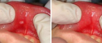
Most expectant mothers are faced with the need to treat their teeth during pregnancy. The growing baby takes everything from the pregnant woman for its development. In particular, it requires a lot of calcium to build its own skeletal system. If the diet contains few foods containing calcium, the fetus will “take” it from the pregnant woman’s body. This can lead to weakened tooth enamel and tooth decay.
Once upon a time, teeth were treated without x-ray examination and the quality of treatment was low. Radiography has taken dentistry to a new level. However, this examination is not safe during pregnancy. Most doctors avoid ordering dental x-rays during pregnancy due to possible adverse effects on the fetus. Therefore, the opinion of experts is divided into pros and cons.
What equipment is used to take an x-ray?
A visiograph is an electronic sensor that perceives x-ray radiation, transforms it into digital form and displays it on a computer screen. This is the most modern radiography system.
The first advantage of a visiograph is a very low radiation dose to the patient compared to a film one. As a result, radiation exposure is reduced to a negligible minimum.
Now the doctor can afford to take as many pictures of the patient at one time as necessary for quality treatment and diagnosis, without fear for the patient’s health.
Summary: important points
As a practicing dentist who knows the system from the inside, I want to draw your attention to the following points that are important for your safety. If the clinic has an X-ray machine, then a license must be obtained for it, the issuance of which presupposes the mandatory presence of a certified radiologist on the staff of the dental clinic. However, in reality, even in large clinics and public clinics, it is not always the case that x-rays will be taken by a trained specialist.
Even if he is, he may go on vacation or get sick, and a regular nurse (dental assistant) will take the pictures instead. This is a gross violation that leads to both the production of low-quality images and an increase in the radiation dose. In small clinics, the risks of receiving a poor-quality x-ray examination are much higher, and the first thing that makes you suspect a forgery is if the picture is taken not by a special employee, but by a nurse from the dentist you came to see.
The second very important point: if you see that the x-ray is not done in a special room, but the x-ray machine is located right next to the dentist’s chair, then you should change the clinic and the doctor. The fact is that all objects in this office (including the dental chair and doctor’s instruments) will have an increased background radiation, and this is no longer safe for health. We hope that our article on the topic: X-ray of the jaw and teeth was useful to you!
Sources:
1. Higher prof. the author’s education in therapeutic and surgical dentistry, 2. Based on personal experience in therapeutic and surgical dentistry, 3. National Library of Medicine (USA), 4. “Digital and film radiography in outpatient dentistry” (Chibisova), 5. “X-ray diagnostics in dentistry" (Lutskaya I.K.).
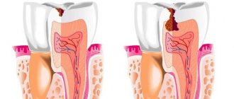
In what cases is it necessary to take dental x-rays?
X-rays should be taken for dental problems such as:
- the presence of foci of infection (chronic periodontitis, loss of bone tissue in the area of the apex (apex) of the root);
- presence of root fractures and cracks;
- pathological periodontal pocket;
- the presence of hidden pocket cavities;
- tooth structure with complex root canal anatomy.
When visiting a dentist in the first trimester of pregnancy, a woman must inform the doctor about this. In the first weeks of development, embryonic cells are in the stage of active division, so it is necessary to eliminate minor negative effects on the body.
What does it show
CT scans provide more detailed images compared to traditional x-rays. This is due to the fact that photographs are taken from different angles and in different sections.
The method allows you to assess the condition of the following structures:
- teeth;
- soft tissues of the face;
- bone structures;
- temporomandibular joints.
Computed tomography helps identify abnormalities:
- inflammatory foci;
- presence of foreign bodies;
- uneven dentition;
- bone damage;
- neoplasms;
- dental damage.
At what time is it permissible to take x-rays?
In modern dentistry abroad, and now in our country, the treatment and preventive direction - dentistry for pregnant women - is becoming increasingly relevant. This is explained by the peculiarities of the functioning of a woman’s body during pregnancy, which requires careful selection of medications and a special approach in choosing methods of dental care.
The most unfavorable time for dental x-ray examination in a pregnant woman is the first trimester, and especially the first two weeks of embryonic development. During this period, the formation of the most important organs and systems occurs. To eliminate the negative impact of radiation on actively dividing embryonic cells, it is better to refuse X-rays at this time.
The second trimester is more favorable for taking x-rays. Because at this time the likelihood of pathologies decreases tenfold.
During the third trimester there is also an increased risk of dental treatment having a negative impact on the pregnancy. The uterus is especially sensitive during this period than in the first weeks, but the risks of premature birth due to stress are undesirable.
Benefits of CT
CT scanning of the jaw has many advantages. That is why this diagnostic method has found wide application in dental practice.
Advantages:
- Safety. Radiation exposure during the study is minimal.
- Information content.
- Image clarity.
- The ability to zoom in on a computer monitor and examine the desired area in detail.
- Fast execution (less than one minute).
- Comprehensive examination of the entire dentofacial apparatus.
- Possibility of saving received data on electronic media.
On the left is a panoramic image of the jaws, on the right is a CT image
How is the imaging procedure performed?
Before the x-ray is taken, the woman must remove all metal jewelry. To protect from X-ray rays, the patient is wearing a lead apron that covers the shoulders, chest, and abdomen. The head and neck are also shielded. Such safety measures completely eliminate the impact of x-ray radiation on the fetus, on the woman’s reproductive organs, as well as other organs and systems.
When ordering an x-ray, specialists at the KIO STOM dental clinic evaluate each case individually from a professional point of view. In this case, the condition of the pregnant patient is taken into account.
Manufacturers of X-ray equipment claim that the resulting radiation cannot have any effect on the child, thereby declaring the complete safety of the procedure.
Doubts of pregnant women
The need for an X-ray is justified for doctors; the image guarantees the accuracy of the diagnosis, eliminates errors in the treatment process and negative consequences for the patient. After making a diagnosis, the dentist develops a treatment regimen taking into account the individual characteristics of the patient: gestational age, drug intolerance. There is a widespread misconception about the harm of x-rays to the body of pregnant women; most women prefer the removal of “problem” teeth. Surgery without the use of anesthesia is impossible; the safety of medications is also accompanied by heated discussions among expectant mothers.
Doubts regarding the safety of x-rays should be discussed with a qualified specialist; refrain from discussing them with friends; treatment by analogy is impossible. Fear for the child negatively affects the well-being of the pregnant woman and the fetus, but the practical experience of dentists eliminates mistakes; it is better not to delay treatment and follow the recommendations.
Features of radiography
A competent treatment plan for a pregnant woman is a guarantee of safety. After examining the patient, the dentist decides on the need for an x-ray, discusses the treatment regimen, the sequence of procedures with the woman, and answers any questions. A comfortable body position is a prerequisite for the procedure; the patient is covered with a protective apron; to provide additional protection, the use of class E film is practiced.
Older models of X-ray machines are safe provided that the process is properly organized and safety rules are followed. Special paper is fixed to the tooth that needs to be treated; the procedure takes less than a minute. Modern equipment simplifies the procedure and reduces time costs. The specialist accurately determines the radiation spectrum taking into account the condition of the tooth; the beam precisely hits the problem area.
Write a comment
Marina
February 21, 2021 at 03:24 pm
Although opinions differ about the harmful effects of x-rays on the fetus, I think the decision should be made by the doctor in a specific case and for a specific pregnant woman. For example, in the seventh month I had a toothache. The dentist removed the nerve and filled the tooth without checking with x-rays. And he said that it would be better to check it later and, if necessary, change it, rather than endanger the child.
Dasha
February 21, 2021 at 10:29 pm
I believe that the dental issue needs to be resolved before pregnancy. Dental treatment in any case involves the use of some medications, drugs, painkillers; all this is not advisable for a pregnant woman. But if this is already necessary, then you need to consume vitamins to the maximum, without them you can’t go anywhere. In general, I think there is nothing wrong with this.
Nastya
February 23, 2021 at 07:22 pm
During pregnancy I was going to get braces. Naturally, it was necessary to take two pictures... I didn’t risk it + I had an ultrasound scan 4 times. Although I remember reading on the Internet, many girls are not afraid - they do it. My friend, a dentist, told me that pregnant women often come with recently taken photographs... I don’t know... here, of course, everyone makes their own choice.
Inna
March 9, 2021 at 03:33 pm
When I was pregnant, I still tried to protect myself from x-rays, the older generation even prohibits the use of cosmetics, and this is an x-ray, no matter how radiation. And as per the law of meanness, it was when I was pregnant that I was diagnosed with a cyst, naturally, that’s why I needed an x-ray. Despite this, the pregnancy went well, the baby was born on time.
Lili
February 8, 2021 at 04:47 pm
This is all nonsense, they are just intimidating naive mothers about how much radiation is needed to affect the fetus... If you listen to the media, you can’t eat meat, drink milk or wash with tap water. They constantly make a big deal out of nothing. I know people who, even if they are not pregnant, are afraid to take an x-ray, and do not yet use a microwave and do not fly on airplanes.
Nastena
February 9, 2021 at 07:41 pm
I’m 5 weeks pregnant, I haven’t gone to the dentist and in fact I only recently found out that I’m pregnant. They sent me to the antenatal clinic to the dentist’s examination room. She scolded me and made me take an x-ray because I had a lot of bad, almost completely destroyed teeth. Well, I’m afraid of dentists; I haven’t been to the dentist for a long time. So I read that x-rays are not allowed, in case there is a miscarriage, what should I do?
Evgeniya A
February 10, 2021 at 09:17 pm
During my second pregnancy, I fell on ice and broke my leg. So at 10 weeks they did an x-ray of my legs! And nothing, ugh, ugh, happened, my son is 8 years old. And here are the teeth... hmm, girls, if you have problems with your teeth, don’t be afraid of this meager radiation exposure, the infection that spreads from a bad tooth throughout the body is much worse. If mommy’s tooth keeps rotting, then the child also comes under the influence of bacteria.
List of abnormalities in the fetus caused by radiation
If a woman had an x-ray in the first 12 weeks of pregnancy , this can cause serious deviations in the development of a number of systems and organs in the fetus:
- spine;
- bronchi;
- hearts;
- faces;
- jaws;
- reduction in the size of the skull and brain.
Radiation contributes to the development of health problems in the unborn child:
- dystrophy;
- blood diseases;
- incurable chronic diarrhea;
- predisposition to cancer.
Also possible:
- frozen pregnancy;
- miscarriage.
But all of the above is unlikely. To cause abnormalities in the fetus, a pregnant woman needs to receive a radiation dose of 3 mSv, and one procedure on modern dental equipment produces a load of 0.02 mSv, and on older devices 0.3 mSv.