The quality of dental treatment depends on the accuracy of the diagnosis. A panoramic photograph of teeth helps to find the cause of dental diseases. However, some patients are concerned about X-rays, fearing harm to their health. In the article we will consider the need for such images, harm to the health of the body and other issues related to this procedure.
What is an orthopantomogram?
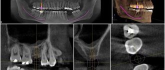
A panoramic photograph of the teeth, or abbreviated as OPTG from the word “orthopantomogram”, is a two-dimensional cross-sectional image of the patient’s jaw. In fact, this is an ordinary x-ray, since it is x-ray radiation and an orthopantomograph x-ray machine that is used to take the picture. This is where patients begin to fear for their health, because X-ray radiation is harmful to the body.
What you can see in the image:
- both jaws;
- all teeth including roots;
- condition of bone tissue;
- jaw joints;
- maxillary sinuses.
The doctor receives a panoramic picture of the patient’s skull and can make the most accurate diagnosis.
However, a panoramic image still differs from the usual x-ray and other hardware diagnostic methods: teleradiography, tomogram.
When an X-ray is taken, a visiograph works. With the help of such hardware research, you can see a maximum of two adjacent molars. That is, a targeted image is obtained in a two-dimensional plane. An x-ray can be taken at any public clinic.
Orthopantomogram
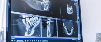
An orthopantomogram is done using a computed tomograph. The result is a panoramic image of the jaw in two dimensions. Diagnostics can be done in any public or private clinic. The price is several times more expensive: from 1000 rubles.
Teleradiography
Teleradiography is also performed on a computed tomograph. However, compared to an orthopantomogram, the doctor already sees the entire skull in front and profile. Accordingly, this is a more informative diagnosis than the first two. The examination method is used to resolve surgical and orthodontic issues. The procedure costs from 2000 rubles.
Tomogram
A tomogram (or computed tomography, CT) is also performed on a CT scanner. However, the doctor already sees a three-dimensional three-dimensional image: not only bone tissue, but also soft tissue. This examination is carried out to resolve issues regarding implantation and surgery. The photo is taken in private clinics, the price for the work starts from 1,500 rubles. You can do a segmental tomogram (a separate section of the jaw): it will be cheaper.
Which method is better
Which examination method is preferable? It depends on the issue that needs to be resolved. To place an implant, it is better to take a 3D image. If only one molar needs to be treated, a simple x-ray examination is sufficient.
In private clinics, patients are offered a panoramic photo at their first appointment. This is necessary to track the dynamics of changes in the condition of the jaw and teeth. The image will be stored along with the patient's record. Sometimes this service is provided free of charge, it all depends on the clinic.
What equipment is used as part of the study?
This is not a new method - by now, OPTG has been actively used in dentistry for decades. And if earlier film cameras were used to obtain a panoramic image, today they have been replaced by more modern digital devices - orthopantomographs. The image is generated in a special computer program and, if necessary, can be printed on film.
Images taken using digital equipment are more accurate and detailed, which virtually eliminates the risk of making an incorrect diagnosis. Answering the question about how long a panoramic photograph of teeth is valid, it must be said that the information received will remain relevant for several months - a maximum of six months. If more time has passed since the examination, the procedure must be repeated.
This is what an orthopantomograph looks like
Minuses
Every medical procedure has its pros and cons. Let's consider them in relation to panoramic diagnostics:
- OPTG does not always provide accurate information, since the image is not three-dimensional, but two-dimensional. In this regard, CT is still much more informative.
- Sometimes image distortion reaches 30%, which negatively affects the diagnostic accuracy.
- OPTG does not provide complete information about the three root teeth, as it produces a two-dimensional image in a slice. The roots of such teeth are located in three planes at once, and the image is only two-dimensional.
Accordingly, many dentists refer to OPTG as a rough diagnosis that only gives a general idea of the condition of the teeth and jaw. When it is necessary to see a crack in a root canal or to fill a curved root canal, they resort to other forms of examination.
Indications
In what cases is it necessary to do without an x-ray? There are several such situations:
- before starting orthodontic treatment;
- for detailed diagnostics of the maxillofacial apparatus and dentition;
- before planning an operation;
- to detect the localization of the focus of the inflammatory process;
- when diagnosing periodontitis;
- in implantology.
Orthodontic treatment
Orthodontic treatment is always preceded by an examination of the jaw apparatus. The image clearly shows the root system and its condition. Based on the information received, the orthodontist will select a bite correction method. A panoramic photograph of the teeth must be taken for children, since the doctor must see the anatomical features of the structure of the jaw system and the rudiments of unerupted molars. It is important to see in advance in which direction the teeth will grow, in what condition the root system is - its stability.
In what cases is a comprehensive diagnosis of the entire dental system performed? This examination method allows:
- identify malocclusion pathologies in young children;
- promptly detect hidden foci of the inflammatory process and unmanifested problems;
- detect the beginnings of the formation of tumors and neoplasms;
- control the quality of filled root canals;
- check the condition of bone tissue;
- see other hidden problems.
It is very important for adult patients to undergo annual examinations and take panoramic photographs in order to promptly identify pathology.
Hidden caries can also be seen on the x-ray.
Before surgery
Radiographs are extremely important before planning dental surgeries. The surgeon needs to see nearby molars, the condition of their roots, and the proximity of the nerve endings of neighboring molars. Also, before the operation, it is necessary to identify the presence of root fractures, foci of the inflammatory process and other nuances. Otherwise, the surgeon will operate “blindly”. So it is in the best interests of the patient to go and take a panoramic photo.
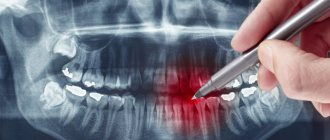
Tumor detection
Detection of the localization of the inflammatory process is of particular interest to the doctor and the patient himself. Because in most cases, patients complain not about a bad tooth, but about nearby healthy molars.
A panoramic photograph is also taken when wisdom teeth are difficult to erupt.
Periodontitis
Periodontitis is a dangerous gum disease that also destroys bone tissue. A panoramic (circular) image allows you to see the depth of tissue damage, the depth of periodontal pockets and make the correct diagnosis.
Implantation
Implantation is a modern method of restoring the smile line and functionality of the jaw system. Panoramic diagnostics (it is better to do a computed tomogram) before implantation will help identify pathology of the maxillary sinuses, in which implantation is strictly prohibited.
CT scans are also performed after implantation to monitor the quality of work. The image shows the condition of the bone tissue, which is especially important when implanting titanium elements.
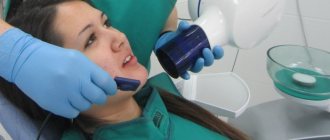
How pictures are taken
The pantomograph diagnostic procedure does not take much time and is easy to perform. A special stick made of medical plastic is placed in the patient’s mouth, which he must squeeze with his teeth. Before the procedure, you must remove all metal accessories: rings, earrings, watches, necklaces. The patient is asked to wear a lead apron, which protects against harmful radiation.
After turning on the device, a digital sensor begins to rotate around the patient’s head, which scans the jaw and temporal area. The whole procedure takes no more than a minute. Then the doctor analyzes the data received, enters the information into a computer or onto a flash card, and takes a picture.
The effectiveness of LPG massage
By awakening “cellular memory” and using the body’s hidden reserves, vacuum LPG massage helps restore the structure of the skin. It stimulates fibroblasts and activates the synthesis of collagen and elastin. As a result, the skin becomes firmer and more elastic, looks healthier and younger.
Vacuum roller massage allows you to achieve the following results:
- skin color and texture improve;
- the “orange peel” effect (the most obvious sign of cellulite) is reduced;
- the skin becomes more elastic and firm;
- extra pounds disappear;
- lymph and blood circulation is normalized;
- well-being and mood improves;
- not only the volume of the body decreases, but also the thickness of the skin-fat fold;
- swelling disappears;
- scars are smoothed out;
- muscle pain and spasms disappear;
- provides a lifting effect.
Vacuum LPG massage reduces the severity of stretch marks, improves posture and coordination of movements due to neurosensory effects. A lasting effect is ensured only in conjunction with following a diet and performing a set of sports exercises.
About the LPG device
To carry out vacuum massage, the CELLU M6 device contains special programs that can independently select the intensity of the effect, taking into account the patient’s goals and test results. There are different attachments for different problem areas. The intensity of the impact can be either mild or aggressive. In the first case, massage promotes relaxation, and in the second, it destroys subcutaneous fat deposits.
The working tool of the device - the manipulator - is a chamber, inside of which there are small rollers. They move in different directions and have a mechanical effect on the epidermis. Thanks to the vacuum, the efficiency of the entire procedure increases many times.
Other types of diagnostics
An image of the jaw is taken using a special device called an orthopantomograph. This device is an X-ray mechanism for scanning the entire jaw and temporal area. There are two types of pantomographs: digital and film (analog). Film mechanisms require a darkroom, while digital mechanisms require a computer. Digital pantomographs are more convenient to use, and their radiation dose is several times lower.
In modern dentistry, film orthopantomographs are used less and less often, since the pictures are unclear and quickly erased. The latest equipment is based on digital technology, and examination methods do not harm the body. The resulting digital image can be printed on photo paper or saved on any medium.
However, some public clinics still have old film-type equipment. If you take a panoramic photograph of your teeth under compulsory medical insurance, be prepared for such a turn of events.
The image can also be obtained using other hardware tools. Therefore, a panoramic test is also done using:
- computed tomography;
- teleroentgenograms.
CT scan
Cone beam tomographs allow you to take panoramic images of the jaw, which facilitates the work of dentists and orthodontists. A panoramic image of the entire oral cavity helps to make a correct diagnosis, prescribe bite correction and implant implants. However, this type of diagnosis is contraindicated for persons suffering from claustrophobia and pathological mobility (cannot sit quietly in one place). Pregnant women should refuse this procedure; breastfeeding women should refrain from breastfeeding for two days after the examination.
Teleradiogram
Teleradiography is prescribed when installing braces or implants, bridges. The procedure is also part of orthodontic treatment and surgery. The radiation exposure during this type of examination is minimal and does not pose a threat to the patient. This procedure is performed even on young children if there is a suspicion of an anomaly in the structure of the dental system.
Types of teleroentgenogram:
- frontal;
- lateral;
- axial
In frontal diagnostics, images are taken from the front and back. A lateral teleroentgenogram is done in case of malocclusion; this procedure is mandatory when installing braces. The chin (axial) radiograph is clarifying (additional). It is necessary for a complete picture of the structure of the patient’s skull in a three-dimensional image. This procedure is mandatory when installing upper jaw implants.
Advantages of teleradiography:
- the resulting image is as close as possible to the actual dimensions of the patient’s skull;
- the result of the study is ready within a few minutes after the image;
- The radiation dose to the patient is negligible.
This type of examination is preferable for patients with claustrophobia who are frightened by the appearance of the tomograph cabin. This type of diagnosis has virtually no disadvantages. However, the doctor’s literacy factor should also be taken into account: you need to be able to correctly decipher an x-ray.
Features of conducting OPTG for children
We figured out what is visible in panoramic photographs and why they are taken, and found out that this is a completely safe diagnostic method. Now it should be noted that an orthopantomogram for children is carried out taking into account some nuances. A radiation exposure of 55 μSv (1 shot) is completely safe for a child, so such a procedure is allowed from 5-6 years of age.
The examination requires the patient to stand still for some time, but for young children this task can be quite difficult. When it comes to small patients, the procedure is carried out in exposure mode - the area of exposure is reduced, and the sensor is adjusted to a trajectory of movement corresponding to the small size of the jaws.
“My son also recently had a panoramic photo taken. He's 10, everything went great. Now there are new technologies everywhere, everything is safe. The procedure took less than a minute, and the doctors assured me that there was nothing wrong with it. How else can you start correcting your bite? The orthodontist prescribed OPTG for us. So far I’ve made the plate, but in a couple of years we’ll get braces. There’s no place here without pictures!”
Lyudmila V., Chelyabinsk, from correspondence on the woman.ru forum
In pediatric dentistry, OPTG is used to detect the rudiments of permanent teeth, assess the correctness of their formation, to diagnose adentia and identify malocclusions. Most often, this procedure is prescribed by an orthodontist before starting to correct defects in bite and teeth position.
The procedure is completely safe for the child
Common Questions
How safe is X-ray diagnostics?
Doctors responsibly declare that this diagnosis can be carried out several times a year without harm to health. A lead apron does not mean that the examination is mortally dangerous for the patient. The apron is worn to protect the thyroid gland from radiation. It is customary to wear aprons in dentistry, traumatology, etc., since the patient receives a dose of radiation annually during a fluorogram.
How often is irradiation allowed?
Since the radiation exposure to the body during an orthopantomogram is small, studies can be done as needed. That is, how much is needed for treatment. However, there is a limitation: no more than 80 images per year. This figure takes into account preventive fluorograms, and not just dental photographs.
How dangerous is x-ray for pregnant women?
X-rays are taken for pregnant women only as a last resort, if it is required to save the life of the woman and the fetus. However, in the first trimester, any manipulations with X-rays are prohibited: this will negatively affect the condition of the embryo. The second trimester is relatively safe, when all the internal organs and systems of the baby are formed. In the third trimester, taking an X-ray is very dangerous, as it can lead to premature birth.
Do children get x-rays?
In dental practice, photographs of baby teeth are taken if a cyst or a hidden source of inflammation is suspected. For diagnosis, a targeted image is sufficient, the radiation dose at which is extremely small. For the body to suffer harm, you need to make 100 such diagnostics in a year, or more than five in a day. Teenagers can already undergo OPTG, as they understand the requirement to stand still and not cry during the procedure.
Making a treatment plan
This procedure is very important, because after receiving the images, the treating doctor can draw up a competent treatment plan that will lead to the fastest possible recovery. In this case, all actions must be indicated with an approximate time frame for their completion. This list mainly consists of the following most important points:
- Treatment of gums and teeth: is it necessary?
- Does the patient need bone augmentation? This procedure is prescribed according to indications when its natural volume is insufficient.
- Selecting the type and model of the implanted implant. This procedure also helps determine the use of other equipment that may be needed during surgery.
- Selection of material, type and model of dental crowns, abutment, prosthesis.
It is also important to note that each of these points must be agreed upon with the patient. The doctor himself can give recommendations. This is all necessary, because depending on the quality and choice of model of implanted parts, the price may vary.
Bottom line
To properly treat teeth, you need to have an idea of the condition of the jaw and skeletal system. This information can be provided by an x-ray, which is taken with a special orthopantomograph or computed tomograph. The picture is taken in every private clinic and government institutions. The cost depends on the equipment and tariffs of private clinics. In a government institution, you can get an X-ray done free of charge upon referral or for a small fee of 300 rubles.
Orthopantomography is a panoramic image of both jaws in a section in a two-dimensional plane. The absence of a three-dimensional image is a common reason for an incorrect diagnosis. Also, a two-dimensional image does not allow one to see microcracks on the roots of teeth, and in three-rooted teeth only 2 roots are visible. The listed disadvantages speak in favor of computed tomography, although it costs several times more.
Panoramic diagnostics are recommended not only during dental treatment and implantation: it must be performed annually for preventive purposes. This will allow timely detection of hidden caries, internal foci of inflammation, cysts, tumors, and neoplasms.
Sources used:
- Abolmasov N. G., Abolmasov N. N. Orthodontics. – 2008.
- American Academy of Implant Dentistry (AAID)
- Distel V. A. Dental anomalies and deformations. Omsk — 2004.
- “Orthopedic dentistry. Textbook" (Trezubov V.N.)
What are the advantages and disadvantages of OPTG
An orthopantogram can significantly improve the quality of diagnosis, and today dental treatment is practically not carried out without radiography, except for minor problems. Among the undeniable advantages of the technique, experts highlight the following points:
- speed of the procedure - literally in a couple of minutes the image is ready for study,
- modern equipment makes it possible to adjust the height and trajectory of the emitter, which made it possible to undergo diagnostics for children and wheelchair users,
- minimal radiation exposure and absolute safety for the body,
- high quality and accuracy of the obtained images,
- the ability to zoom in on the area under study and study it in detail on a computer monitor.
An orthopantomogram uses minimal radiation exposure.
If we talk about the disadvantages, the main disadvantage of OPTG is obtaining a flat two-dimensional image. As a result, the image may slightly distort the dimensions of teeth, root canals and bone tissue. Such deviations can vary from 15 to 30%. To obtain a more accurate image, it is better to resort to computed tomography.