OPTG or orthopantomogram is a panoramic x-ray of the entire dentofacial apparatus, including the sinuses and soft tissues. Without this research it is impossible to imagine modern dental treatment.
Unlike targeted radiography of an individual tooth, OPTG gives the specialist a complete picture of the condition of both jaws of the patient. Based on this knowledge, further treatment tactics are selected.
The whole procedure takes 10-15 seconds, after which the image is obtained on digital or analogue media. The image quality is high, with good resolution, the doctor can examine the details and notice the smallest changes.
Modern diagnostic methods
Effective treatment is impossible without correct diagnosis.
Such procedures allow you to determine an accurate diagnosis and find the best option for eliminating the disease. At the same time, modern medicine does not stand still. Every year, new analysis methods appear that make it possible to determine an accurate diagnosis. Thus, one of the most effective options is the use of digital equipment that provides panoramic images.
For this reason, such methods have almost completely replaced conventional computer diagnostics. OPTG of the dentition is especially popular. This analysis is deservedly considered the most effective. In this case, the inspection is carried out using the most modern equipment, which provides an accurate result!
Content
- What is OPTG in dentistry?
- Main types of procedure
- Diagnostic procedure
- Features of the procedure
- Main areas of use
- Children's examinations
- Equipment Features
- Indications and contraindications
- The advantages of an orthopantomogram and what is the difference between it and CT
- How is a dental x-ray done?
- Decoding the received data
- Why do dental x-rays take place?
- How often can children’s teeth be x-rayed without harming their health?
- Orthopantomogram or CT?
- Where can I do it?
Safety and radiation exposure –
There are a lot of untruths written on the Internet regarding X-ray examinations in dentistry. You may find that such a picture can be taken only once a year; on other sites they say that it can be taken even once a day. All this happens because most “dental” sites on the Internet are written not by dentists, but by ordinary programmers for the purpose of placing advertising on their site.
Radiation exposure is measured in microsieverts (µSv) or millisieverts (mSv). According to the Ministry of Health of Russia dated December 21, 2012, the radiation dose when performing 1 orthopantomogram for a patient over 15 years old should be 55 μSv (=0.055 mSv), and for a patient under 15 years old – 24 μSv (=0.024 mSv). The indicated radiation doses are given only by digital orthopantomographs; the radiation exposure of film devices will be higher.
Conclusions: the maximum permissible radiation dose for the population (according to SanPiN 2.6.1.1192-03) is a dose not exceeding 1000 μSv per year. Thus, you should not take such a picture more than once a month, because... You must take into account that the body is also affected by normal background radiation.
Risks when performing an orthopantomogram –
X-ray irradiation has a more negative effect on a weakened body, especially if such studies are repeated.
Even a small dose of radiation can stimulate genetic disorders and the appearance of malignant tumors. In children, the risks are always higher, because Children's bodies have increased radiosensitivity. Therefore, in no case should you exceed the permissible radiation dosages that we described above. When contacting a clinic, you should definitely ask what type of orthopantomograph is installed in this clinic, and what radiation exposure will be received for 1 image. The fact is that clinics often purchase equipment that is outdated in Europe, and the radiation exposure from outdated models can be 2-3 times higher - from the recommended radiation dose of 55 μSv, normally received for 1 panoramic image.
According to the medical literature, a rough estimate of the risk of malignant oncology after an orthopantomogram is 1 case in 20,000,000, for children aged 1 to 10 years - 1 case in 10,000,000. In general, these are very low rates (compared to many other types of X-rays), although they also seem dangerous.
Comparison of the radiation exposure of an orthopantomograph with targeted images - the radiation exposure from targeted images will be much less, and in 1 day of treatment you can safely take 4-5 such images, but no more than 100 images per year. The usual radiation dose for 1 digital intraoral dental x-ray will be (if modern rather than outdated equipment is used):
- lower jaw – 2.0 µSv (patients under 15 years old – 1.0 µSv),
- upper jaw – 5.0 µSv (patients under 15 years old – 3.0 µSv).
What is OPTG in dentistry?
Similar methods are used in various medical fields. For example, this diagnosis is carried out before the appointment of therapeutic, surgical and orthopedic procedures.
An OPTG image of teeth is prescribed in cases where conventional X-ray analysis is not enough. It gives the doctor a circular image of the oral cavity, which is unfolded in a plane. Such images show not only teeth and crowns, but also roots, canals and tissues.
Thus, such a procedure allows us to obtain maximum information about the condition of the dentofacial region. In some cases, making a correct diagnosis is only possible after obtaining a survey image.
In this case, the image is automatically transferred to the computer in digital format. The use of special programs allows the doctor, if necessary, to enlarge individual parts of the image or set a certain viewing angle.
Description / what dental x-rays show
When performing a pantomogram, the recording program includes not only both dentitions, but also the bones of the nose, lower jaw, both mandibular joints and adjacent soft tissues.
Dental X-ray determines:
- Position of tooth roots.
- Defects in the contact surfaces of crowns.
- Hidden carious spots and cavities.
- Cystic and other neoplasms.
- The course of teeth eruption and growth in childhood.
- Presence of undetected teeth.
- Condition of surrounding tissues (periodontal).
- Defects in jaw closure.
- Bone destruction, arthrosis of the jaw joints.
- Quality of fillings, sealed canals.
- Inflammation of the facial nerves, nasal sinuses.
The image is prescribed for prosthetics, orthodontics, complex cases of surgical treatment, for maxillofacial injuries, and ENT diseases.
Main types of procedure
At the moment, there are three options for carrying out this procedure. Their varieties depend on the models of equipment used. Thus, a digital orthopantomogram is used for diagnosis. It is more expensive, but does not require expenses for consumables and photo laboratory services. At the same time, it provides high image quality.
In addition, it is possible to use film equipment. In this case, the level of radiation will be significantly higher. In this case, the accuracy of the result will be slightly lower, and the procedure itself will take longer.
Another popular option is a 3D orthopantomogram. For this diagnosis, a special computed tomograph is used, which allows you to examine the tooth from all sides.
In this case, the image consists of a number of projections that provide a three-dimensional model of the desired area. This method is considered the most effective.
Diagnostic procedure
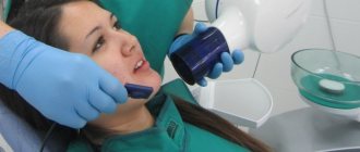
Many patients who encounter this procedure are interested in the question - what is an orthopantomogram and how is it done? It is worth noting that such diagnostics do not require complex and lengthy preparation. Moreover, the examination takes only a couple of minutes, after which the doctor has a ready-made image in his hands. The procedure itself consists of the following steps:
- The patient removes all metal jewelry and objects - chains, earrings, piercings, etc. Their presence may distort the results.
- The patient stands in front of the device and puts on a protective apron.
- After this, you need to hold the plastic holder between your teeth.
- The X-ray emitter then begins to rotate around the patient's head until a complete image is obtained.
The finished image is automatically transferred to the doctor’s computer, after which the specialist interprets it. If necessary, the photo can be printed or sent to another medium.
Features of the procedure
Such a study can be called basic, since it allows us to assess the condition of the dentofacial region as a whole. For this reason, it often makes it possible to detect diseases that have not yet manifested themselves with pronounced symptoms, but could lead to serious consequences.
Many patients are interested in the question: what does an orthopantomogram of teeth show? It gives a complete picture of the condition of the oral cavity. So, the picture shows:
- jaws;
- maxillary sinuses;
- joints;
- roots;
- teeth, crowns, fillings;
- fabrics;
- bones.
For this reason, this diagnosis makes it possible to detect a disease that is associated with the dentoalveolar region. Therefore, this procedure is universal and can be used to detect any problem. At the same time, it guarantees the most accurate result. These features explain the high popularity of OPTG, which is only growing every year.
How does the procedure work?
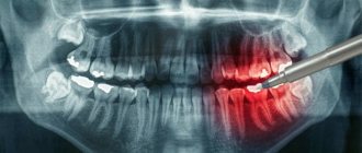
An orthopantomogram (abbreviated as OPTG) is an overview circular photograph of the teeth, which shows the entire jaw, unfolded in a plane. In addition to teeth, roots, canals, crowns and fillings, such an image can also be used to study the condition of periodontal tissues, maxillary sinuses, and sinuses.
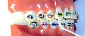
A panoramic photograph of teeth also helps to identify hidden carious cavities, cysts, inflammations, periodontal pockets, abscesses, and to analyze the exact condition of the roots and periodontal tissues, the temporomandibular joint, and the maxillary sinuses. In addition, OPTG is indispensable if orthodontic treatment with aligners or braces is planned.
In the process of preparing for dental implantation, using a panoramic image, the doctor can determine whether there is enough bone tissue for implantation or a sinus lift is required, how many implants are needed and what their optimal sizes are, as well as what the dynamics of their engraftment will be.
It is extremely important to do an orthopantomogram to find out the position and condition of the wisdom teeth. That is why a competent doctor makes a decision whether to remove or cure the “eight” only after analyzing a panoramic image.
Main areas of use
There are a number of diseases for which the appointment of such diagnostics is the optimal or even the only solution. In addition, it can be used in various interventions, operations and procedures aimed at recovery. If patients are wondering why OPTG is needed, it is worth checking out the following list:
- Allows you to detect carious lesions, neoplasms and cysts.
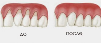
- Makes it possible to identify cases of periodontal disease - this disease has no noticeable symptoms in the early stages.
- Helps detect bone diseases.
- Simplifies orthodontic correction, implantation and prosthetics.
OPTG also allows you to control the quality of operations performed, for example, installing a filling in a root canal. Thus, such diagnostics are multifunctional and help in carrying out a number of procedures. In addition, with the help of such images, a specialist can determine the density and height of the tooth, which is important when making prosthetics and installing fillings.
Children's examinations
The level of radiation exposure when creating an image is only 10-40 μSv. Thus, you can undergo the procedure up to a hundred times a year without harm to your health. It is also worth noting that this process is safe for children.
This level of radiation cannot harm the body. For this reason, OPTG is often used to detect permanent tooth buds. In addition, the method allows you to notice possible developmental anomalies in time. Indeed, in this case, the full row of teeth, including the roots, is visible. This uses an exposure mode that reduces the level of radiation exposure.
How often can you do it
According to sanitary standards, the total radiation exposure per person for 1 year should not exceed 1000 μSv. During orthopantomography, the patient is exposed to a dose of 20-30 μSv. Using simple calculations, we calculate that it is possible to take a photo 50 times in a year!
The answer to the question of how often an orthopantomogram can be done is as determined by the attending physician. Calculations show that radiation during OPTG is almost safe. It is allowed to be carried out during pregnancy, bypassing the 1st trimester. In terms of workload, the study is comparable to a 3-4 hour plane flight.
Indications and contraindications
A number of factors may be the reason for conducting a survey. These include the occurrence of inflammation, pain and swelling of the gums. In addition, the procedure is performed before implantation, tooth extraction or filling. Another prerequisite for OPTG is the occurrence of a cyst. The method has also shown high efficiency in diagnosing infectious diseases.
It is believed that there are no specific contraindications for the examination. However, this method is not recommended during the first trimester of pregnancy, or when feeding the baby.
The advantages of an orthopantomogram and what is the difference between it and CT
This procedure has a number of undeniable advantages. These include high image clarity and low radiation levels. In addition, OPTG allows you to zoom in on the image for a detailed inspection of individual areas. The information received is archived and can be reused if necessary. Another advantage is the speed of the process and its safety for the body.
For this reason, computed tomography is inferior to OPG in a number of indicators. In the first case, a separate area of the dentoalveolar region is examined. For this reason, problems such as cysts, granulomas and impacted teeth are difficult to identify. The difference between an orthopantomogram and a computed tomography scan is that the first option provides extensive information about the patient's condition. Meanwhile, who has worse accuracy and may not detect a number of important symptoms.
Sign up for a study by phone
+7 (812) 332-52-54
How to take a panoramic photograph of the jaw -
Devices for obtaining panoramic images are called orthopantomographs. Technically, the procedure is as follows: the patient is placed in the center of the device and allowed to bite a special plate between the teeth to achieve immobility of the jaws. Next, the moving part of the orthopantomograph moves around the patient’s head, which consists of an x-ray source on one side, and x-ray film or a digital sensor on the other.
The entire procedure lasts from 8 to 20 seconds, which depends on the equipment, and the shorter the irradiation time, the better. Depending on what the image is recorded on (film or digital media), orthopantomographs are divided into film and digital. Let's say right away that film orthopantomographs provide a fuzzy, blurry image - especially in the area of the central teeth of the upper and lower jaw, and therefore are frankly outdated.
Digital orthopantomographs are of great value for diagnostics due to the fact that a special computer program for such devices improves the quality of the image, which can then be viewed in detail on a computer screen. In addition, if necessary, a digital image can also be printed later on film or photo paper (if necessary), but it is still best to have a digital medium.
Panoramic shot of the jaw: video
An interesting point is that a panoramic photograph of teeth can be obtained not only by using an orthopantomograph. Modern devices for computed tomography (for example, the Sirona Galileos tomograph) allow the patient to simultaneously receive a three-dimensional computed tomography image and a two-dimensional flat panoramic image of the teeth. In some cases, this is very convenient for diagnosis and does not require additional radiation exposure.
Panoramic photo of teeth: disadvantages
Considering that a person’s jaws are curved and not flat, the orthopantomograph has to combine several images (taken in different planes) into one flat two-dimensional image (24stoma.ru). There is a big disadvantage in this process: different people have different jaw shapes and curvatures, but the path of the radiation tube around a person’s head during the procedure is always standard.
This leads to the fact that any panoramic image will always not accurately convey the dimensions of teeth, root canals, and bone tissue dimensions. The distortion from the actual size of these objects from different manufacturers is approximately from 15 to 30%. Therefore, any panoramic photograph of teeth (both film and digital) is always only an approximate diagnostic method, allowing you to see only the general picture of the condition of the teeth and jaws.
Important: since orthopantomographs take an image in only one section on a certain area of the jaw, the root canals of three-rooted teeth in the upper jaw (6-7-8) will always turn out a little blurry (both on digital and film panoramic images). This is because the three roots will always be in different slices/planes and therefore the orthopantomograph will not be able to focus well on each of the roots.
Therefore, only single-rooted, as well as double-rooted teeth of the upper and lower jaws (the two roots are in the same plane) will be clear - and only on digital photographs. But the anterior teeth of both jaws on conventional film orthopantomographs will always be very blurred (Fig. 5) - due to the strong bending of the anterior parts of the jaws, as well as the technical imperfections of the old film equipment.
Comparison of digital and film quality -
Advantages of digital panoramic photographs -
- reducing the time and dose of radiation to the patient,
- higher image clarity,
- the image can be subjected to graphic processing (for example, enlarged on a computer screen to see small details),
- ability to write to a flash drive or disk.