CLASSIFICATION
Odontogenic infections (infections of the oral cavity), depending on the anatomical location, are divided into truly odontogenic
associated with damage to tooth tissue (caries, pulpitis);
periodontal
, associated with damage to the periodontium (periodontitis) and gums (gingivitis, pericoronitis), surrounding tissues (periosteum, bone, soft tissues of the face and neck, maxillary sinus, lymph nodes);
non-odontogenic
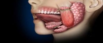
, associated with damage to the mucous membranes (stomatitis) and inflammation of the major salivary glands.
These types of infections can cause serious life-threatening complications from the cranial cavity, retropharyngeal, mediastinal and other localizations, as well as hematogenously disseminated lesions of the heart valve apparatus, sepsis.
Purulent infection of the face and neck
may be of non-odontogenic origin and includes folliculitis, furuncle, carbuncle, lymphadenitis, erysipelas, hematogenous osteomyelitis of the jaws.
Specific infections (actinomycosis, tuberculosis, syphilis, HIV) can also be observed in the maxillofacial area.
MAIN PATIENTS
Infections of the oral cavity are associated with the microflora that is constantly present here. Usually this is a mixed flora, including more than 3-5 microorganisms.
With truly odontogenic
infections, along with facultative bacteria, primarily viridans streptococci, in particular (
S.mutans, S.milleri
), anaerobic flora is released:
Peptostreptococcus
spp.,
Fusobacterium
spp.,
Actinomyces
spp.
In case of periodontal infection, five main pathogens are most often isolated: P.gingivalis, P.intermedia, E.corrodens, F.nucleatum, A.actinomycetemcomitans
, less commonly
Capnocytophaga
spp.
Depending on the location and severity of the infection, the patient’s age and concomitant pathology, changes in the microbial spectrum of pathogens are possible. Thus, severe purulent lesions are associated with facultative gram-negative flora ( Enterobacteriaceae
spp.) and
S.aureus
.
In elderly patients and hospitalized patients, Enterobacteriaceae
spp also predominate.
In the conditions of domestic bacteriological laboratories, it is quite difficult to isolate a specific pathogen of a certain odontogenic infection. However, it seems possible to localize pathogens that play a major role in the development of oral infections in supragingival and subgingival plaque.
PRINCIPLES OF TREATMENT OF INFECTIONS OF THE ORAL CAVITY AND MAXILLOFACIAL AREA
Treatment of odontogenic infection
often limited to local therapy, including standard dental procedures.
Systemic antibacterial therapy is carried out only when odontogenic infection spreads beyond the periodontal period (under the periosteum, into the bones, soft tissues of the face and neck), in the presence of elevated body temperature, regional lymphadenitis, and intoxication.
When choosing antimicrobial therapy in a hospital setting, in cases of severe purulent infection, it is necessary to take into account the possibility of the presence of resistant strains among anaerobes such as Prevotella
spp.,
F. nucleatum
to penicillin, which determines the prescription of broad-spectrum drugs, amoxicillin/clavulanate in monotherapy or a combination of fluoroquinolone with metronidazole.
Due to increasing levels of resistance by Streptococcus
spp.
to tetracycline and erythromycin, these drugs can only be used as alternatives. Since metronidazole has moderate activity against anaerobic cocci ( Peptostreptococcus
spp.), simultaneous administration of β-lactam antibiotics is necessary to increase effectiveness.
Preventive systemic use of AMPs when performing dental procedures in the oral cavity or periodontium does not guarantee a reduction in the incidence of infectious complications in somatically healthy patients. Convincing data on the sufficient effectiveness of topical antibiotics for oral infections have not been obtained. At the same time, oral bacteria are a reservoir of determinants of resistance to AMPs. Excessive and unjustified use of antibiotics contributes to their appearance and the development of resistance of pathogenic microflora.
ODONTOGENIC AND PERIODONTAL INFECTION
PULPITIS
Main pathogens
Viridans streptococci ( S. milleri
), non-spore-forming anaerobes:
Peptococcus
spp.,
Peptostreptococcus
spp.,
Actinomyces
spp.
Choice of antimicrobials
Antimicrobial therapy is indicated only in case of insufficient effectiveness of dental procedures or spread of infection into surrounding tissues (periodontal, periosteal, etc.).
Drugs of choice
: phenoxymethylpenicillin or penicillin (depending on the severity of the disease).
Alternative drugs
: aminopenicillins (amoxicillin, ampicillin), inhibitor-protected penicillins, cefaclor, clindamycin, erythromycin + metronidazole.
Duration of use
: depending on the severity of the current (at least 5 days).
PERIODONTITIS
Main pathogens
Microflora is rarely detected in the periodontal structure and is usually S.sanguis, S.oralis, Actinomyces
spp.
In periodontitis in adults, gram-negative anaerobes and spirochetes predominate. P.gingivalis, B.forsythus, A.actinomycetemcomitans and T.denticola
are highlighted most often.
In juveniles, there is rapid involvement of bone tissue in the process, with A. actinomycetemcomitans
and
Capnocytophaga
spp.
P.gingivalis
is rarely isolated.
In patients with leukemia and neutropenia after chemotherapy along with A. actinomycetemcomitans
C.micros
is isolated , and in prepubertal age -
Fusobacterium
spp.
Choice of antimicrobials
Drugs of choice
: doxycycline; amoxicillin/clavulanate.
Alternative drugs
: spiramycin + metronidazole, cefuroxime axetil, cefaclor + metronidazole.
Duration of therapy
: 5-7 days.
For patients with leukemia or neutropenia after chemotherapy, cefoperazone/sulbactam + aminoglycosides are used; piperacillin/tazobactam or ticarcillin/clavulanate + aminoglycosides; imipenem, meropenem.
Duration of therapy
: depending on the severity of the course, but not less than 10-14 days.
PERIOSTITIS AND OSTEOMYELITIS OF THE JAWS
Main pathogens
With the development of odontogenic periostitis and osteomyelitis, S. aureus
, as well as
Streptococcus
spp., and, as a rule, anaerobic flora prevails:
P.niger, Peptostreptococcus
spp.,
Bacteroides
spp.
Specific pathogens are less commonly identified: A.israelii, T.pallidum
.
Traumatic osteomyelitis is often caused by the presence of S.aureus
, as well as
Enterobacteriaceae
spp.,
P.aeruginosa
.
Choice of antimicrobials
Drugs of choice
: oxacillin, cefazolin, inhibitor-protected penicillins.
Alternative drugs
: lincosamides, cefuroxime.
When P.aeruginosa
, antipseudomonas drugs (ceftazidime, fluoroquinolones) are used.
Duration of therapy
: at least 4 weeks.
ODONTOGENIC MAXILLARY SINUSITIS
Main pathogens
The causative agents of odontogenic maxillary sinusitis are: non-spore-forming anaerobes - Peptostreptococcus
spp.,
Bacteroides
spp., as well as
H.influenzae, S.pneumoniae
, less often
S.intermedius, M.catarrhalis, S.pyogenes
.
Isolation of S.aureus
from the sinus is characteristic of nosocomial sinusitis.
Choice of antimicrobials
Drugs of choice
: amoxicillin/clavulanate. For nosocomial infection - vancomycin.
Alternative drugs
: cefuroxime axetil, co-trimoxazole, ciprofloxacin, chloramphenicol.
Duration of therapy
: 10 days.
Odontogenic infections.
Patients are often interested in what an odonotogenic infection is.
Odontogenic infections are diseases that, due to improper or untimely treatment of caries, pulpitis, periodontal disease, inflammation of the bone tissue of the upper and lower jaws.
It is difficult to say who is more to blame for the occurrence of such diseases. In some cases, they are provoked by the patients themselves: poor oral care, rare visits to the dentist, self-medication.
Odontogenic infections are truly odontogenic and the main causative factor in this case is the affected tooth. Nonodontogenic infections are infections that develop when the oral mucosa is damaged.
Periodontal infections - the causes of which are lesions of the gums, periodontium, periosteum, and soft tissues of the neck.
The main pathogens are viridans streptococci, spirochetes, and non-spore-forming anaerobes.
In most cases, odontogenic infections develop when the patient pays insufficient attention to his own health. Also, the infectious process can be triggered by poor-quality canal filling, tooth extraction with traumatic consequences, inadequate surgical intervention, i.e. have an atrogenic character.
Odontogenic infections can spread through lymphatic and blood vessels into nearby tissues. An important factor in the spread and severity of odontogenic infections is the low resistance of the immune system. Under such conditions, a complicated inflammatory process can transform into a peripharyngeal abscess or phlegmon of the cervical region. It is necessary to note the groups of people most susceptible to infection. This :
- patients with cancer;
- patients with hepatitis and carriers of HIV infections;
- children;
- patients over 60 years of age;
- patients with an advanced form of somatic pathology (heart disease, kidney disease, liver disease);
- patients suffering from diabetes;
- patients who received radiation. Chemotherapy;
- persons using drugs that suppress the immune system;
Symptoms of the pathology are quite difficult to determine, especially since patients often do not associate pain in the neck or facial area with the affected tooth and turn to a general surgeon. In this case, therapy will be incomplete without sanitation of the main source of the disease.
Some symptoms of odontogenic infections should be highlighted:
- signs of general intoxication in the patient (fever, weakness);
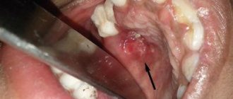
- pain in the area of purulent focus;
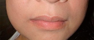
- redness and swelling of the affected area;
- swollen tongue, slurred speech, difficulty swallowing. Sometimes the patient requires intubation because... due to severe edema, the flow of air into the larynx and trachea is disrupted.
The causative tooth, at any localization of the process, gives symptoms of pain on palpation and percussion. The gums around it are hyperemic and swollen.
To identify phlegmons and abscesses, ultrasound and computed tomography are prescribed. The latter procedure is especially effective because helps to diagnose the focus of odontogenic infection in the chest area, which is inaccessible to external examination.
Odontogenic infections tend to quickly spread in the maxillofacial area, moving into the stage of a purulent process. Complications may be as follows:
- purulent inflammation of the soft tissues of the neck and face, mouth;
- diffuse purulent inflammation;
- blockage of facial veins and inflammation of their walls;
- damage to the lymph nodes;
- sinusitis;
- odontogenic sepsis.
Treatment begins with sanitation of the oral cavity and opening of the abscess. Anaerobic bacteria have their own characteristics. Usually such wounds have a foul odor. Adequate outflow is important for therapy, which provides an influx of oxygen, which is harmful to microorganisms. Each location of the abscess has its own method of opening. It takes into account the topographic anatomy of the face, neck and parapharyngeal space. Large vessels and nerve trunks are located in the area of surgical intervention. Nowadays, ultrasound-guided puncture is actively used in cases where the abscess is located deep in the tissues. The operation should be accompanied by the administration of antibiotics. Without them, the effect will not be achieved. Treatment in this case is combined. Typically, first-line drugs (antibiotics) are combined with antimicrobial drugs (metronidazole, tinidazole), and in parallel, general restorative therapy and desensitization are carried out.
In order to protect yourself from odontogenic infections, careful oral care and regular visits to the dentist are required.
Dentist-therapist - Volobueva Raisa Evgenievna.
PURULENT INFECTION OF SOFT TISSUE OF THE FACE AND NECK
Main pathogens
Purulent odontogenic infection of the soft tissues of the face and neck, tissue of deep fascial spaces is associated with the release of polymicrobial flora: F. nucleatum
, pigmented
Bacteroides, Peptostreptococcus
spp.,
Actinomyces
spp.,
Streptococcus
spp.
ABSCESSES, PHLEGMONS OF THE FACE AND NECK
Main pathogens
With an abscess in the orbital area in adults, a mixed flora is released: Peptostreptococcus
spp.,
Bacteroides
spp.,
Enterobacteriaceae
spp.,
Veillonella
spp.,
Streptococcus
spp.,
Staphylococcus
spp.,
Eikenella
Streptococcus
spp.,
Staphylococcus
. predominate in children .
The causative agents of abscesses and phlegmon of non-odontogenic origin, most often caused by minor skin lesions, are S.aureus, S.pyogenes
.
With putrefactive-necrotic phlegmon of the floor of the mouth, a polymicrobial flora is released, including F.nucleatum, Bacteroides
spp.,
Peptostreptococcus
spp.,
Streptococcus
spp.,
Actinomyces
spp.
S.aureus
can be isolated in patients with severe disease (more often in patients suffering from diabetes and alcoholism).
Choice of antimicrobials
Drugs of choice:
inhibitor-protected penicillins (amoxicillin/clavulanate, ampicillin/sulbactam), cefoperazone/sulbactam.
When P.aeruginosa
- ceftazidime + aminoglycosides.
Alternative drugs:
penicillin or oxacillin + metronidazole, lincosamides + aminoglycosides of the II-III generation, carbapenems, vancomycin.
Duration of therapy:
at least 10-14 days.
BUCCAL CELLULITE
Main pathogens
Usually observed in children under 3-5 years of age. The main pathogen is H. influenzae
type B and
S. pneumoniae
.
In children under 2 years of age, H. influenzae
is the main pathogen, and bacteremia is usually observed.
Choice of antimicrobials
Drugs of choice
: amoxicillin/clavulanate, ampicillin/sulbactam, third generation cephalosporins (cefotaxime, ceftriaxone) IV, in high doses.
Alternative drugs
: chloramphenicol, co-trimoxazole.
Duration of therapy
: depending on the severity of the current, but not less than 7-10 days.
LYMPHADENITIS OF THE FACE AND NECK
Main pathogens
Regional lymphadenitis in the face and neck is observed with infection in the oral cavity and face. Localization of lymphadenitis in the submandibular region, along the anterior and posterior surfaces of the neck in children aged 1-4 years, is usually associated with a viral infection.
Abscess formation of lymph nodes is usually caused by a bacterial infection. With unilateral lymphadenitis on the lateral surface of the neck in children over 4 years of age, GABHS and S.aureus
.
Anaerobic pathogens, such as Bacteroides
spp.,
Peptococcus
spp.,
Peptostreptococcus
spp.,
F.nucleatum, P.acnes
, can cause the development of odontogenic lymphadenitis or inflammatory diseases of the oral mucosa (gingivitis, stomatitis), cellulite.
Choice of antimicrobials
Drugs of choice: AMPs corresponding to the etiology of the primary source of infection.
Treatment of odontogenic inflammatory diseases of the maxillofacial area
Important information about the treatment of odontogenic inflammatory diseases of the maxillofacial area:
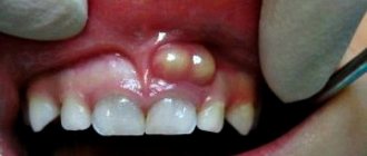
- Purulent periostitis
Immediate consultation with a specialist requires acute purulent periostitis - inflammation of the periosteum, which is manifested by severe, sometimes throbbing pain, swelling and fever. The disease is dangerous because it can develop into acute purulent osteomyelitis of the jaw - an infectious-inflammatory process that affects all structural components of the jaw bone and leads to osteonecrosis.
- Abscess and phlegmon
The most common odontogenic inflammatory disease of the maxillofacial area is an abscess - a local purulent inflammation of the tissues of the gums, tongue, palate or cheek. This condition can occur as a result of complicated dental pathology; its development is also possible due to a violation of the integrity of the mucous membrane or in common infectious diseases. In the absence of timely treatment, the abscess can become chronic and also cause the development of severe complications that pose a threat to life (phlegmon, sepsis, mediastenitis). Therefore, at the first symptoms of the disease (pain, swelling, fever), it is important to immediately consult a specialist.
- Emergency help
The Ilyinskaya Hospital is ready around the clock to provide emergency care to patients with acute odontogenic inflammatory diseases of the maxillofacial area. The patient will urgently undergo all necessary tests and diagnostic studies - computed tomography, ultrasound, etc. A surgeon with extensive experience in providing emergency surgical care is on duty at the hospital at any time of the day. If the pathology requires highly specialized care, for example, a maxillofacial surgeon is needed, such a specialist will be able to arrive at the operating room within one hour. During this time, the surgeon on duty will relieve the life-threatening condition and prepare the patient for specialized intervention. To learn more.
- Diagnostics
To diagnose inflammatory diseases of the maxillofacial area, X-rays, computed tomography, cone-beam computed tomography, and ultrasound are used. Ilyinskaya Hospital is equipped with the most modern radiation diagnostic devices. High-tech equipment is extremely important for diagnosing diseases. But even more important is the expert radiologist who interprets the images obtained with this equipment and answers the questions posed to him by the oral surgeon. Our radiologists work closely with surgeons, provide them with accurate interpretation of images, and in complex cases participate in consultations with surgeons, neurologists, orthodontists, the patient's family doctor and other specialists. All acquired images are stored in the hospital’s unified electronic system and are always available. In an emergency situation, this allows surgeons and other specialists to quickly evaluate the images obtained and quickly make the right decisions. More information about the radiology department of the Ilyinskaya Hospital can be found here.
- Surgery
Limited and diffuse purulent-inflammatory diseases of the maxillofacial area require emergency surgical intervention. The operation is performed under local anesthesia, the surgeon opens the purulent focus, carries out drainage, and ensures the outflow of discharge. For diffuse purulent diseases (phlegmon), the operation is performed under general anesthesia. Cellulitis refers to diffuse purulent-inflammatory diseases that require prompt opening of the purulent focus and adequate drainage (creation of outflow from the source of infection). Surgical procedures are complemented by the administration of active antibacterial and anti-inflammatory therapy. Due to extensive tissue damage and in order to avoid further occurrence of gross aesthetic defects, after the initial surgical intervention the wound is not sutured, but is treated openly. For this reason, the patient requires daily postoperative dressings, and often secondary surgical treatment of tissue, which will prevent the formation of a rough scar. More information about the operating unit of the Ilyinskaya Hospital can be found here.
- Prevention
Prevention of inflammatory diseases of the oral cavity consists of timely sanitation of foci of chronic infection. As well as carrying out regular preventive examinations at the dentist - the frequency of visiting the dentist is once every six months. To avoid possible problems, it is important for the patient to strictly follow the recommendations of the treating dentist for oral care. It is important to use the correct technique for brushing your teeth, use toothpastes with the prescribed composition to clean your teeth, and rinses to care for the oral mucosa.
NON-ODONTOGENIC AND SPECIFIC INFECTION
NECROTIC STOMATITIS (VINCINS ULCER-NECROTIC GINGIVOSTOMATITIS)
Main pathogens
Fusobacterium is concentrated in the gingival sulcus
, pigmented
Bacteroides
, anaerobic spirochetes. With necrotizing stomatitis, there is a tendency for infection to quickly spread into surrounding tissues.
The causative agents are F. nucleatum, T. vinsentii, P. melaninogenica, P. gingivalis and P. intermedia
.
In patients with AIDS, C. rectus
.
Choice of antimicrobials
Drugs of choice:
phenoxymethylpenicillin, penicillin.
Alternative drugs:
macrolide + metronidazole.
Duration of therapy:
depending on the severity of the current.
ACTINOMYCOSIS
Main pathogens
The main causative agent of actinomycosis is A.israelii
, association with gram-negative bacteria
A. actinomycetemcomitans
and
H. aphrophilus
, which are resistant to penicillin but sensitive to tetracyclines, is also possible.
Choice of antimicrobials
Drugs of choice
: penicillin at a dose of 18-24 million units/day, with positive dynamics - transition to step-down therapy (phenoxymethylpenicillin 2 g/day or amoxicillin 3-4 g/day).
Alternative drugs
: doxycycline 0.2 g/day, oral drugs - tetracycline 3 g/day, erythromycin 2 g/day.
Duration of therapy
: penicillin 3-6 weeks IV, drugs for oral administration - 6-12 months.