Oral tuberculosis is an infectious disease that, like other types of tuberculosis, is caused by mycobacteria. This pathology is classified as chronic. As a rule, the inflammation characteristic of tuberculosis, affecting the lining of the mouth and the red border around the lips, is secondary: that is, it occurs as a consequence or the most characteristic manifestation of other forms of tuberculosis. For example, these include tuberculous lesions of the lymph nodes and bones.
The mucous membrane of the oral cavity is far from the most favorable environment for the development and reproduction of mycobacteria that cause tuberculosis. As a rule, when entering this environment, mycobacteria die. However, in the presence of favorable circumstances (microtraumas, mechanical damage in the oral cavity, opening the gates for infection), bacteria penetrate inside and provoke the formation of a tuberculosis ulcer.
Oral tuberculosis is a relatively rare form of tuberculosis, usually diagnosed only in children. This is due to the characteristics of teething in children.
In an infant, such a disease can be extremely severe and includes the generalization of tuberculosis infection. Secondary tuberculosis infection begins to develop as tuberculous lupus or miliary ulcerative tuberculosis.
Tuberculosis is transmitted by airborne droplets. The incubation period lasts from eight to thirty days, and after this the formation of an uneven, blurred ulcer begins, which is severely painful. After a few days it begins to gradually increase. At the same time, the adjacent lymph nodes swell. There are no other signs of inflammation yet
Classification of types of oral tuberculosis
Oral tuberculosis is a disease that appears and develops mainly in patients with reduced immunity. The disease is caused by Koch's bacillus. The source of inflammation located in the patients' oral cavity is secondary in most cases. It develops as a result of the spread of infection from the main focus, and spreads through the circulatory or lymphatic systems. In the case when the patient is affected by the pulmonary form of tuberculosis, the infection spreads to the oral cavity due to the penetration of mycobacteria contained in the sputum.
There are four forms of oral tuberculosis. Among them:
- Primary tuberculosis of the oral cavity. Transmitted by the respiratory or fecal-oral route, this disease almost never occurs in adults. Most patients with this diagnosis are infants.
- Tuberculous lupus. It is the lupus form of oral tuberculosis that is most often encountered in dental practice. Erosive elements are localized on the mucous membrane of the gums. If treatment is not started on time, these tumors can develop into a malignant form.
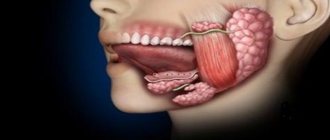
- Miliary-ulcerative form. This form usually develops in patients who have already been weakened by tuberculosis. Along with their cough, they produce sputum, which is a source of bacteria that cause oral tuberculosis. The lesions are on the palate and tongue, in rare cases - in the area of marginal gums or on the side of the cheek.
- Scrofuloderma. This form of the disease is diagnosed in most cases in children. The clinical signs of scrofuloderma are somewhat different from those of other forms of oral tuberculosis. In the first stages - the appearance of not a tubercle, but a rather large nodule. After its softening and necrosis occurs, the stage of formation of fistula tracts begins. The healing process of the surface of the resulting ulcers takes a long time, and after it characteristic fringed scars form.
Types of tuberculosis
- Open form - the disease is clearly expressed, bacteria are easily detected in sputum and feces. The patient himself poses a danger to others, since the infection is transmitted by airborne droplets. Microbacteria can be found in sputum, urine, and feces.
- Closed form - not dangerous to others. Characterized by the difficulty of detecting infection in sputum. The most common tuberculosis is pulmonary tuberculosis, but this infection can also affect bones, joints, genitourinary system, intestines, peritoneum, meninges, central nervous system, peripheral lymph nodes, skin
Diagnostics
Primary tuberculosis of the oral cavity is diagnosed during an external examination. Thus, it is characterized by the presence of lacerations and ulcers in the corners of the mouth with yellow, gray and bluish layers. Areas with ulcers tend to gradually enlarge.
At the same time, the lymph nodes begin to enlarge.
Diagnosis is carried out on the basis of the collected anamnesis, as well as using histological and bacterioscopic studies. During examination, dense painful lumps or ulcers are found on the mucous membrane, depending on the severity of the condition.
There is minor inflammation. The process of formation of adhesions begins between the lymph nodes and the surrounding surface.
Perforation of the hard palate in tuberculosis
Tuberculosis is a chronic granulomatous disease caused by Mycobacterium tuberculosis. WHO reports more than 8 million new cases each year and approximately 3 million deaths from the disease worldwide. So recently, antibiotic resistance of bacteria has become a huge problem. Oral tuberculosis is quite rare and accounts for less than 1% of all cases. With the increasing incidence of the disease, rare and unusual forms are increasingly observed, which often become malignant. In this clinical case, we describe a rare case of perforation of the hard palate during the development of tuberculosis.
Description of a clinical case
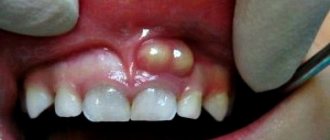
A 7-year-old boy sought help because he had problems swallowing solid food, low-grade fever, and weight loss for the last six months. His mother was on anti-tuberculosis treatment due to pulmonary tuberculosis. There was no history of cough, abdominal pain, vomiting, diarrhea, or urinary problems. On examination, the pulse was 104 beats/min, the respiratory rate was 28/min. and blood pressure 98/60 mm Hg. The child's weight is 13 kg and his height is 104 cm (both figures are below the norm for this age and gender). There was slight pallor of the skin. Examination of the oral cavity revealed a perforation of the hard palate measuring 3x3 cm with irregular, torn edges and necrotic contents (Photo 1). Bilateral lymphadenopathy of the cervical lymph nodes was noted. Auscultation of the lungs revealed crepitus on the right side above. The remaining organ systems are without pathology. The Mantoux test is positive (28 mm). Laboratory examination: hemoglobin 8.8 g/dl, total leukocyte count 9000/cumm (neutrophils 40%, lymphocytes 60%), ESR 80 mm/h. Liver and kidney function is not impaired. Serological test for HIV is negative. Urine and blood cultures revealed no growth of microorganisms. A CT scan of the oral cavity revealed erosion and a defect in the posterior part of the hard palate on the right (Figure 2). X-ray examination of the chest revealed a homogeneous opacity in the upper right zone. Lymph node puncture showed granular caseous necrosis and granuloma composed of epithelioid cells and histiocytes (Figure 3). The child's parents did not consent to a biopsy of the palate lesion. However, taking into account the clinical manifestations, as well as the presence of pulmonary tuberculosis and caseous necrosis in the lymph nodes, it is highly likely that perforation of the palate also occurred due to tuberculosis. The patient was prescribed antituberculosis therapy (isoniazid, rifampicin, pyrazinamide and ethambutol). The boy was registered with maxillofacial surgeons for subsequent elimination of the perforation of the palate.
Photo 1: Examination of the oral cavity, revealing a perforation of the hard palate measuring 3x3 cm with uneven, torn edges and necrotic contents
Figure 2: Axial CT scan with 3D reconstruction showing erosion and defect in the right posterior hard palate
Figure 3: Biopsy of cervical lymph nodes showing granular caseous necrosis and garnulloma consisting of epithelial cells and histiocytes.
Discussion
Tuberculosis in the oral cavity can be either primary or, more often, secondary to pulmonary tuberculosis. With secondary damage to the oral cavity, bacteria reach the mucosa by hematogenous or lymphogenous route. In primary oral tuberculosis, mycobacterium penetrates the mucosa directly due to disruption or loss of the natural barrier due to injury, inflammation, leukoplakia, tooth extraction or poor hygiene. Other predisposing factors include odontogenic cysts, periapical granulomatosis, gingival abscesses, periodontitis and jaw fractures. Abbot was able to detect mycobacteria in oral lavage from 44.9% of patients with active pulmonary disease, confirming the importance of intact mucosa for oral resistance to tuberculosis infection. The incidence of tuberculosis in the oral cavity varies from 0.8% to 3.5%. Oral involvement is rare, even in populations with a high incidence of tuberculosis. Factors responsible for oral resistance include the protective properties of saliva, the presence of saprophytes, muscle resistance to microbial invasion, and the thickness of the protective epithelial cover. The most common location of tuberculous lesions in the oral cavity is the tongue. Other localizations may include the soft and hard palate, lips, cheeks, tonsils, gums, floor of the mouth, uvula and alveolar mucosa. Tuberculosis in the oral cavity usually appears as single, painless ulcers with hardened, irregular edges and necrotic contents. The lesions may also appear as nodules, grooves, plaque, vesicles, tubercles, or granulomas. Tuberculosis of the palate can be expressed in granulomatosis, ulceration or perforation, and damage to the hard palate occurs somewhat more often than the soft palate. According to Baruah, palate involvement occurs in patients with a reactive immune response and hypersensitivity to acidic bacteria that cause tissue destruction.
In addition to tuberculosis, cases of perforation of the hard palate can be associated with various infections (syphilis, leprosy, leishmaniasis or fungal infection), granulomatosis, sarcodia, neoplasms (glandular and epithelial tissue), drug use (cocaine) and median granuloma. In this case, the differential diagnosis was based on clinical manifestations, the presence of pulmonary and lymphatic lesions by mycobacteria, and improvement in condition with anti-tuberculosis therapy. Treatment of palatal tuberculosis should be carried out on the basis of instructions for the treatment of any form of extrapulmonary tuberculosis.
Conclusion
Tuberculosis of the palate is a relatively rare phenomenon, but should be included in the differential diagnosis in the presence of perforation of the hard palate. A thorough examination of the patient is also necessary to determine the primary source of mycobacterial infection.
Authors: Syed Ahmed Zaki, Swapnil Bhongade, Shailesh S. Vartak Department of Pediatrics, Lokmanya Tilak Municipal General Hospital and Medical College, Sion, Mumbai, India
Treatment of oral tuberculosis
Treatment of this form, like all others, is carried out in specialized institutions - tuberculosis dispensaries. First of all, the underlying disease is treated. To prevent the bacterial infection from spreading further, antiseptic baths are prescribed.
After the acute condition has been relieved, it is necessary to take care of timely and high-quality sanitation of the oral cavity. It is important to promptly consult a specialist at the first symptoms: advanced oral tuberculosis leads to serious consequences for the patient’s health.
Symptoms of tuberculosis
Common symptoms of active pulmonary TB are cough, sometimes with sputum and blood, chest pain, weakness, weight loss, fever and night sweats. The patient may complain of a prolonged cough that lasts more than three weeks, chest pain, loss of appetite and weight loss, night sweats, general malaise and weakness, periodic increases in body temperature, and the appearance of blood in the sputum. Some people infected with Koch's bacillus do not show symptoms of the disease and the disease proceeds in the so-called “closed” form.
Tuberculosis of the meninges is characterized by impaired blood supply to the brain, increased intracranial pressure, and cerebral edema. The patient becomes irritable, apathetic, quickly gets tired, and suffers from headaches. If mycobacteria have invaded the intestines, the person may complain of diarrhea, abdominal pain, and bloating. In the future, intestinal obstruction or intestinal bleeding may develop.
Of the entire musculoskeletal system, the vertebrae and pelvic bones are most often affected. When tuberculosis spreads to the joint and surrounding tissues, constant pain in the joint and limited mobility occur.
Symptoms of tuberculosis of the kidney, urinary tract and genital organs are similar to the symptoms of the inflammatory process of the genitourinary system: heaviness in the lower abdomen, itching, burning, pain when urinating.
Skin tuberculosis is manifested by changes in the color and texture of the skin, purulent non-healing wounds on the skin, and enlargement of the lymph nodes located near the site of infection.
Development of laryngeal tuberculosis
The tuberculous process in the larynx takes various forms. Mycobacteria invade the mucous tissue of the larynx and infect the human body.
Formation of infiltrate
In infiltrative forms of laryngeal tuberculosis, the mucous membrane of the larynx thickens. The affected epiglottis swells and hangs over the entrance to the larynx in the form of a turban, closing the passage. Complaints at this stage are rare.
Ulcer formation
With the progression of infiltrative tuberculosis of the larynx, inflammation appears, turning into ulcers, constantly increasing in size.
Cartilage damage
As the disease progresses, a pathological process occurs with damage to cartilage and muscles. In some cases, the epiglottis can be completely destroyed.
General information
Oral tuberculosis is a specific infectious disease that develops against the background of reduced resistance when Mycobacterium tuberculosis is introduced into the body. Tuberculous lupus is the most commonly diagnosed form of oral tuberculosis. It is detected in 18-35% of patients. As a rule, with tuberculous lupus, combined lesions of the mucous membrane, skin of the perioral area, and lips occur. Miliary ulcerative tuberculosis of the oral cavity is diagnosed in 1% of patients. In other cases, primary oral tuberculosis and scrofuloderma are detected. The disease is more often found in males. A decrease in the body's resistance contributes to the activation of opportunistic microflora of the oral cavity, which often results in additional bacterial infection of the ulcerative surface. In 10% of cases, tuberculous ulcerations become malignant.
In accordance with the phase of the tuberculosis process
you can determine the period of illness:
- at the stage of infiltration, the mucous layer of the pharynx begins to thicken, small tubercles appear;
- at the ulceration stage, foul-smelling tumor ulcers with copious bloody discharge are formed;
- at the stage of decay, a cough with mucopurulent sputum, gurgling wheezing in the lungs, hemoptysis and the release of MVT appears;
- then comes the compaction stage. If compaction does not occur, the disease is in remission;
- Scarring leads to a persistent narrowing of the lumen of the larynx.
In some patients, cases of rapid healing of laryngeal tuberculosis have been observed with timely treatment. After the course, young connective tissue begins to grow, and tuberculous changes completely disappear.
Treatment of laryngeal tuberculosis
Correct diagnosis of this disease is very difficult. But in medicine, thanks to modern high-precision equipment for diagnosing and treating tuberculosis, such difficulties do not arise. Therefore, doctors can make the correct diagnosis, even at an early stage of the disease, and promptly eliminate foci of the disease.
Doctors select individual treatment for each patient. Thanks to the professional and attentive attitude of the doctors of the Clinic K+31, after a course of treatment, patients regain their voice and respiratory function and return to normal life.
Diagnosis of tuberculosis
As with any disease, diagnosis begins with collecting anamnesis and analyzing the patient’s complaints, studying the medical history. To confirm or refute the diagnosis, a number of examinations are carried out:
- The Mantoux test or Pirquet test is a common test to determine the presence of infection. A tuberculin test shows probable contact with Koch's bacillus, but does not mean confirmation of the disease.
- The Diaskin test also refers to skin tests, complementing tuberculin diagnostics using the Mantoux reaction method. Being a more specific test, it detects a reaction only to mycobacterium tuberculosis;
- Quantiferon test or ELISA is an enzyme-linked immunosorbent diagnostic test recommended for patients with an allergy to tuberculin, as well as when it is necessary to differentiate a false-positive reaction of the body to the Mantoux test and the Diaskin test after BCG vaccination. Recommended for identifying latent and extrapulmonary forms of the disease;
- Smear microscopy;
- PCR allows you to determine the presence of mycobacterial DNA in various biological fluids;
- Histological analysis is carried out after a biopsy and is prescribed in situations where it is impossible to confirm the diagnosis by analyzing biological fluids, in particular, in cases of indolent tuberculous lesions of bone tissue.
Using radiography and fluorography, the presence of foci of inflammation in the lung tissues is determined.
In accordance with the localization and prevalence of the process in the larynx
lesions noted:
- epiglottis. The patient's fingertips begin to turn blue, and cyanosis appears around the mouth due to lack of oxygen;
- subvocal space. This lesion negatively affects sound production. The vocal folds lose their elasticity, thicken and shorten;
- laryngeal ventricles. This defeat does not manifest itself for a long time;
- vestibular folds. Their mobility is limited, so breathing becomes difficult;
- interarytenoid space. Characterized by sharp pain when swallowing;
- arytenoid cartilages. This damage makes it difficult to breathe in and out.
Monochorditis also appears - voice fatigue. Its timbre changes, and over time it becomes difficult for the patient to speak.