22.11.2019
Dental phlegmon is an acute infectious disease of the soft tissues of the oral cavity and face. The pathology is not only dangerous to health, but also poses a threat to human life. Therefore, if symptoms appear that even vaguely resemble phlegmon, you should immediately consult a dentist. And we offer you to read an article from which you will learn all the signs of this dangerous pathology and ways to combat it.
What is the disease
This infectious disease is caused by microbes and pathogenic microorganisms, in particular Staphylococcus aureus and Streptococcus, which, under the influence of certain factors, cause the development of an acute purulent inflammatory process, initially starting in the subcutaneous fatty tissue, and then spreading to all nearby areas. The process of development of phlegmon is also called diffuse, which indicates that the infection does not have clearly defined boundaries and localization, or a protective capsule. That is why its course is almost always accompanied by intoxication of the body.
As a rule, pathology occurs against the background of reduced immunity and the presence of injury or damage to the skin or mucous membrane of the oral cavity. The infection can enter the body through traces of injections and injections, abrasions and cuts, wounds, damaged areas of the mucous membrane, gums, and also develop as a result of neglected conditions of the teeth. for example, with untreated pulpitis or periodontitis.
On a note! Phlegmon develops rapidly and can greatly worsen a person’s condition in a short time. The speed of its spread is influenced by many factors: age, condition of the oral cavity, teeth and immune system, taking medications. Depending on the intensity of the process, the pathology can spread to a small volume of tissue (about 2-4 centimeters in circumference) or affect large areas.
Causes and pathogenesis of abscesses and phlegmons
Most purulent lesions of the maxillofacial area are odontogenic in nature. This means that the disease is formed due to the spread of infection from the inflamed root or periodontium. In such cases, streptococci and staphylococci from the primary lesion penetrate through the lymphatic vessels into the deep layers of the soft tissues of the face.
The state of immunity is of great importance in the development of the purulent process. A decrease in the body's protective abilities is a powerful predisposing factor for tissue suppuration.
Which areas are most often affected by phlegmon?
Compared to an abscess, phlegmon has no definite boundaries. It can spread not only in the oral cavity and maxillofacial part of the face, but also spread to the neck, cheekbones, temples, and under the eyes. The most common locations are the area under the tongue, including the root of the tongue, the area of the lower jaw and chin, or both at the same time.
Good to know! The rate of spread of phlegmon can be very rapid. In 2-3 days, the inflammatory process can not only spread to nearby areas, but also change the serous form to a purulent one and cause necrotic changes in the affected areas.
What is phlegmon
This is purulent necrotization of soft tissues, which has no clear boundaries. Unlike an abscess, it tends to affect adjacent intercellular spaces. It can occur anywhere in the human body. But the head and neck area is the most dangerous, due to the presence of a large number of blood vessels through which the infection can spread to the brain.
The main reasons for the development of pathology
The main cause of phlegmon is the penetration of microbes into certain tissues of the body. This can occur in the presence of untreated dental diseases - odontogenic type, or through lymph and blood flow due to injuries to the mucous membrane - non-odontogenic type. The disease is provoked by the following factors:
- poor or complete lack of oral care, especially after tooth extraction and other situations associated with surgery. That is, most often phlegmon is a complication of the underlying disease, including its treatment,
- advanced diseases of the teeth, gums and oral cavity: caries, pulpitis, periodontitis, gingivitis, periodontitis, stomatitis,
- problematic eruption of wisdom teeth,
- injury to soft tissues after tooth extraction: for example, on the first day the patient began to actively rinse the wound and washed out the protective clot, or chewed on the side of the jaw where the operation had recently been performed,
- ignoring the dentist’s recommendations by the patient after tooth extraction or other intervention,
- failure to comply with aseptic standards when performing dental procedures by a doctor,
- the presence of boils, pustules, ulcers on the oral mucosa or skin,
- injuries to the oral mucosa: for example, with regard to complex tooth extraction, the doctor could carry out the procedure with unnecessary trauma.
Of the microorganisms, the most common agents that cause the development of cellulitis are staphylococcal and streptococcal bacteria.
Characteristic symptoms of the pathology
At the beginning of its development, phlegmon may not manifest itself in any way. Symptoms of the pathology become obvious when it enters the active stage:
- it becomes difficult to open your mouth,
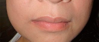
- swelling of the affected area, face, neck, cheekbones appears: it all depends on the area in which the infectious process is localized. This is usually the most noticeable symptom - part of the face suddenly swells,
- pain syndrome occurs
- bad breath bothers you,
- problems with speech, chewing and swallowing cause discomfort,
- body temperature rises to 39–41˚С and above,
- A local increase in temperature occurs: there may be a feeling that the skin in the area of localization of inflammation is warmer than usual or even “burns” or “bakes”.
As the disease progresses, breathing problems arise, a grayish coating appears on the tongue, and the mucous membrane at the site of inflammation becomes red and swollen. If phlegmon is localized in the zygomatic region, discomfort occurs when blinking, swelling and cyanosis of the eyelid, pain in the eyes, and blurred vision.
Putrefactive-necrotic phlegmon of the mouth, or Ludwig's angina
This pathology is similar in appearance to the previous disease, but its main difference is that with soft tissue necrosis there is no accumulation of pus. Symptoms of Ludwig's angina are very specific. This disease carries a huge danger, since sepsis can develop if you do not consult a doctor in a timely manner. Fortunately, the incidence is very low.
The specificity of Ludwig's angina is that basically only the muscles are the main point of damage. The perimuscular tissues may be in a state of inflammation, but it is the muscles that take the main “blow”. Their consistency becomes very dense and woody.
With this disease, patients complain of headaches and high fever. Moreover, in the first two days it may not reach high levels. However, every day the patient gets worse. Externally, a person’s face looks asymmetrical, the chin and neck are swollen. It is impossible to remove the tongue inside as the swelling prevents this from happening. There is a putrid odor and abundant gas formation from the source of infection.
What types of diseases exist
Phlegmon is classified according to several criteria.
By localization
- superficial: local symptoms predominate - pain, swelling, fever up to 39˚C, chills,
- deep: general symptoms are more pronounced - the temperature rises sharply to 42˚C, the heart rhythm is disturbed, blood pressure drops, and difficulties with breathing and urination occur.
By stages of development
- acute: the temperature rises sharply, swelling and hyperemia of the soft tissues are pronounced,
- chronic: the infectious focus is compacted, it is characterized by cyanosis, pain when pressing,
By type of exudate (accumulated liquid)
- serous,
- purulent,
- necrotic.
Classification
For a more complete and better assimilation of the studied purulent processes of soft tissues, it is advisable to once again return to their classification, which is based, as already indicated, on the principles of anatomical and topographic localization, which to a certain extent is conditional, since there are communications between the areas of the face and neck. On the other hand, specification of the localization determines the clinical and diagnostic features of phlegmon (abscess), the choice of surgical approach and possible ways of spreading the process.
By localization:
1. Perimaxillary superficial and deep phlegmons and abscesses.
2. Perimandibular superficial and deep phlegmons and abscesses.
3.Phlegmons and abscesses of the floor of the mouth, tongue and root of the tongue.
4. Common phlegmons of the face and neck.
Why is dental phlegmon dangerous?
Tooth phlegmon leads to serious consequences for the body. Among its complications are meningitis, phlebitis, sepsis, asphyxia and respiratory failure, and chest inflammation. In severe cases, the disease can lead a person to a wheelchair and even death.
We must not forget that the presence of an advanced disease in the oral cavity can at any time provoke a massive proliferation of microorganisms and intoxication of internal organs. It is important not only to stop this process and carry out treatment on time, but not to prevent it by contacting the dentist in a timely manner. That is why in no case should the manifestations of phlegmon be ignored; it is necessary to consult a doctor in time and cure the pathology.
Differential diagnosis
When making a diagnosis, doctors differentiate the disease from furunculosis, erysipelas, cysts, and inflammation of the salivary glands. As a rule, an experienced dentist can easily recognize phlegmon by visual signs and complaints. However, to clarify the localization of pus, its volume, and the degree of tissue damage, a computed tomography or ultrasound examination is required. In addition, the contents of the phlegmon are taken for bacterial culture. This makes it possible to determine the type of infectious agent and prescribe the optimal treatment, i.e. choose the right and most effective antibiotic.
Clinical picture of the disease
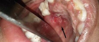
In the initial stages, abscesses and phlegmons adjacent to the lower jaw are manifested by compaction and progressive swelling of the soft tissues of the face. The skin over the purulent focus is often hyperemic. The key symptom of suppuration is fluctuation - the feeling of fluid in a confined space.
The course of phlegmon is accompanied by general intoxication, in which the patient presents the following complaints:
malaise and feeling of chronic fatigue; increase in body temperature; fatigue and joint pain.
Abscesses, as a rule, do not cause such symptoms due to the limited nature of the pathological process.
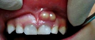
Abscess, phlegmon of the maxillofacial area in children is characterized by an acute and severe course of the disease. Diffuse purulent lesions of soft tissues are formed as a result of the imperfection of the children's immune system.
Pathology treatment methods
Treatment of dental phlegmon depends on its spread and stage of inflammation. If the pathology is not advanced and the patient is referred at an early stage, conservative methods can be used. Often the disease requires surgical intervention in an inpatient setting.
Use of drugs
At the very beginning of the disease, treatment is possible by taking medications:
- antibiotics: to select the most suitable drug, it is necessary to first conduct a bacterial study. If it is impossible to take material for bacterial culture, it is recommended to take medications with a wide spectrum of action,
- analgesics: in severe cases, prescribed in injection form,
- antiseptics for washing and rinsing, for example, “Furacilin”,
- vitamins.
The complex of therapeutic measures necessarily includes eliminating the cause of the inflammatory process.
Surgery
In difficult cases, hospitalization and surgical treatment are indicated. The operation is performed under anesthesia - general or local - depending on the person’s well-being, how the dental phlegmon looks, the presence or absence of problems with the heart, breathing and other factors. The surgeon opens the infectious focus, excises the affected tissue, and, if necessary, performs a resection of the tooth root (if it has not previously been removed). To ensure the drainage of pus, drainage is installed into the incision, after which sutures and a sterile bandage are applied to the wound.
On a note! The operation to remove dental phlegmon is carried out strictly along the lines of the physiological folds of the patient’s face. The intervention requires high professionalism from the doctor, since there is a possibility of damage to the nerve endings in the operated area.
After the operation, the patient is prescribed antibacterial drugs, antihistamines, medications to support the heart, analgesics, and vitamins. The wound is regularly treated with special compounds to speed up healing.
Physiotherapy methods
In addition to conservative treatment, as well as after surgery, physiotherapeutic procedures may be prescribed, including ultraviolet, ultrasound or magnetic therapy. As a rule, the course consists of 5-7 procedures. Physical methods activate local immunity, promote tissue regeneration, normalize their functions, and reduce the duration of the recovery period. Relief of the condition, disappearance of swelling and pain becomes noticeable after the first sessions.
Topographic anatomy
Region boundaries. The zygomatic region corresponds to the location of the zygomatic bone (os zygomaticum), the edges of which are the boundaries of the region (Fig. 43): upper - the lower-outer edge of the orbit (above and posteriorly there is the anterior-inferior section of the temporal region, above and anteriorly - the orbit), lower - the lower edge the zygomatic bone and its temporal process (the buccal region is located below), the anterior border is the zygomaticomaxillary suture (the infraorbital region is located anteriorly), the posterior border corresponds to the temporomygomatic suture (the parotid-masticatory region is located posteriorly).
In the subcutaneous tissue of the zygomatic region, the initial fibers of the zygomatic muscle (m. zygomaticus) can be traced. Sensitive innervation is provided by the branches of n. zygomaticus (from the second branch of the trigeminal nerve), motor - by the same branches of the facial nerve. The blood supply is carried out by the zygomatic orbital artery (a. zygomatocoorbitalis), which arises from the transverse artery of the face.
Will traditional medicine methods help?
The use of traditional methods to treat the disease is possible only in agreement with a doctor and simultaneously with taking prescribed medications, as well as after surgery to remove the source of inflammation. Under no circumstances should you self-medicate at home - it is important to remember the negative consequences of phlegmon.
Traditional medicine is mainly used in the postoperative period. The oral cavity can be rinsed with decoctions and water infusions of chamomile, eucalyptus, yarrow, sage, and honey solution. Such rinses eliminate inflammation, pain, swelling, increase local immunity, and have a wound-healing and antiseptic effect.
Category Uncategorized Posted by Mister stomatolog