Lip cancer is a severe, progressive oncological disease during which a malignant tumor forms on the upper or lower lip from epithelial cells located in the red border area. As the tumor grows, it penetrates other anatomical structures, causing the patient to have difficulty eating. Often the course of the disease is complicated by infectious inflammation. In the absence of timely medical care, tumor tissue begins to disintegrate over time, and the patient’s face becomes disfigured. Metastases spread most often through the lymphatic system.
Kinds
According to localization they distinguish:
- cancer of the lower lip, accounting for about 95-98% of all cases;
- Cancer of the upper lip, which occurs in no more than 2-5% of patients, but has a more aggressive and rapid course, affects mainly women.
According to histological classification, lip cancer is a squamous cell tumor, represented by two types:
- keratinizing – the most common, accounting for up to 95% of cases, which is characterized by a slow course with moderate germination into other tissues and slight metastasis;
- non-keratinizing - rarer and significantly more malignant, quickly growing into nearby tissues (usually into the anatomical structures of the jaw), forming ulcers and metastasizing relatively early through the lymph and blood flow. Metastases usually affect the lymph nodes under the jaw, in the chin and jugular vein, as well as lung tissue.
In addition, there are four clinical forms of the tumor: ulcerative, ulcerative-infiltrative, papillary and warty. The first two have a more malignant course.
Etiology and factors predisposing to lip cancer
- The influence of climatic and meteorological factors that negatively affect human mucous membranes and skin (solar insolation, low and high humidity, air temperature, sudden changes in such parameters). There is a direct relationship between the incidence rate and the geographic latitude of residence, which is due to the high level of insolation (solar exposure).
- Long-term and frequent mechanical trauma to the epithelial layer of the lips: damage from dentures, cuts during shaving, piercing of the upper/lower lip, atrophic processes in the skin in the elderly.
- Periodically appearing extensive lesions of the lips with the herpes virus, papilloma.
- Tobacco smoking, especially using a pipe and low-grade tobacco, chewing dry plant mixtures, abuse of low-quality alcohol-containing drinks, eating very spicy, spicy and hot food.
- Background pathological processes: cracked lips, flat leukoplakias, chronic ulcers, cheilitis.
- Poor oral health: presence of carious teeth.
- Occupational hazards: working in close contact with chemical carcinogens.
Symptoms
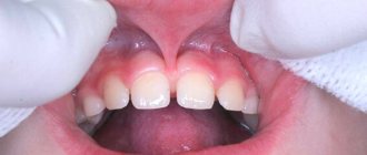
Many patients mistake the first signs of lip cancer for a rash characteristic of an exacerbation of herpes: a small seal forms on the lip, in which a slight itching is felt. At the same time or even a little earlier, the lymph nodes of the chin enlarge. As the tumor develops, the following appear:
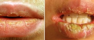
- small painless ulcers, covered with a crust, with a bleeding, tuberous base;
- cracks and crusts along the red border of the lip;
- a compaction that grows in size over time and becomes crusty;
- pain in affected soft tissues;
- increased secretion of saliva;
- difficulty eating, deterioration of diction.
If left untreated, the tumor penetrates the tissues of the jaw and begins to destroy them. Simultaneously with the local symptoms of lip cancer, manifestations common to all types of cancer develop: general weakness, decreased performance, decreased appetite, unmotivated weight loss.
What precedes cancer
The lips are muscles covered with tissue and skin, which is called the “red border”. The inner part, covered with the mucous membrane, anatomically belongs to the vestibule of the oral cavity, and tumors arising there are no longer considered labial. A malignant tumor of the lip can arise out of nowhere - practically out of nowhere, and since this disease is considered to belong to a not very socially prosperous population, cancer in this localization is often preceded by skin diseases of the red border of the lips. These pathological conditions are classified as precancerous, although not all of them become fertile ground for the development of a malignant process.
Precancerous processes are similar in appearance, but differ in cellular structure. Precancerous lips are characterized by hyperplasia - excessive cell growth, frequent cell division, however, within the strictly prescribed framework of nature, and not endlessly, as in a malignant process. Cells of irregular shape appear, prone to rapid keratinization, which is manifested by hyperkeratosis - scaly dry skin. All lip diseases are seriously treated surgically or with close-focus radiotherapy.
- Previously, Bowen's disease was classified as an obligate precancer - a condition that, if persisted for a long time, was highly likely to develop cancer. In reality, cancer develops at the site of this “sore” in approximately every sixth patient. Today the disease is already considered cancer in situ - stage 0 cancer. With Bowen's disease, a spot with small nodules and papillae, velvety or smooth, sometimes with superficial ulcers - erosions, lives and grows on the lip for a long time.
- Erythroplasia Keira, a bright red lump with clear contours that rises above the skin, is no less likely to become a cause for the development of lip cancer. Over time, the lump ulcerates and is also considered stage 0 cancer.
- Manganotti's abrasive cheilitis is a pathology in which most often polished erosions with raised edges appear in the center, become covered with crusts and even heal on their own, but certainly recur.
- In young men, a bulging area with white scales surrounded by inflamed tissue may appear on the lower lip - this is limited hyperkeratosis of the red border of the lips.
- The lip is also affected by a variety of leukoplakia, often with small warts, plaques and erosions, which threatens every fourth patient with lip cancer.
- Keratoacanthoma is a semicircular fat cyst covered with scales, with a depression in the center similar to a volcanic crater. The pathology affects the lower lip, as a rule, of rural men, and in the elderly it is single, and in the young it consists of several nodules.
Causes and risk factors
The main cause of lip cancer is a malignant modification of cells located at the border of the transition of the skin epithelium to the mucous membrane. As a result, cells begin to actively divide, which leads to tumor growth and displacement of healthy tissue. What becomes the trigger for the reformatting of the original healthy cell into a cancerous one has not yet been established, but the factors that increase the likelihood of developing pathology are well known:
- prolonged exposure to the open air, due to which the lips are constantly chapped and cracked;
- diseases of the digestive tract, metabolic disorders;
- habit of biting lips;
- unbalanced diet, chronic vitamin deficiency;
- regular consumption of strong drinks that burn the mucous membrane of the lips;
- smoking and microburns of the lips caused by it, the habit of chewing tobacco;
- irritation due to poorly fitting dentures or broken edges of a tooth;
- poor oral hygiene.
Among patients with lip cancer, rural residents who constantly work outdoors predominate. Men get sick two to three times more often than women; the main age group at risk is people over 60 years of age.
Etiology of neoplasms
External factors remain the main reasons for the development of lip cancer in patients of all age groups. The pathological process develops against the background of regularly repeated mechanical, chemical, temperature and meteorological influences.
The most common mechanical injuries are damage to the red border of the lips during shaving, systematic removal of keratinized epithelium (biting with teeth or tearing off with fingers). Often the cause of damage to the lips is cuts that appear due to low-quality dentures or sharp edges of damaged teeth.
Oncologists include smoking and drinking excessively hot food and drinks as dangerous thermal effects. Regular interactions with carcinogens during the patient’s professional activities are included in the group of chemical factors. Dangerous weather conditions include excessive amounts of ultraviolet radiation, high humidity and low air temperatures. In 10–15% of cases, cancer of the lower or upper lips develops against the background of human infection with the herpes simplex virus type 1.
Stages
There are four stages of lip cancer.
- The size of the neoplasm is small and does not exceed 2 centimeters; the malignant tissue is concentrated within the lip. Lymph nodes are not affected, there are no metastases.
- The pathological tissue increases in size up to 4 centimeters, but the lymph nodes are not affected and there are no metastases.
- The tumor exceeds 4 centimeters in diameter and affects one or two regional lymph nodes.
- Malignant tissue spreads to the jaw bones, tongue, maxillary sinus, or penetrates the masticatory muscles and pterygoid process, reaches the carotid artery and grows into the base of the skull.
Forecast
Fortunately, lip cancer remains one of the most curable malignancies of the head and neck region.
Average survival after 10 years in developed countries can reach 98%, and disease-free survival can be more than 90%.
This is due, first of all, to the fact that the lips are always visible, and early detection of damage to them is not difficult.
Hidden tumors have a worse prognosis and gradually grow into the mentum skin, alveolar mucosa, mandible, floor of the mouth and tongue, as well as form regional nodal and distant metastases.
Diagnostics
External signs of lip cancer, as a rule, are not enough to accurately determine the diagnosis, so the oncologist prescribes a series of laboratory and instrumental tests for the patient.
- A smear or scraping from the surface of the ulcer to perform a cytological analysis of cells and determine the type of cancer.
- Tissue biopsy followed by histological examination of the removed sample to confirm malignant changes.
- Ultrasound of the neck to identify affected lymph nodes.
- X-ray of the lower jaw, chest to identify metastases or ensure their absence.
- CT scan of the facial skeleton to detect metastases in them.
Attention!
You can receive free medical care at JSC “Medicine” (clinic of Academician Roitberg) under the program of State guarantees of compulsory medical insurance (Compulsory health insurance) and high-tech medical care.
To find out more, please call +7, or you can read more details here...
Diagnostic methods
To detect such a pathology, an x-ray of the lower jaw and chest organs is taken. If there is a suspicion that the tumor has grown into the bone, a CT scan of the facial bones is performed.
The X-ray technique is based on the fact that X-ray radiation is attenuated differently when passing through different tissues of the human body. The result is a summation image. CT provides a layer-by-layer image of internal structures, so the extent of the tumor process can be analyzed more accurately.
If the doctor suspects that there are metastases in the bones, he gives a referral for osteoscintigraphy. For the study, a radiopharmaceutical is injected into the patient and then its distribution and accumulation in the bones is recorded using a gamma camera.
To diagnose lymph nodes, an ultrasound of the soft tissues of the neck is performed. This safe and affordable method produces transverse images using high-frequency sound waves produced by the transducer. When the sound wave returns to the sensor, it takes digital form and appears on the monitor as dots or echoes.
Images can be obtained in any plane and appear in real time during the examination. To confirm the diagnosis, a smear or scraping is taken from the tumor ulcer, which is sent for cytological examination to the laboratory. Biomaterial obtained by puncture from metastatic lymph nodes is also sent for cytology. For research, the material is prepared in a special way and stained, after which it is examined under a microscope. In this way, the pathologist can determine the type of tumor.
In preparation for surgery, in order to clarify the functional status of the body, additional examination may be prescribed, because surgical intervention is always a risk for the patient.
Treatment
After diagnostic confirmation of lip cancer, treatment must begin immediately, since the sooner it is carried out, the less traumatic and more organ-preserving the methods used will be.
- Surgery. At the first and second stages, in the absence of metastases, surgery, as a rule, serves as the main method of treatment. The affected tissue is removed, and reconstructive lip surgery is performed at the same time. Patients in the third and fourth stages can be operated on depending on the local spread of the tumor and the expected volume of tissue to be removed. At the same time, the affected lymph nodes in the neck must be removed. If the tumor has spread to vital organs, surgical treatment is not performed.
- Radiation therapy. As an independent method, irradiation is used for small tumor sizes and few metastases; chemotherapy can be used to enhance the effect. After surgery, radiation exposure is necessary to avoid relapse of the disease and destroy residual cancerous foci.
- Chemotherapy. For inoperable cancer and distant metastases, chemotherapy is prescribed to inhibit the growth of malignant tissue and prolong the patient's life. Courses are prescribed, depending on the patient’s condition and the characteristics of the tumor, simultaneously or alternately with radiation.
- Cryogenic method. Low-temperature destruction of tumor tissue is in some cases used in the initial stages to avoid surgery and chemoradiotherapy.
- Photodynamic method. Therapy for limited superficial oncological pathologies consists of the introduction of a photosensitizer and subsequent laser treatment.
Therapeutic measures for a confirmed diagnosis
The treatment strategy for lip cancer is determined by the oncologist, taking into account the stage of development of the malignant neoplasm. A significant factor is the patient's chronic diseases and the presence or absence of a secondary infection.
Stage one lip cancer allows for surgery, during which doctors excise the affected areas. Sometimes oncologists refer patients for radiotherapy. The second stage of the pathological process forces doctors to pre-irradiate the tumor. After this, radical surgery is performed. Stage three cancer will require long-term radiotherapy. The affected areas are the red border of the lips and the lymph nodes affected by metastases. The remains of the tumor and lymph nodes are excised during surgery.
Stage four cancer requires more complex treatment. The patient is undergoing radiotherapy and chemotherapy. After this, surgeons perform a wide excision of the affected tissue. At the terminal stage of the pathological process, people suffering from a malignant tumor of the lips receive palliative treatment (courses of chemotherapy and radiotherapy).
Diagnosis and treatment of lip cancer in Moscow
If you find signs of lip cancer, contact the Meditsina clinic for a comprehensive examination and confirmation or refutation of suspicions. At your service:
- exclusive diagnostic equipment;
- own laboratory performing all types of analyses;
- oncologists of the highest category who will conduct treatment;
- a well-equipped inpatient department;
- medical service of international level.
Call us to find out the details you are interested in and schedule a consultation.
Treatment in Belgium
In clinics in Belgium, for oncological pathologies such as lip cancer, treatment is concentrated on preserving the patient’s quality of life after surgical or therapeutic removal of the tumor.
For this purpose, innovative treatment methods are being introduced into clinical practice, with the help of which it is possible to remove the tumor and preserve the functional and aesthetic parameters of the lips.
Hyperfractionated radiotherapy
This is a type of radiation therapy in which the total dose of radiation is divided into smaller fractional doses, and treatment sessions are carried out more than once a day. This approach makes it possible to reduce radiation damage to healthy tissue and avoid complications such as radiation necrosis and mucositis.
Local chemotherapy
Chemotherapy is a cancer treatment that uses drugs to stop the growth of cancer cells by killing the cells or stopping the cells from dividing. When chemotherapy is taken orally or injected into a vein or muscle, the drugs enter the general bloodstream and have systemic toxic side effects in addition to treating cancer cells. As a result, the dosage of the therapeutic agent must be strictly limited.
With regional chemotherapy, drugs are injected directly into the tumor and primarily affect cancer cells. This is more effective, since the dosage of the drug in the tumor can be maximum.
Hyperthermic therapy
Hyperthermia therapy is a treatment in which body tissue is heated above normal temperature to damage and kill cancer cells or to make cancer cells more sensitive to the effects of radiation and certain cancer drugs.
Experimental biological treatment
As part of clinical research programs in oncology clinics in Belgium, studies are being conducted on the local effect on basal cell carcinoma and squamous cell carcinoma of a complex of the genetically modified T-VEC virus and an intracellular chemical therapeutic agent.
Complex functional and aesthetic reconstructive facial plastic surgery
Aesthetic and reconstructive surgeons in multidisciplinary treatment centers in Belgium are present in the medical team already at the planning stage of tumor removal surgery. This makes it easier for them to plan their own surgery for functional and aesthetic reconstruction, which improves the quality of the recovery and makes it less traumatic.
Questions and answers
What does lip cancer look like?
In terms of external signs, lip cancer at the initial stage resembles a herpetic rash: a small swelling appears, which later turns into one or several ulcers covered with crusts. A thickened area with redness of the skin may form. Unlike a herpetic infection, ulcers do not heal after two to three weeks, but on the contrary, increase in size.
How to recognize lip cancer?
If a lump or swelling appears on one of your lips and does not go away within two to three weeks, you should definitely contact an oncologist to check for lip cancer. It is impossible to independently determine cancer by external signs; laboratory tests are required - histological and cytological examination of tumor tissue, biochemical blood tests, examinations using a tomograph and other types of medical equipment.
How long do people live with lip cancer?
A malignant tumor on the lip is one of the least aggressive oncological pathologies and is most often detected at an early stage. More than 90% of people who develop it live after diagnosis for more than 10 years, i.e. are completely cured of cancer.
Attention! You can cure this disease for free and receive medical care at JSC "Medicine" (clinic of Academician Roitberg) under the State Guarantees program of Compulsory Medical Insurance (Compulsory Medical Insurance) and High-Tech Medical Care. To find out more, please call +7(495) 775-73-60, or on the VMP page for compulsory medical insurance
Prevention of lip cancer
- Preventive examinations of the population, clinical examination of persons constituting a high-risk group, sanitary and educational work.
- Carrying out hygienic measures, sanitation of the oral cavity, adequate prosthetics.
- Treatment of underlying diseases, precancerous changes in the mucous membrane: the use of indifferent ointments and hygienic lip protection products in persons whose profession is associated with harmful effects on the lips and are exposed to prolonged adverse meteorological factors;
- giving up bad habits (smoking, eating chewing mixtures that irritate the mucous membrane of the lips) and eliminating harmful environmental factors;
- normalization of gastrointestinal tract function, increased immunity.
What are the types of squamous cell carcinoma?
Malignant neoplasms of this histological type are found on different parts of the body. Depending on the location, their properties, approaches to diagnosis and treatment, and prognosis for the patient may differ slightly.
Skin cancer
Malignant skin tumors are represented by squamous cell carcinoma in approximately 20% of cases. Much more often, patients suffer from basal cell carcinoma, which originates from cells located in the lower layer of the epidermis.
Squamous cell carcinoma is more aggressive than basal cell carcinoma. It is more likely to grow into the deeper layers of the skin and spread throughout the body with the formation of distant metastases. However, this happens quite rarely. Most often, the tumor can be detected and removed at an early stage.
As a rule, squamous cell carcinoma occurs on the skin of the face, ears, neck, back of the hands, and less commonly in the genital area. Often, a neoplasm develops where scars and chronic injuries are located.
Squamous cell carcinoma of the red border of the lips
Malignant lip tumors account for no more than 1–3% of all cancers. In most cases (95%) they are represented by squamous cell carcinoma, which comes in two types:
- Squamous cell keratinizing carcinoma does not behave as aggressively, grows slowly, and rarely forms distant metastases.
- Nonkeratinizing squamous cell carcinoma grows rapidly, ulcerates earlier, and metastasizes more often.
Research shows that this type of cancer is 3 to 13 times more common in men than in women. This is probably due to the fact that males are more often exposed to sunlight at work, and smoking and drinking alcohol are more common among them.
Oral cancer
Oral cancer is a malignant tumor that occurs on the mucous membrane of the lips, cheeks, gums, the anterior two-thirds of the tongue, the palate, and the floor of the mouth (located under the tongue). In 90% of cases they are represented by squamous cell carcinoma, of which 5% are keratinizing squamous cell carcinoma, which is less aggressive, less likely to grow into surrounding tissues, spread to lymph nodes and metastasize.
Esophageal carcinoma
The mucous membrane of the esophagus is lined with stratified squamous epithelium, and squamous cell carcinoma can develop from it. Most often, such tumors are located in the cervical esophagus and the upper two-thirds of the thoracic region. In the lower third of the organ, adenocarcinomas, malignant tumors of glandular cells, are more common.
Book a consultation 24 hours a day
+7+7+78
Laryngeal cancer
In laryngeal cancer, the tumor almost always develops from squamous epithelium and is a squamous cell carcinoma. Typically, the appearance of a tumor is preceded by precancerous changes - dysplasia. The cells that are located in the lesion do not look like normal ones, but they also differ from cancer cells. In some cases, dysplasia does not lead to the development of cancer and even goes away on its own, especially if its cause is eliminated, for example, a person quits smoking. But in some people, precancerous changes lead to “cancer in situ” and then an invasive tumor.
Trachea and bronchus cancer
Squamous cell carcinoma is the most common type of malignant tumor in the trachea. It usually occurs in the lower part of the trachea, grows quite quickly, invades its wall, leading to ulceration and bleeding. This is a rare type of cancer and its main cause is smoking.
The most common lung cancer is non-small cell cancer - it occurs in 80% of cases and in 30% of cases it is squamous cell carcinoma. Often these tumors are located in the bronchi.
Cervical cancer
The cervix consists of two parts. The exocervix is located outside, in the vagina, this is what the gynecologist sees during the examination. The endocervix is the canal of the cervix, it connects the uterus to the vagina. Normally, the exocervix is lined with squamous epithelium, and the endocervix is lined with glandular epithelium. The place where they meet is called the transformation zone.
Squamous cell carcinoma represents 90% of malignant tumors of the cervix. Most often, the neoplasm occurs in the area of the transformation zone. Cancers that develop from the glandular cells of the endocervix are called adenocarcinomas.
In rare cases, glandular squamous cell carcinoma occurs in the cervix.
Vulvar cancer
The vulva is the name given to the external female genitalia: the vestibule of the vagina, the labia majora and minora, and the clitoris. The majority of cancer types that develop in this area are squamous cell carcinoma (70–90%). They are divided into two groups:
- A large group are tumors whose origin is unknown. Most often they are diagnosed in older women.
- A smaller group are malignant tumors caused by the human papillomavirus.
Rectal cancer
In most cases, malignant tumors of the rectum are represented by adenocarcinomas - glandular cancer. Squamous cell carcinoma in this organ is very rare and accounts for 10 to 25 cases for every 100 thousand cases of colorectal cancer.
Squamous cell carcinoma accounts for 90% of all malignant neoplasms of the anal canal, the narrow passage that connects the rectum to the anus.
Tonsil cancer
A person has four types of tonsils: palatine (when they become inflamed, tonsillitis develops), tubal (located in the pharynx near the openings of the auditory tubes), lingual (behind the tongue) and pharyngeal (in children this causes adenoids). Most often, malignant tumors develop in the tonsils. In most cases it is squamous cell carcinoma. It is difficult to diagnose, so it is often detected in late stages.
Prevention
Basic measures to prevent squamous cell carcinoma:
- Quitting smoking and drinking alcohol.
- Protecting your skin from ultraviolet rays is the most important measure for preventing skin cancer. You should not visit solariums or go to the beach from 10.00 to 16.00, when solar activity is highest. Clothes with long sleeves and trousers, a wide-brimmed hat, and sunglasses help protect yourself.
- Preventing infection with HPV, which leads to the development of cancer: you need to avoid promiscuity and use condoms. There is currently a vaccine against human papillomavirus infection. It is recommended that all adolescents be vaccinated before becoming sexually active.
| More information about treatment at Euroonco: | |
| Oncologist consultation | from 5100 rub. |
| Chemotherapy appointment | 6900 rub. |
| Radiologist consultation | 10500 rub. |
| Palliative care in Moscow | from 40200 per day |
Book a consultation 24 hours a day
+7+7+78
Causes of squamous cell carcinoma
The causes of squamous cell carcinoma are the same as for other types of malignant tumors. Certain mutations occur in cells that lead to malignant degeneration. “Incorrect” cells lose the external features and functions of normal ones, begin to multiply uncontrollably, and acquire the ability to spread throughout the body.
The main risk factors for squamous cell carcinoma:
- On the skin, such tumors often arise due to the action of ultraviolet rays. Exposed areas of the body are the most vulnerable.
- Squamous cell carcinoma of the genitals, head and neck is caused by certain types of human papillomavirus.
- The risk of developing squamous cell carcinoma is increased in smokers and people who drink a lot of alcohol.
- The likelihood of developing cancer increases with age as mutations accumulate in the cells of the body.
- Scars, burns, chronic inflammation.
- Exposure to certain carcinogenic substances, for example, if a person works in an industrial environment and comes into contact with chemicals.
- Decreased immunity.
None of these factors is guaranteed to lead to the disease - each of them only increases the likelihood to a certain extent.
Treatment of squamous cell carcinoma
Treatment depends on the location, stage of cancer, the general condition of the patient, the presence of concomitant diseases and other factors.
Radiation therapy
Ionizing radiation damages tumor and other rapidly multiplying cells. This type of treatment for squamous cell carcinoma can be prescribed before or after surgery, or in advanced stages for palliative purposes.
Surgery
Radical operations are possible if there are no metastases and the cancer has not grown strongly into surrounding tissues. In some cases, only surgical treatment is indicated for such patients, in others it is supplemented with antitumor drugs and radiation therapy - this helps reduce the risk of relapse.
For advanced squamous cell carcinoma, palliative surgery can be performed to eliminate symptoms and restore the patency and function of the affected organ.
Drug treatment of squamous cell carcinoma
Chemotherapy for squamous cell carcinoma can be adjuvant (after surgery), neoadjuvant (before surgery), or used as a stand-alone treatment in advanced stages.
If the tumor has certain molecular genetic characteristics, targeted therapy is prescribed. Targeted drugs target molecules that help cancer grow and maintain its vital functions.
Doctors at Euroonko use original antitumor drugs of the latest generation for squamous cell carcinoma, prescribing them in accordance with modern international protocols.
Symptomatic treatment for squamous cell carcinoma
Treatment for squamous cell carcinoma and any other malignant neoplasms should be aimed not only at fighting the tumor itself, but also at relieving symptoms and improving the patient’s condition. At Euroonko, the patient can receive all types of symptomatic therapy for cancer:
- Relief of pain syndrome in accordance with the WHO three-step scheme.
- Restoration of patency of the esophagus, intestines, and respiratory tract.
- Elimination of bleeding, if necessary, blood transfusion.
- Relief of nausea.
- Removing tumor compression of internal organs, nerves, and blood vessels.
- Treatment of emergency conditions in an intensive care unit equipped with modern equipment.
- Monitoring and correction of nutritional status.
- Maintenance therapy helps you comfortably endure chemotherapy and prevent and manage side effects.