Palate cancer is a rare cancer. It accounts for approximately 2% of all malignant tumors of the head and neck. Men are affected 4 times more often than women; in most cases, the disease is diagnosed at the age of 40–60 years.
Our expert in this field:
Sergeev Pyotr Sergeevich
Oncologist, surgeon, chemotherapist, Ph.D.
Call the doctor Reviews about the doctor
Anatomically, the palate is divided into two parts. The anterior 2/3 is located on the hard palate. It is a bony septum between the nasal and oral cavities, formed by the palatine processes of the upper jaws, covered with mucous membrane. The posterior third, the soft palate, is a freely hanging fold of the mucous membrane. It covers the nasal cavity during swallowing and is involved in the formation of sounds during conversation.
Malignant tumors that arise in the hard and soft palate differ somewhat in their histological structure.
Cancer of the soft palate in most cases (80%) is represented by squamous cell carcinomas. In case of cancer of the hard palate, they account for 53%, the remaining cases occur in other types of malignant tumors:
- adenoid cystic carcinomas - 15%;
- mucoepidermoid carcinomas - 10%;
- adenocarcinomas and anaplastic carcinomas - 4% each;
- others - 14%.
Next to the molars, there are holes in the hard palate through which nerves and blood vessels pass. This creates the preconditions for the spread of malignant tumors into the nasal cavity.
There are minor salivary glands in the mucous membrane of the palate. Some malignant tumors originate in them. Most often these are adenoid cystic carcinomas, mucoepidermoid carcinomas, adenocarcinomas, and mixed malignant tumors.
Sometimes patients use the not entirely correct term “cancer of the upper palate.” In fact, there is no “lower palate”; there is only the floor of the mouth.
Classification of oncological pathology of the palate
In half of all cases, malignant tumor of the hard palate is squamous cell carcinoma.
Nonsquamous cells, including minor salivary gland cancer, sarcoma, and melanoma, account for the other half of hard palate neoplasms. In 80% of cases, a tumor of the soft palate is squamous cell carcinoma. The remaining 20% are benign neoplasms.
The histological distribution of malignant neoplasms of the hard palate is as follows:
- squamous cell carcinoma - 53%;
- adenoid cystic carcinoma - 15%;
- mucoepidermoid carcinoma - 10%;
- adenocarcinoma - 4%;
- anaplastic carcinoma - 4%;
- other - 14%.
The histological types and frequency of neoplasms of the minor salivary glands of the palate are as follows:
- Benign - 26%
- Malignant - 74% overall Adenoid cystic carcinoma - 30%
- Mucoepidermoid carcinoma - 16%
- Adenocarcinoma - 18%
- Malignant mixed tumor - 8%
- Other - 2%
Causes of palate cancer
The causes of this disease have not been established, but it is known that certain factors may increase the risk of developing malignant tumors of the palate.
Smoking and chewing tobacco
Active smoking, especially unfiltered cigarettes and cigars, at least doubles the risk of developing malignant neoplasia of the oral cavity. Their development is also facilitated by the use of chewing tobacco mixtures - betel nut, gutka, snus, etc.
Alcohol
Strong alcohol is a recognized risk factor for palate cancer. This is mainly associated with the direct damaging effect of ethyl alcohol on the oral mucosa.
Human papillomavirus
HPV infection has been significantly associated with an increased risk of developing oral squamous cell carcinoma in recent studies.
Chronic inflammatory diseases of the oral cavity
Sluggish inflammatory processes, such as chronic gingivitis or periostitis, increase the likelihood of the formation of palate tumors.
Precancerous diseases
In the presence of pathologies of the mucous membrane such as leukoplakia or erythroplakia, the likelihood of developing cancer becomes extremely high - up to 90%.
Forecast
The prognosis for RPR depends on the stage and location of the tumor. In most cases, the stage of the tumor is related to the time of diagnosis. The earlier the diagnosis is made, the lower the stage of the tumor, and, therefore, the better the survival rate.
Other factors must also be taken into account, such as the location of the tumor, the patient's general health, age, tobacco use, and the presence of human papillomavirus (HPV).
Based on 2021 data from EU countries, the overall estimated 5-year survival rate for RCC is approximately 66%.
Lip carcinomas generally have the best 5-year survival rate (88%), while floor of the mouth cancers have the worst (54%).
Tumor staging has been shown to be the best prognostic factor for lip carcinoma, whereas transcriptionally active HPV status is considered an important prognostic factor for RCC.
Those with HPV-positive tumors tend to respond better to chemotherapy or radiation therapy compared to those with HPV-negative tumors.
According to the US National Cancer Institute, the five-year survival rates for RCC are as follows:
- 83%, for localized cancer (that has not spread);
- 64%, for cancer that has spread to nearby lymph nodes;
- 38%, for cancer that has spread to other parts of the body.
Symptoms of the disease
Squamous cell carcinoma appears as ulcerative superficial lesions. However, there are often no signs of palate cancer in the initial stages. The first manifestations appear, as a rule, already in the advanced stages of the disease.
Palate cancer - symptoms and first signs:
- bleeding;
- difficulty swallowing;
- speech disorders;
- bad breath;
- wounds in the mouth that do not heal for a long time;
- loosening of the teeth of the upper jaw;
- pain when swallowing;
- weight loss;
- ear pain;
- swelling of the neck;
- white spots in the mouth that do not disappear for a long time.
A swelling on the roof of the mouth, bleeding, foul odor, or loose teeth may be symptoms in patients with hard palate cancer. Patients with advanced soft palate cancer may have difficulty swallowing, altered speech, trismus, or neck swelling.
Because the area is easily visualized, tumors are often discovered in the early stages by chance by the patient or physician.
Survival prognosis
When oncologists talk about the prognosis for certain types of cancer, most often they refer to the five-year survival rate. It refers to the percentage of patients alive five years after diagnosis:
| Cancer of the gums and other parts of the mouth, including the hard palate, excluding the tongue, lips and floor of the mouth | Cancer of the oropharynx, including the soft palate | |
| Stage I | 81% | 56% |
| Stage II | 62% | 58% |
| Stage III | 45% | 55% |
| Stage IV | 40% | 43% |
You need to understand that these data are statistics for previous years, and they give only a very rough idea of the prognosis for a particular patient. In addition to the stage, other factors play a role, such as the resection margin after surgery, age, general condition of the patient, his concomitant diseases, etc.
We will call you back, leave your phone number
Message sent!
expect a call, we will contact you shortly
At the international clinic Medica24, patients with cancer of the soft and hard palate receive the most modern types of treatment in accordance with the latest versions of international recommendations. We take on any complex cases, our experienced oncologists will definitely try to help. Contact us.
The material was prepared by the deputy chief physician for medical work of the international clinic Medica24, candidate of medical sciences Sergeev Petr Sergeevich.
Sources:
- Choinzonov Evgeny Lkhamatsyrenovich, Podvyaznikov S.O., Minkin A.U., Mudunov A.M., Azizyan R.I., Pustynsky I.N., Tabolinovskaya T.D., Brzhezovsky V.Zh., Alieva S.B. . Clinical recommendations. Diagnosis and treatment of oropharyngeal cancer // Siberian Journal of Oncology. 2021. No. 1.
- Kropotov M.A., Yakovleva L.P., Dronova E.L., Alieva S.B., Allahverdieva G.F. An integrated approach to the treatment of locally advanced oropharyngeal cancer // Head and neck tumors. 2016. No. 2.
- Kropotov M. A., Epikhina A. V. Surgical aspects of the treatment of oropharyngeal cancer // Head and neck tumors. 2011. No. 2.
Stages of development of palate cancer
The stages of malignant neoplasms of the palate are determined based on the international TNM system. With its help, the characteristics of the tumor are described according to three main criteria.
Primary tumor stage (T)
- TX—primary tumor not evaluable
- T0 - no evidence of a primary tumor (T - carcinoma in situ.)
- T1—tumor 2 cm or less in greatest dimension
- T2 - tumor larger than 2 cm but not larger than 4 cm in greatest dimension
- T3 - tumor larger than 4 cm in greatest dimension
- T4 - Tumor invades adjacent structures (eg, cortical bone, soft tissue of the neck, maxillary sinuses)
Regional lymph node involvement (N)
- NX—regional lymph nodes not evaluable
- N0 - Absence of regional metastasis in the lymph node
- N1 - Metastasis in one ipsilateral lymph node, 3 cm or less in greatest dimension
- N2 - metastasis in one ipsilateral lymph node, more than 3 cm, but not more than 6 cm in greatest dimension; in several ipsilateral lymph nodes, not more than 6 cm in greatest dimension; or in bilateral or contralateral lymph nodes, not more than 6 cm in greatest dimension
- N3 - Metastasis in a lymph node greater than 6 cm in greatest dimension
Distant metastasis (M)
- MX - The presence of distant metastases cannot be assessed
- M0 - no distant metastases
- M1 - distant metastases
Based on the TNM characteristics, the so-called clinical stage is determined.
| Stage | Characteristics |
| 0 | Tis N0 M0 |
| I | T1 N0 M0 |
| II | T2 N0 M0 |
| III | T3 N0 M0 |
| (T1, T2, T3) N1 M0 | |
| IVA | T4a ( N0 or N1) M0 |
| (T1, T2, T3 or T4a) N2 M0 | |
| IVB | Any T N3 M0 |
| T4b Any N M0 | |
| IVC | Any T Any N M1 |
Folk remedies
For treatment, you can use traditional medicine. The following juices, decoctions, oils and infusions have proven themselves well.
You can gently lubricate the affected areas of the mucous membrane with juices and oils. Solutions and decoctions of herbs are used for regular rinsing of the mouth.
Important! Before using traditional medicine, you should consult a doctor. Only a specialist can determine the advisability of their use without harm to health. The use of folk remedies does not cancel the main therapy, but only complements its effect.
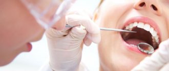
Diagnosis of palate oncology
In most cases, detection of oncological pathologies of the palate occurs during a routine examination by a dentist. The diagnosis is then confirmed by biopsy findings.
However, to assess the extent of the tumor in the structures of the upper jaw, pharynx, etc. Additional diagnostic tools are required:
- MRI (magnetic resonance imaging) - to detect tumor growth in the sinuses of the upper jaw or the nasal cavity.
- PET (positron emission tomography) - to detect metastasis in regional lymph nodes and separated metastasis.
Treatment of the disease
Radiation therapy
Radiation therapy to treat palate cancer can be used in several ways.
- As the primary treatment for small, early-stage tumors.
- As an adjunct to surgery or ongoing chemotherapy. In this case, it is possible to use adjuvant radiation therapy as after surgery to destroy tumor remnants. So is the use of radiation before surgery to shrink the tumor and make it easier to remove—neoadjuvant radiation therapy.
- Radiation therapy can also be used to relieve symptoms of advanced, inoperable cancer, such as pain, bleeding, problems swallowing, and problems caused by bone metastases.
Modern radiotherapy for palate cancer in Belgium
Radiation in cancer centers in Belgium is carried out using techniques that help doctors focus the radiation more precisely.
There are two main such methods.
- Three-dimensional conformal radiation therapy (3D-CRT);
- Intensity modulated radiation therapy (IMRT).
These methods rely on the results of imaging tests, such as MRI, and special computer programs to pinpoint the location of the tumor. The beams are then shaped and directed at the tumor from multiple directions, reducing damage to healthy nearby tissue.
Another modern way of delivering radiation is to place radioactive materials directly into the tumor. This is called brachytherapy.
To do this, a small grain of radioactive material is placed into the tumor. In this case, the radiation spreads only over a very short distance and is limited to the tumor.
Surgery
The type of operation is selected depending on the location and extent of the tumor.
For small to medium-sized tumors up to T3, an oral approach is often used without trauma to the mandible. For most tumors that do not have regional spread, surgery to remove the hard or soft palate is used - palatectomy.
For tumors that have grown into the paranasal sinuses or nasal cavity, a unilateral or bilateral maxillectomy (removal of the upper jaw) may be required.
Chemotherapy
Treatment of palate cancer with chemotherapy is considered as an additional method in combination with radiation therapy and surgery.
The chemotherapy drugs most often used for cancer of the oral cavity and oropharynx are:
- Cisplatin;
- Carboplatin;
- 5-fluorouracil (5-FU);
- Paclitaxel (Taxol);
- Docetaxel (Taxotere).
Targeted and immunotherapy
As a rule, targeted and immunological drugs for this disease are used for metastatic inoperable forms, or for relapses.
Cetuximab (Erbitux®) is a monoclonal antibody that targets the epidermal growth factor receptor (EGFR), which helps cells grow and divide. Palate cancer cells often have higher than normal amounts of EGFR. By blocking EGFR, cetuximab can help slow or stop cell growth.
Pembrolizumab (Keytruda®) and nivolumab (Opdivo®) are drugs that target the PD-1 checkpoint. It is a protein on the membranes of T cells that normally helps keep these cells from attacking other cells in the body.
By blocking PD-1, these drugs boost the immune response against cancer cells, which can shrink some tumors or slow their growth.
Diagnosis of cancer of the soft and hard palate
The resulting cancerous tumor of the palate is difficult to determine independently in the early stages. If the pathological process has affected large areas of the soft or hard palate, a preliminary diagnosis can be made after a visual examination of the oral cavity.
To confirm the diagnosis, oncologists at the Yusupov Hospital perform the following diagnostic procedures:
- X-ray – finds pathological changes in bone tissues adjacent to the oral cavity;
- Biopsy – taking a piece of tissue for histological examination (analysis is necessary to identify altered tumor cells and its type);
- Blood tests - signs of anemia;
- Radioisotope examination - allows you to examine the structure of the neoplasm.
Ultrasound examination is carried out to detect cancer metastases in distant organs. Patients of the Yusupov Hospital can undergo complex diagnostic procedures in partner clinics and receive advice from leading dental oncologists in Moscow.
Rehabilitation after treatment
Treatment of malignant tumors of the palate is always very traumatic. The consequence of the operation is, at a minimum, a violation of the process of swallowing and speech. In multidisciplinary clinics in Belgium, rehabilitation doctors and speech therapists begin working with the patient immediately after the operation - this greatly shortens the rehabilitation period and increases its effectiveness
Reconstructive surgery - palate surgery in Belgium
Palatectomy and maxillectomy operations require subsequent prosthetics.
Small defects of the hard palate can be covered with local flaps from the rest of the hard palate or from the buccal mucosa. Larger defects are adequately and effectively treated with obturators. Because the hard palate is not dynamic, obturators are very effective and well tolerated.
The soft palate is a dynamic structure and its optimal functioning requires muscular action to lift and contract the palate during swallowing and relax it during nasal breathing. Therefore, any reconstruction of the soft palate using flaps and prosthetics does not reproduce its functionality - it will be limited even with the most successful outcome.
About the oral cavity
Figure 1. Oral cavity
The oral cavity (or simply the mouth) is used for speech, chewing, swallowing, and breathing. The oral cavity consists of:
- lips;
- anterior two-thirds of the tongue;
- palate (hard and soft);
- floor of the mouth (under the tongue);
- lining of the inner surface of the cheeks (buccal mucosa);
- gums;
- a small area behind the wisdom teeth on the lower jaw (retromolar triangle).
to come back to the beginning
Complications
Complications of surgical resection of the soft palate may include:
- velopharyngeal insufficiency (most often);
- hypernasal speech;
- dysphagia (impaired swallowing);
- effusion in the middle ear due to scarring at the opening of the eustachian tube.
The extent and likelihood of these complications depend on the extent of the resection, the size of the defect, and the method of reconstruction. The larger the resection and defect, the greater the likelihood of these complications occurring.
With soft tissue resection preserving only the bony palate and soft palate, the resulting defect heals with granulation and epithelialization, and no complications are expected. If patients have previously had radiation therapy in this area, healing may be delayed.
Palate cancer and its treatment can sometimes cause problems such as loss or changes in taste, dry mouth, or even tooth loss. This can make it difficult to eat, leading to weight loss and weakness due to poor nutrition.
Radiation therapy may also cause long-term or permanent side effects.
- Damage to the salivary glands. It may cause dry mouth. This leads to problems with eating and swallowing. Lack of saliva can also lead to tooth decay (cavities).
- Damage to the jaw bone. This problem, known as osteoradionecrosis of the jaw, can be a serious side effect of radiation therapy. This condition is difficult to treat. Osteoradionecrosis is manifested by pain in the jaw, and sometimes by fractures, which require surgical treatment.
Newer radiation treatments such as IMRT may help reduce this side effect. The drug amifostine (Ethyol®) may also help reduce this side effect by limiting radiation damage to normal tissue. It is injected into a vein over 15 minutes before each radiation treatment.
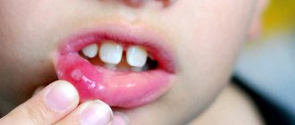
How to treat stomatitis at home?
Effective treatment of stomatitis involves a set of measures aimed at achieving five goals:
- relief of the inflammatory process;
- elimination of pain;
- maintaining oral hygiene;
- speedy healing of the lesion;
- strengthening the immune system.
For this, the patient is prescribed anti-inflammatory, painkillers, disinfectants, antibacterial, antihistamines and other drugs. The choice of remedies directly depends on the form and type of disease.
Regardless of the type of stomatitis, treatment of the disease should begin with sanitation of the oral cavity - a procedure for thoroughly cleansing the mucous membrane. For this purpose, you can use rinses using various solutions based on the following agents.
- Asepta rinse aid.
- Lugol.
- Malavit.
- Hydrogen peroxide.
- Rotokan.
- Chlorhexidine.
- Chlorophyllipt.
- Furacilin.
- Humer.
- Ketotifen.
- Loratodine.
- Tavegil.
- Amiksin.
- Anaferon.
- Immunal.
- Laferobion.
- Imudon.
- Levamisole.
- Alizarin ointment.
- Acyclovir.
- Bonafton.
- Interferon ointment.
- Oxaline ointment.
- Tebrofen ointment.
- Zovirax.
- Inhalipt.
- Metrogyl denta.
- Sodium tetroborate.
- Azithromycin.
- Amoxicycline.
- Ampiox.
- Augmentin.
- Gentamicin.
- Doxycycline.
- Kanamycin.
- Clarithromycin.
- Lincomycin.
- Sumamed.
- Flemoxin solutab.
- Ecolink.
- Hexoral tabs.
- Solcoseryl.
- Karotolin.
- Vinilin (Shostakovsky balm).
- Aekol.
- Diflucan.
- Candide.
- Clotrimazole.
- Levorin.
- Nystatin (tablets) or nystatin ointment.
- Pimafucin.
- Vitaon.
- Flemoxin.
- Boric acid.
- Methylene blue solution.
- Furacilin.
The further choice of remedies and medications directly depends on the type of stomatitis and its form.
Treatment of allergic stomatitis
To effectively treat stomatitis of an allergic nature, antihistamines are actively used.
Taking immunostimulating drugs is useful.
Important! Before starting to take antihistamines and immunostimulating drugs, you should consult your doctor.
Herpetic or cold sore
When treating herpes stomatitis, it is necessary to take antiviral drugs. As a rule, these are ointments that are used to lubricate the affected area of the mucous membrane or tablets for oral administration.
Important! Antiviral drugs are taken only as prescribed by a doctor.
Traumatic (bacterial)
Treatment of this type involves taking antibacterial agents. The following medications may be recommended to the patient.
For ease of use and to achieve a speedy recovery, the specialist determines the advisability of using one or another form of the drug - gel, solution or tablets. In case of severe disease, the patient may be prescribed antibiotics.
Important! Self-medication with antibiotics is strictly prohibited! Drugs in this group are taken only as prescribed by a doctor!
Catarrhal and catarrhal-hemorrhagic
To treat catarrhal stomatitis, anti-inflammatory drugs and agents that accelerate the healing of ulcers are used.
Important! Drugs for speedy healing can be used for all types of stomatitis.
Candidiasis (fungal)
When treating candidiasis, special attention is paid to the choice of antifungal drugs.
Also, during the treatment of a fungal disease, it is advisable to use the following anti-inflammatory drugs.
To timely remove plaque that forms, you can use the following products.
Use a cotton pad soaked in the solution to regularly and carefully remove accumulated plaque.
Treatment of ulcerative stomatitis
Treatment of ulcerative forms is carried out comprehensively and includes the following measures.
- Sanitation of the oral cavity.
- Taking antibiotics, antihistamines and painkillers.
- Using remedies to speed up the healing of ulcers.
If the temperature rises, take antipyretics.
Aphthous stomatitis
To effectively dry the resulting canker sores, you can use the following means.
- Iodinol.
- Burnt alum.
- Iodine solution.
- Potassium permanganate solution.
- Fukortsin.
- Tantum Verde.
- Lidocaine.
- Hexoral.
- Asepta gel.
- Kamistad
- Kalgel.
- Lidochlor.
- Holisal.
After removing dry crusts and disinfecting the oral cavity, you can use drugs that accelerate the healing of ulcers.
Painkillers
For severely painful ulcers, painkillers are used. To ensure their uniform application to the mucous membrane, you can choose a spray.
Aerosols are no less effective in eliminating pain.
You can lubricate the affected areas with anesthetic gels.
Important! It is not possible to get rid of stomatitis using painkillers. But they make it easier to eat and carry out medical procedures.
Prevention
Limit smoking and alcohol
Tobacco and alcohol are the most important risk factors for these cancers. Not starting to smoke is the best way to limit your risk of cancer. Quitting tobacco also significantly reduces the risk of developing these cancers, even after years of use. The same applies to alcohol - limiting its amount helps reduce the risk of developing palate cancer.
Preventing HPV infection
Vaccination against HPV reduces the risk of mouth and throat cancer. But vaccines are only effective if they are given before someone gets HPV, given at a young age, before they become sexually active.
Correctly selected dentures and proper oral hygiene
Avoiding sources of oral irritation (such as dentures that don't fit properly) can help reduce your risk of developing oral cancer.
Care after surgery for oral cancer
Changes in diet
You will be able to drink fluids immediately after surgery. Your doctor will tell you when you can start taking pureed foods. You can start eating soft foods when your doctor allows, as long as you can tolerate them.
Read the resource Recommendations for Pureeed and Mechanically Gentle Diets for nutritional recommendations. You can also take liquid supplements that are high in protein and calories. For example, Ensure®, Boost® and Carnation Instant Breakfast®. Do not eat regular solid foods until your doctor tells you to.
If you have had surgery on the lining of your mouth or lower gum, chew food on the other side of your mouth until you have your first doctor's appointment after surgery.
Oral care
- Your doctor will tell you whether you can brush your teeth after surgery.
- Keep your mouth clean with rinses or an oral irrigation device. Your nurse will give you an oral irrigation device and teach you how to use it.
- To make a rinse solution, mix 1 quart (250 ml) warm water with 1 teaspoon salt and 1 teaspoon baking soda. You can store it at room temperature. Do not use mouth rinses that contain alcohol. They can cause oral irritation and slow healing.
Caring for the incision
- Do not wet the stitches on your lip or neck for the first 48 hours (2 days). After 48 hours, you can shower as usual. Do not direct the water stream towards the cut itself. Instead, let the water run down the cut and pat it dry with a clean towel.
- Apply bacitracin ointment twice daily if your doctor tells you to do so. Before you leave the hospital, you will be given bacitracin if you need it.
- The absorbable sutures will loosen and fall out 6 to 8 weeks after surgery. When you feel them in your mouth, simply spit them out. As long as they stay in place, they don't require much care other than keeping your mouth clean.
- Non-absorbable sutures will be removed at your first appointment with your doctor after surgery.
- If you have had lip surgery, avoid stretching your lip, such as when smiling, until the area has healed.
Speech changes
- If you have had surgery on your tongue, floor of your mouth, or soft palate, your speech may change. Your tongue may become swollen and numb, and you may not be able to move it freely. This will subside as the area heals. If you have significant changes in your speech, you will be referred to a speech therapist for further help.
Anesthesia
- Most people experience pain or discomfort after surgery. You will be given a prescription for pain medication before you leave the hospital. Take it as prescribed.
- If pain medication doesn't help, call your doctor.
- Painkillers may cause constipation (having bowel movements less frequently than usual). To prevent constipation, take a stool softener such as docusate sodium (Colace®) 3 times daily. If this does not help, take a laxative (eg 2 Senokot® tablets) before bed. Both drugs are available without a prescription. If constipation persists after taking these medications, call your doctor. For more information, read the resource Constipation.
Follow-up appointment
The day after surgery, call your doctor to make a follow-up appointment.
to come back to the beginning