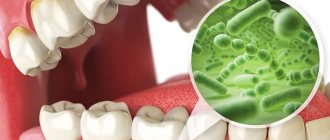
Symptoms and their manifestation
Symptoms characteristic of diseases in this category, in most cases, allow one to independently diagnose the presence of deviations from the norm. Notable changes include:
- The occurrence of itching and burning.
- The appearance of pain syndrome.
- Formation of tissue edema.
- Formation of ulcers and ulcers.
- Bleeding gums.
- Violation of the enamel structure.
- Constant unpleasant odor.
- Feeling weak and tired.
At the first signs of a pathological condition, it is recommended to seek help from specialists. Thus, in Dentika dental clinics you can undergo comprehensive diagnostics, which guarantees the accuracy of the diagnosis and the timely initiation of therapeutic measures.
Types of pathologies
In accordance with the ICD-10 classification for dentistry, treatment of diseases of the oral mucosa is carried out in four categories. There are inflammatory, infectious, viral and fungal diseases. The treatment plan depends both on the severity of negative symptoms and on the individual characteristics of the patient’s body, which are manifested, among other things, in the reaction to certain groups of medications.
Infectious viral processes
The form of development is directly related to the source of occurrence - thus, the following varieties are characteristic of pathological cycles provoked by a viral infection:
- Catarrhal is a condition characterized by severe swelling and rashes, on the surface of which a gray coating forms.
- Aphthous - damage to the oral mucosa, associated with the formation of painful blisters, leaving behind areas of erosion.
- Ulcerative – similar in symptoms to the first option, but differs in a more pronounced pain syndrome.
Let's consider the main diseases classified as infectious.
Pharyngitis
An inflammatory process affecting the pharynx area, caused both by the introduction of the pneumococcal virus and by systematic inhalation of smoke, polluted or cold air. It is accompanied by discomfort when swallowing, increased sensitivity and a persistent feeling of tickling, as well as general malaise and fever. Diagnosed by visual examination and by examining a smear sample. In difficult cases, it is treated with antibiotics, but in most situations a therapeutic course, including regular inhalations, rinses and compresses, is sufficient.
Glossitis
A disease of the mucous surface of the tongue, leading to a change in its structure and shade. The likelihood of infection increases with the systematic consumption of hot or alcoholic drinks and spicy food, non-compliance with hygiene standards, as well as burns and mechanical damage to tissues. Primary symptoms are characterized by discomfort and a burning sensation. As the pathology develops, areas of redness form, the intensity of salivation increases, and taste perception worsens.
Prolonged absence of medical intervention leads to extensive swelling, which makes it difficult to perform normal functions, as well as to the formation of growths. The drug course of treatment for glossitis involves a special dietary regimen that excludes solid foods from the diet.
Gingivitis
An inflammatory disease of the gum mucosa, most often diagnosed in adolescence, as well as in pregnant patients. Various forms of manifestation determine the presence of an additional classification:
- Ulcerative-necrotic - affects the tissues of the oral cavity, provokes enlargement of the lymph nodes and is characterized by severe pain.
- Catarrhal and atrophic - cause swelling, itching, bleeding, an acute reaction to temperature changes, as well as weakening of the structure.
- Hypertrophic – manifested by a noticeable increase in the size of the gingival papillae, accompanied by bleeding and pain.
The timing of treatment for gingivitis depends on the stage at which the pathology is detected. The dentist’s priority task is to remove plaque and relieve inflammatory processes.
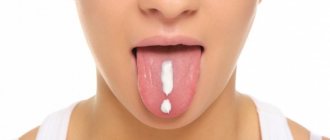
Stomatitis
Infection is introduced in different ways, most often as a result of mechanical damage to tissue. As it develops, characteristic round ulcers are formed, localized on the inside of the lips and cheeks, as well as in the area of the root of the tongue.
When diagnosing, it is recommended to treat the oral mucosa, having previously excluded products containing sodium lauryl sulfate from everyday use, since the active component provokes additional irritation. Anesthetics help relieve pain, and antiseptic and anti-inflammatory compounds are used to remove the film.
| — Why are diseases of the oral mucosa dangerous? — Diagnosis of diseases of the oral mucosa — What provokes diseases of the oral mucosa? — Common diseases of the oral mucosa | — Herpetic stomatitis — Recurrent aphthous stomatitis — Leukoplakia — Lichen planus |
Why are diseases of the oral mucosa dangerous?
In the process of eating, food begins to break down in the mouth, under the action of enzymes contained in saliva. Inflammation of the soft tissues of the oral cavity leads to impaired fermentation, which causes problems in the gastrointestinal tract.
Diseases of the oral mucosa have the form of purulent formations that cause itching, burning, and aching pain. They are often accompanied by halitosis. Halitosis is bad breath that brushing your teeth cannot prevent.
The inflammatory process that damages the mucous membrane and soft tissues is caused by poor oral hygiene, smoking (due to the high tar content), abuse of alcohol, hot foods and sweets, as well as increased acidity in the oral cavity.
Such deviations and bad habits create favorable conditions for the development of pathogenic bacteria, and increased acidity irritates the mucous membrane due to the reaction of the acid to the alkaline environment of the oral cavity.
Diagnosis of diseases of the oral mucosa
The variety of types of diseases of the oral mucosa creates certain difficulties for diagnosis. Since the same symptoms can often serve as indicators of completely different diseases, an accurate diagnosis is often the result of long-term observations of the dynamics of the disease.
Methods for studying and diagnosing diseases of the oral mucosa are examination, observation, smears, scrapings. Bacterial cultures and allergy tests.
It is very important to take into account the fact that any inflammatory process in the body (and diseases of the respiratory system are nothing more than an inflammatory process) is a factor that provokes the development of cancer.
The patient can be advised to trust an experienced specialist, be patient and scrupulously follow all the recommendations and prescriptions of the attending physician while waiting for a correct diagnosis, and then take all possible measures to, with the help of a highly qualified dentist, fully and completely recover and avoid consequences.
What causes diseases of the mucous membranes?
An impressive list of reasons is involved in the emergence of diseases of the oral mucosa:
- diabetes mellitus: high blood sugar levels cause decay of mucous tissues, and low levels cause bleeding wounds;
- lack of calcium, phosphorus, fluorine makes tooth enamel and pulp capillaries fragile;
- colds of various etiologies;
- some types of bacteria, fungi and viruses;
- low hemoglobin due to iron deficiency;
- decreased immunity;
- lack of oxygen in soft tissues;
- vitamin deficiency, which destroys capillary walls and leads to the appearance of microthrombi in soft tissues;
- immune and autoimmune diseases (arthritis, HIV);
- vein diseases;
- cancer;
- allergic reactions;
- stress.
The most common diseases of the oral mucosa.
Among the variety of oral mucositis diseases, dentists highlight those that occur most frequently.
The most common are: acute and chronic herpetic stomatitis, recurrent aphthous stomatitis, leukoplakia, lichen planus, trauma to the oral mucosa.
The most common disease of the oral mucosa is herpetic stomatitis
. It progresses in the autumn-winter periods, when the body is susceptible to hypothermia and suffers from changes in temperature.
With herpetic stomatitis, the infection is transmitted by both non-contact airborne droplets and contact. The causative agent of the disease is the herpes virus. The disease is contagious and easily transmitted.
Visually, the disease manifests itself as rashes on the face, especially around the mouth, and on the oral mucosa in the form of single or grouped small blisters (papules) filled with exudate.
Over time, the contents of the vesicle become cloudy, it bursts and an aphtha is formed - a small ulceration of the mucous membrane. In the lip area, the blisters dry out, forming hemorrhagic crusts.
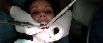
For treatment to be effective, patients must urgently consult a doctor who will conduct an examination and additional diagnostics and prescribe antiviral, antihistamines and vitamins. Additionally, it is necessary to treat the affected elements with leukocyte interferon and perform oral baths with antiseptic solutions. In case of severe pain, the oral cavity can be treated with local anesthetics as prescribed by the dentist. On the fifth day, it is recommended to apply an oil solution of vitamin A, rosehip oil or dental solcoseryl to the aphthae.
To relieve pain, prevent new rashes and shorten the healing time of already formed lesions, experts recommend the procedure of biostimulation with a diode laser at a low power therapeutic mode in the dentist's chair. The procedure is comfortable for the patient, does not require pain relief, and lasts four minutes.
It is also necessary for the first three days after the rash to exclude all planned manipulations in the oral cavity for dental treatment to prevent damage to adjacent areas of the patient’s mucosa and infection of medical personnel.
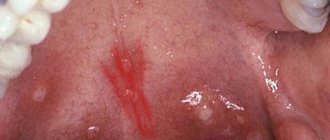
Erosion on the mucous membrane of the lower lip after opening of the vesicles.
Herpetic eruptions on the skin of the lower lip.
Recurrent aphthous stomatitis
accompanied by constant aching pain in the oral cavity, aggravated by speech and chewing loads. Aphthous stomatitis is an infectious-allergic, bacterial disease.
The causes contributing to the occurrence of aphthous stomatitis include atopy, trauma, endocrinopathy, diseases of the gastrointestinal tract, malnutrition, stress, and food allergies.
The formation of painful recurrent aphthae is preceded by the appearance of a pinhead-sized area of mucosal necrosis in the subcutaneous layer on a reddish base.
Gradually, the focus of necrosis increases, and the aphtha acquires a typical appearance: a round or oval epithelial defect measuring 0.3-0.5 cm, located in the inflamed area. The aphthae is covered with purulent plaque. Most often, aphthae are single and form once or twice a year. Small aphthae heal in two weeks on their own and do not leave scars.
It is impossible to effectively cure aphthous stomatitis, but some improvement is still observed when local anti-inflammatory drugs, Tantum Verde, Cholisal, as well as epithelium-restoring drugs, for example, dental solcoseryl, are applied to the lesion.
Good results are obtained by a single treatment of aphthae with a diode laser in therapeutic mode. Patients who underwent this procedure noted a decrease in pain the very next day after treatment, and the healing time of ulcers was reduced.
The positive effect of the laser is associated with its biostimulating properties. It consists in normalizing trophism and metabolic processes in the tissues of the working state of capillaries, improving microcirculation and regeneration.
Aphtha surrounded by a belt of hyperemia.
Afta before laser treatment.
Afta after laser treatment.
Leukoplakia
It is a white plaque on the oral mucosa that cannot be separated by friction. The disease is more common in men aged 45 to 65 years.
Leukoplakia develops against the background of trauma (sharp edges of carious teeth, overhanging edges of fillings, pathological occlusion), galvanosis, nutritional disorders, excessive consumption of spices, alcohol, smoking, adverse meteorological factors, hormonal imbalance, vitamin deficiency, gastritis, colitis, diabetes mellitus, hereditary and other unfavorable factors.
The size, location of the affected area, and other symptoms may vary. Most often, leukoplakia is visible on the mucous membrane of the cheeks along the line of closure of the teeth, in the area of the corners of the mouth, on the back of the tongue, hard palate, and sometimes on the alveolar process.
The clear edges of the keratinizing spot do not protrude above the level of the surrounding tissue. The surface of the affected area can be smooth and uniform, thin and vulnerable, folded, warty, granular or spotty, and the color can be whitish, gray or brown.
Leukoplakia is benign in 80% of cases; in the rest, dysplasia is noted, that is, a precancerous condition, or transformation into cancer. Within five years, leukoplakia transforms into cancer in 4-6% of patients.
When treating leukoplakia, the causes of irritation of the mucous membrane (nicotine, tars, essential oils formed during the combustion of tobacco, spicy foods) are first eliminated. Sanitation of the oral cavity with subsequent clinical observation, diagnosis and treatment of diseases of internal organs that reduce the resistance of the oral mucosa to the effects of adverse external factors are mandatory. Local applications are made with a solution of retinol acetate and a solution of tocopherol. B vitamins are prescribed internally. A biopsy is performed if the disease does not subside.
Leukoplakia as a result of traumatic tooth brushing.
Leukoplakia of the floor of the mouth and lower surface of the tongue.
Lichen planus
– another common disease of the oral mucosa, accompanied by damage to the mucous membranes. This disease occurs as a result of immune disorders.
Those with an easily excitable nervous system, as well as patients with the hepatitis C virus, are especially predisposed to this disease. Most patients are women over 40 years old. Lichen planus is characterized by a long course of the disease with remissions and exacerbations.
In patients with characteristic dark red, irregularly shaped, itchy nodules on the skin, lesions of the oral mucosa are usually detected.
The typical form is expressed by small grayish nodules on the unchanged mucous membrane, merging into a mesh. Patients complain of roughness, tightness of the oral mucosa and pain.
Treatment of the disease depends on the form. In the typical form, a change in the usual lifestyle is recommended, since eliminating the causes of stress can significantly alleviate the disease. In the chronic erosive form, the effect is achieved by local use of glucocorticoids and epithelizing agents (vitamin A oil solution). Complex treatment of the disease includes tranquilizers, sedatives, immunocorrective therapy, and vitamins. Correction of personal hygiene, sanitation of the oral cavity, elimination of galvanosis and mechanical traumatic factors are mandatory.
Typical form of lichen planus.
Council of the dentist of the Family Dentistry Network "RODNIA".
As soon as you experience discomfort in your mouth in the form of burning, itching and pain, you should immediately consult a dentist, as there is a high risk that you may have herpetic rashes, and herpes is easily transmitted to loved ones, especially children. Parents kiss their children, drink from the same cup, use the same towel, and the infection is transmitted very easily.
The consequences of untreated inflammatory diseases of the oral cavity are loss of teeth, spread of inflammation to the organs of the gastrointestinal tract and upper respiratory tract, and oncology.
Only a doctor will be able to carry out the necessary diagnostics, establish the cause, make the correct diagnosis and prescribe adequate treatment.
Treatment of oral diseases in the RODNYA Family Dentistry Network is performed by periodontists and dental surgeons:
Dentists-therapists of the Family Dentistry Network "RODNIA": | ||
| Mukhambetova Elena Nikolaevna Counterpart, Astrodent | Podarvanova Svetlana Vasilievna Counterparts | Volkolup Ekaterina Igorevna Astrodent |
| Morozov David Iskandyarovich Counterpart, Astrodent | Nogina Anna Yurievna Counterparts | Hovhannisyan Teresa Lazarevna T-med clinic |
| Aleksanyan Hrachya Karapetovich T-med clinic | Baishev Marat Failievich Smile aesthetics | Sargsyan Ruzanna Vladimirovna Rimma-Dent |
| Tarvi Ekaterina Vladimirovna Rimma-Dent | ||
Welcome to RODNYA! Make an appointment (calls from any phone number are free)